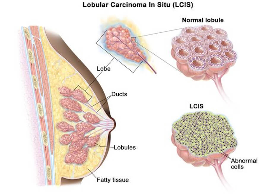
Lobular carcinoma in situ (LCIS) is a non-invasive neoplastic proliferation of dyscohesive epithelial cells within the lobules and terminal duct lobular units (TDLUs) of the breast. Unlike ductal carcinoma in situ (DCIS), LCIS is not considered a true cancer but rather a high-risk indicator lesion and a non-obligate precursor to invasive breast cancer. It is typically asymptomatic, usually discovered incidentally in breast biopsies performed for other reasons, and is associated with a substantially elevated lifetime risk of developing invasive breast cancer in either breast. The management of LCIS has evolved significantly, shifting away from surgical excision toward surveillance, risk assessment, and chemoprevention, with treatment decisions heavily dependent on the histologic subtype and individual patient risk factors.
📋 ICD-10 Classification
In the International Classification of Diseases, Tenth Revision (ICD-10), LCIS is classified under D05.0 — Lobular carcinoma in situ of breast. The category is further specified by laterality: D05.00 (unspecified breast), D05.01 (right breast), and D05.02 (left breast). The parent code D05 encompasses carcinoma in situ of the breast, which also includes intraductal carcinoma in situ (DCIS, D05.1) and other specified types (D05.7). In the WHO Classification of Tumours, LCIS is grouped under the umbrella term lobular neoplasia, which also includes atypical lobular hyperplasia (ALH). The term lobular intraepithelial neoplasia (LIN) has been proposed as an alternative classification, though it is not yet universally adopted.
🧬 What Causes LCIS?
The exact cause of LCIS remains incompletely understood, but it is fundamentally driven by the loss of E-cadherin expression, a key cell adhesion protein encoded by the CDH1 gene on chromosome 16q22.1. This loss of E-cadherin leads to the dyscohesive growth pattern characteristic of lobular lesions and is a defining molecular feature that distinguishes LCIS from ductal lesions. The pathogenesis involves additional genetic alterations, including loss of heterozygosity at 16q and amplification of 1q, which are early events in the progression toward invasive lobular carcinoma (ILC).
Risk factors for LCIS include a family history of breast cancer, BRCA1 and BRCA2 pathogenic variants, older age, and hormonal factors such as exposure to combined hormone replacement therapy. LCIS is almost exclusively a lesion of the female breast and is typically diagnosed in women around the time of menopause, with a mean age of approximately 50 years for classic LCIS and slightly older for variant forms. The overall incidence of LCIS in the United States is estimated at approximately 3.19 per 100,000 women.
🛡️ How to Avoid and Prevent LCIS
Because LCIS is primarily a risk marker rather than a direct precursor in most cases, prevention strategies focus on reducing the subsequent risk of invasive breast cancer. Risk-reducing lifestyle modifications—including maintaining a healthy body weight, limiting alcohol intake, and engaging in regular physical activity—are generally recommended for overall breast health, though their specific impact on LCIS progression is not precisely quantified. For women with LCIS, chemoprevention is the most evidence-based risk-reduction strategy. Tamoxifen (20 mg daily for 5 years) is recommended by the American Society of Clinical Oncology (ASCO) for premenopausal women with LCIS to reduce the risk of estrogen receptor-positive invasive breast cancer. Raloxifene and aromatase inhibitors (such as anastrozole or exemestane) are options for postmenopausal women. Chemoprevention has been shown to reduce the risk of developing invasive breast cancer by approximately 56% in women with LCIS. Bilateral prophylactic mastectomy is a more aggressive risk-reducing option that reduces breast cancer risk by approximately 90%, but it is typically reserved for women at very high risk (e.g., BRCA mutation carriers) and is not routinely recommended for LCIS alone.
🔬 Types of LCIS
LCIS is classified into three main histologic subtypes, which differ in their clinical behavior and management:
Classic LCIS (C-LCIS) is the most common form, characterized by small, uniform cells (type A) or slightly larger cells with mild atypia (type B) filling and distending the acini of the TDLU. It is typically an incidental finding, often multifocal (in 90% of cases in some series) and bilateral (in 20–60% of cases). Classic LCIS carries a 7- to 12-fold increased risk of subsequent invasive breast cancer, but the risk is equally distributed between both breasts, and the lesion itself does not require surgical excision with negative margins. The upgrade rate to invasive cancer or DCIS upon excision is approximately 4–16%.
Florid LCIS (F-LCIS) is a non-classical variant characterized by markedly distended acini and ducts with little intervening stroma, sometimes forming a mass-like architecture. It often shows comedonecrosis and calcifications, and it has a higher upgrade rate (approximately 33%) and a stronger association with invasive carcinoma compared to classic LCIS. The mean age at diagnosis is in the late 50s to early 60s.
Pleomorphic LCIS (P-LCIS) is another non-classical variant characterized by significant cytologic atypia and pleomorphism, resembling high-grade DCIS. It is frequently associated with microcalcifications and is detected on screening mammography. P-LCIS has an upgrade rate of approximately 35% and is considered a more aggressive lesion, with a higher risk of progression to invasive lobular carcinoma. Both florid and pleomorphic LCIS are managed more aggressively than classic LCIS, often with surgical excision to clear margins, and they are classified as B5a lesions (high-risk) in the AGO guidelines.
🩺 Symptoms and Early Signs
LCIS is almost always asymptomatic and does not produce a palpable mass, skin changes, or nipple discharge in the vast majority of cases. It is most commonly discovered incidentally during a breast biopsy performed for another indication, such as a mammographic abnormality or a palpable mass that turns out to be a different lesion. When symptoms do occur—such as bloody nipple discharge or a red, swollen breast—they are typically attributable to a concurrent invasive cancer rather than to LCIS itself. On mammography, classic LCIS is usually not visible or appears as nonspecific findings; however, the variant forms (florid and pleomorphic LCIS) may present with suspicious microcalcifications, a mass, or architectural distortion, making them detectable on screening.
🖥️ Diagnosis: Imaging and Biopsy
The diagnosis of LCIS is histologic and requires tissue sampling, as imaging alone cannot reliably identify the lesion. Mammography is the primary screening modality, but its sensitivity for classic LCIS is low; the lesion is often mammographically occult or associated with incidental calcifications. Digital breast tomosynthesis (3D mammography) may improve detection of associated calcifications, with a reported sensitivity of approximately 85% for some lesions. Breast ultrasound is frequently used as an adjunct to evaluate mammographic abnormalities and to guide biopsies, but it is not a primary diagnostic tool for LCIS. Breast MRI has high sensitivity for lobular lesions and is considered by some to be the imaging modality of choice for evaluating the extent of LCIS, particularly when associated with invasive lobular carcinoma; on MRI, LCIS most commonly appears as non-mass enhancement.
Core needle biopsy (stereotactic or ultrasound-guided) is the standard diagnostic procedure. The pathology report should confirm the diagnosis and specify the subtype (classic, florid, or pleomorphic) because this determines management. Immunohistochemistry is essential: LCIS characteristically shows loss of E-cadherin expression (sensitivity 94%, specificity 98% for distinguishing lobular from ductal lesions) and positive p120 catenin staining. Hormone receptor testing typically shows ER positivity in 80–100% of cases, while HER2 amplification is rare in classic LCIS (0–11%) but more common in pleomorphic LCIS (up to 31–41%). When LCIS is found on core biopsy, surgical excision is recommended to rule out an adjacent invasive component or DCIS, particularly for non-classical variants or when imaging and pathology are discordant.
💊 Treatment Types
The management of LCIS is individualized and differs fundamentally from that of DCIS. Classic LCIS does not require surgical excision with negative margins because it is a marker of bilateral risk rather than a focal lesion that progresses locally. Surveillance is the standard of care for classic LCIS, consisting of clinical breast examination every 6–12 months and annual mammography (with consideration of MRI in high-risk patients). For florid and pleomorphic LCIS, surgical excision with negative margins is recommended by NCCN guidelines due to their higher upgrade rates and association with invasive carcinoma.
Chemoprevention with tamoxifen (for premenopausal women) or raloxifene or aromatase inhibitors (for postmenopausal women) should be discussed with all patients with LCIS to reduce the risk of subsequent invasive breast cancer. Tamoxifen 20 mg daily for 5 years is the most established regimen, and low-dose tamoxifen (5 mg daily) is an emerging option with a better tolerability profile. Chemoprevention has been shown to reduce breast cancer risk by approximately 50–56%.
Bilateral prophylactic mastectomy is an option for a small subset of women with LCIS who are at very high risk (e.g., BRCA carriers) or who have severe cancer-related anxiety. It reduces breast cancer risk by approximately 90%, but it is not routinely recommended for LCIS alone and should be preceded by thorough counseling about risks, benefits, and impact on quality of life. Radiation therapy and chemotherapy have no role in the treatment of pure LCIS, as the lesion is non-invasive and does not require cytotoxic or locoregional therapy beyond excision for variant subtypes.
📊 Stages and Metastasis
LCIS is classified as Stage 0 (Tis, N0, M0) in the TNM staging system, identical to DCIS, because it is a non-invasive lesion. However, the clinical meaning of Stage 0 differs: LCIS is not a direct precursor that inevitably progresses to cancer at the same site, but rather a risk factor for developing invasive breast cancer in either breast over time. Pure LCIS cannot metastasize because the neoplastic cells are confined within the lobules and terminal ducts and have not breached the basement membrane to access lymphatic or vascular channels. When invasive breast cancer subsequently develops in a woman with LCIS, it is most often invasive ductal carcinoma (IDC) rather than invasive lobular carcinoma (ILC), despite the lobular origin of the risk marker; the cumulative risk of invasive cancer is approximately 1% per year, reaching 25–35% at 20–30 years after diagnosis.
📈 Cure Rate, Survival Rate, and Life Expectancy
The prognosis for women diagnosed with LCIS is excellent. LCIS itself is not a life-threatening condition, and the 10-year breast cancer-specific survival (BCSS) is 98.9%, while the 20-year BCSS is 96.3%. Population-based data from the SEER database show estimated overall survival rates of 99.7% at 1 year, 96.7% at 5 years, and 91.7% at 10 years (reflecting competing causes of mortality in an older population). The life expectancy for a woman diagnosed with LCIS at age 50 is estimated at approximately 32.8 years, and interventions such as chemoprevention or prophylactic mastectomy provide only modest gains in life expectancy (on the order of months) while potentially reducing quality-adjusted life years. Without treatment, the natural history is one of elevated but non-obligate risk: most women with LCIS will not develop invasive breast cancer, but the risk persists for decades and is bilateral, necessitating lifelong surveillance.
🕊️ Palliative Care
Palliative care is not applicable to pure LCIS, as it is a non-invasive, non-life-limiting risk lesion. Palliative care is relevant only if a woman with a history of LCIS subsequently develops metastatic invasive breast cancer, at which point symptom management and supportive care would be integrated according to the stage and biology of the invasive disease. For women living with LCIS, supportive care—including psychological support to address cancer-related anxiety, counseling about risk-reduction strategies, and shared decision-making about surveillance and chemoprevention—is an important component of comprehensive care.
❓ Five Questions and Answers
Basics, Terminology, & Biology
1. What is Lobular Carcinoma In Situ (LCIS)? LCIS is a non-invasive neoplastic proliferation of dyscohesive cells within the lobules and terminal duct lobular units (TDLUs) of the breast; it is best understood as a risk indicator and non-obligate precursor rather than a true cancer.
2. Is LCIS considered true cancer or a benign non-cancerous condition? LCIS is not true cancer; it is a neoplastic proliferation that serves as a marker of increased risk for developing invasive carcinoma in either breast.
3. Why does LCIS have the word “carcinoma” in its name if it is not invasive cancer? The term is historical; the cells appear malignant cytologically and fill the lobular acini, but they remain confined by the basement membrane and do not invade.
4. What does “in situ” mean in pathology? “In situ” means the neoplastic cells are confined to their site of origin and have not breached the basement membrane to invade surrounding tissue.
5. Where in the breast does LCIS originate (milk-producing lobules)? LCIS originates in the terminal duct lobular units (TDLUs), which are the milk-producing lobules and their terminal ducts.
6. What is the difference between LCIS and Ductal Carcinoma In Situ (DCIS)? DCIS is a non-invasive cancer confined to ducts and is a direct precursor treated with surgery; LCIS involves lobules, is usually an incidental risk marker, and is managed with surveillance for classical subtypes.
7. Why is DCIS managed as a non-invasive cancer precursor, whereas LCIS is managed primarily as a risk marker? DCIS has a higher and more direct progression risk to invasive cancer at the same site; classical LCIS increases risk equally in both breasts and often does not progress to cancer at the biopsy site.
8. What is the difference between LCIS and Invasive Lobular Carcinoma (ILC)? LCIS is non-invasive and confined to lobules; ILC has broken through the basement membrane and invaded breast stroma.
9. Does LCIS directly turn into invasive lobular carcinoma? LCIS is a non-obligate precursor; it can be clonally related to subsequent ILC, but most LCIS does not progress directly, and invasive cancer often develops elsewhere.
10. Is LCIS a precursor lesion, a non-obligate precursor, or a risk factor for both breasts? It is best considered a non-obligate precursor and a risk factor that elevates risk in both breasts.
11. What is Lobular Neoplasia? Lobular neoplasia is an umbrella term encompassing the spectrum of atypical lobular hyperplasia (ALH) and LCIS.
12. What is the difference between Atypical Lobular Hyperplasia (ALH) and LCIS? ALH partially involves a lobule, whereas LCIS fills and distends the entire lobule.
13. How do pathologists distinguish ALH from LCIS under a microscope (based on lobular unit distension)? LCIS fills and expands >50% of the acini in a TDLU; ALH involves only a portion.
14. What is the Page classification system for lobular neoplasia? The Page classification distinguishes ALH from LCIS based on the extent of lobular involvement and distension.
15. What is the WHO classification for LCIS? The WHO classifies LCIS under lobular neoplasia and recognizes classic, pleomorphic, and florid subtypes.
16. How common is LCIS in the general population? LCIS is found in approximately 0.5–3.6% of otherwise benign breast biopsies.
17. What percentage of routine breast biopsies yield an unexpected finding of LCIS (~1–4%)? About 1–4% of biopsies performed for other indications reveal incidental LCIS.
18. What is the cell of origin in LCIS? The cell of origin is the epithelial cell of the terminal duct lobular unit, which has lost E-cadherin–mediated cohesion.
19. What is E-cadherin (CDH1), and why is its loss central to LCIS biology? E-cadherin is a calcium-dependent cell adhesion molecule; its loss, due to CDH1 inactivation, causes dyscohesion and is the defining molecular feature of lobular lesions.
20. How does complete loss of membrane E-cadherin cause LCIS cells to become non-cohesive? Without E-cadherin, cells cannot adhere to one another, leading to the dyscohesive growth pattern characteristic of LCIS.
21. What immunohistochemistry (IHC) stain confirms E-cadherin negativity? E-cadherin IHC shows absent or markedly reduced membrane staining in >95% of LCIS cases.
22. What is p120-catenin, and how does its cytoplasmic translocation confirm a lobular phenotype? p120-catenin normally binds E-cadherin at the membrane; when E-cadherin is lost, p120 shifts to the cytoplasm, confirming lobular differentiation.
23. What are catenins (alpha, beta, gamma) and their role in cell-cell adhesion? Catenins link E-cadherin to the actin cytoskeleton; their loss alongside E-cadherin contributes to dyscohesion.
24. What is Classical LCIS (Classic LCIS)? Classic LCIS is the most common subtype, composed of small, uniform, dyscohesive cells filling and distending lobular acini.
25. What do classical LCIS cells look like under the microscope (small, uniform, round, discohesive)? They are monomorphic, small, round cells that do not stick together, with type A (hyperchromatic) or type B (vesicular) nuclei.
26. What is Florid LCIS (FLCIS)? Florid LCIS is a nonclassic variant with marked distension of ducts and lobules, often forming a mass-like architecture, and may show central comedo-like necrosis.
27. What features define Florid LCIS (marked distension of lobules, central comedo-like necrosis)? Marked lobular distension, confluence, and central necrosis with calcifications.
28. What is Pleomorphic LCIS (PLCIS)? Pleomorphic LCIS is a nonclassic variant with large cells showing marked nuclear pleomorphism and prominent nucleoli, resembling high-grade DCIS morphologically.
29. What features define Pleomorphic LCIS (large, pleomorphic nuclei, prominent nucleoli)? Significant nuclear atypia, enlarged cell size, and prominent nucleoli.
30. Why is Pleomorphic LCIS considered biologically more aggressive than classical LCIS? It has higher upgrade rates to invasive cancer, more frequent HER2 amplification, and is managed more like DCIS.
31. What is Apocrine LCIS? Apocrine LCIS is a rare variant with cells showing abundant eosinophilic cytoplasm and androgen receptor positivity.
32. What is Signet-Ring Cell LCIS? A rare variant with cells containing large cytoplasmic vacuoles that push the nucleus to the periphery.
33. How does Pleomorphic LCIS resemble High-Grade DCIS? Both show marked nuclear atypia and pleomorphism; E-cadherin negativity distinguishes PLCIS from DCIS.
34. Why is Pleomorphic LCIS often managed more like DCIS (with surgical excision and clear margins)? Because of its high upgrade rate (approximately 35%) and association with invasive carcinoma.
35. Are classical LCIS cells hormone receptor-positive? Yes, classical LCIS is typically strongly ER-positive.
36. What percentage of LCIS cases are Estrogen Receptor-positive (ER+) (~90–95%)? Approximately 90–95% of classical LCIS cases are ER-positive.
37. What percentage of LCIS cases are Progesterone Receptor-positive (PR+)? Most ER-positive LCIS cases are also PR-positive, though rates vary.
38. What is HER2 status in classical LCIS (typically HER2-negative)? Classical LCIS is typically HER2-negative.
39. What is HER2 status in pleomorphic LCIS (often HER2-positive or overexpressed)? Pleomorphic LCIS frequently shows HER2 amplification or overexpression.
40. What is the Ki-67 proliferation index in classical LCIS (typically low, <5–10%)? Ki-67 is typically low in classical LCIS.
41. What is the Ki-67 index in pleomorphic LCIS (often elevated)? Ki-67 is often elevated in pleomorphic LCIS.
42. Is classical LCIS considered a fast-growing condition? No; classical LCIS is generally slow-growing and indolent.
43. Can LCIS cells cross the basement membrane of the lobule? No; by definition, LCIS cells remain confined within the lobule and do not cross the basement membrane.
44. Why can pure LCIS NOT spread to lymph nodes or distant organs? Because the cells are confined by the basement membrane and lack access to lymphatic or vascular channels.
45. What happens if LCIS is found alongside invasive breast cancer? The invasive cancer dictates staging and treatment; LCIS is noted as an associated finding.
46. What happens if LCIS is found alongside DCIS? Management is driven by the DCIS, which is a more direct precursor requiring surgical clearance.
47. Is LCIS more commonly bilateral (present in both breasts) than DCIS? Yes; LCIS is frequently bilateral (20–60% of cases), whereas DCIS is usually unilateral.
48. What percentage of women with LCIS have bilateral involvement (~30–50%)? Bilateral involvement is reported in approximately 30–50% of cases.
49. Is LCIS often multifocal and multicentric within the same breast? Yes; multifocality is common, reported in up to 85–90% of cases in some series.
50. What is terminal duct lobular unit (TDLU) involvement in LCIS? LCIS fills and distends the acini of the TDLU, which is the functional milk-producing unit.
51. What is pagetoid spread of LCIS into adjacent ducts? It is the extension of LCIS cells along ductal epithelium without invasion.
52. How do pathologists distinguish pagetoid LCIS from DCIS in a milk duct? E-cadherin and p120 IHC help confirm lobular lineage despite intraductal spread.
53. Does classical LCIS cause microcalcifications inside the lobule? Classical LCIS usually does not produce calcifications; calcifications are more common in florid and pleomorphic variants.
54. Why do Florid and Pleomorphic LCIS frequently produce microcalcifications, unlike classical LCIS? Central comedo-like necrosis and cellular debris calcify, similar to high-grade DCIS.
55. What germline gene mutations are linked to LCIS (CDH1, BRCA2, CHEK2)? Pathogenic variants in CDH1, BRCA2, CHEK2, and PALB2 have been associated with lobular neoplasia and ILC.
56. What is Hereditary Diffuse Gastric Cancer (HDGC) syndrome, and how does it link to LCIS? HDGC is caused by CDH1 mutations and is associated with lobular breast cancer and LCIS.
57. What is the somatic mutation landscape of LCIS (PIK3CA, CBFB, RUNX1)? Somatic alterations commonly involve PIK3CA, CBFB, and RUNX1, among others.
58. Why are PIK3CA mutations common in both LCIS and ILC? PIK3CA mutations are early events in lobular neoplasia and persist in invasive lobular carcinoma.
59. What is the clonal relationship between LCIS and subsequent invasive lobular carcinoma? Synchronous LCIS and ILC are often clonally related, supporting LCIS as a non-obligate precursor.
60. Is the invasive cancer that develops after LCIS always lobular, or can it be ductal? It can be either; subsequent invasive cancers are often ductal, despite the lobular risk marker.
61. What percentage of subsequent invasive cancers following LCIS are ductal vs lobular (~50% IDC, ~50% ILC)? Approximately half are invasive ductal and half are invasive lobular.
62. Does an invasive cancer always occur in the exact same breast where LCIS was found? No; the risk is bilateral and equal, so cancer can develop in either breast.
63. Why is LCIS called an “equal-opportunity” risk marker for both breasts? Because the increased risk applies equally to the ipsilateral and contralateral breast.
64. What is the average age at LCIS diagnosis (typically premenopausal women aged 40–50)? LCIS is often diagnosed in women around age 50, frequently before menopause.
65. Can postmenopausal women get LCIS? Yes; LCIS can be diagnosed at any age, including postmenopause.
66. Can men get Lobular Carcinoma In Situ? Male LCIS is extremely rare but can occur.
67. Why is male LCIS extremely rare (due to lack of mature lobular development in male breast anatomy)? The male breast lacks the mature lobular development that characterizes the female breast, making lobular lesions exceedingly uncommon.
68. Does LCIS cause physical tissue destruction or desmoplasia? No; LCIS does not incite a desmoplastic stromal response, which is why it is not palpable.
69. Why does lack of desmoplastic stromal response make LCIS impalpable? Without fibrosis and stromal reaction, LCIS does not form a firm mass and is usually microscopic.
70. What is flat epithelial atypia (FEA), and how does it relate to lobular neoplasia? FEA is a columnar cell lesion with mild atypia that can coexist with lobular neoplasia.
71. What is the “Rosen Triad” (tubular carcinoma, LCIS, and columnar cell lesions)? The Rosen triad is the pathological association of tubular carcinoma, columnar cell lesions, and LCIS.
72. How do estrogen and progesterone drive lobular cellular proliferation? Hormones stimulate ER-positive lobular cells, contributing to the high ER positivity of LCIS.
73. What is the role of the microenvironment in keeping LCIS confined within lobules? The intact basement membrane and myoepithelial cell layer contain the neoplastic cells.
74. What is the rate of apoptosis in classical LCIS cells? Apoptosis is generally low, contributing to the accumulation of cells within lobules.
75. Can LCIS disappear or regress spontaneously? Spontaneous regression is uncommon but has been reported.
76. Is LCIS graded like invasive cancer (Grade 1, 2, 3)? Classical LCIS is not graded using the Nottingham system; however, nuclear grade (low/intermediate/high) is described for variants.
77. Why is formal Nottingham grading NOT applied to classical LCIS? Because classical LCIS is not an invasive carcinoma, and its biology differs from invasive cancer.
78. How does loss of E-cadherin alter signaling through the Wnt/beta-catenin pathway? Loss of E-cadherin releases β-catenin, which can translocate to the nucleus and affect transcription.
79. What is p120-catenin’s role in activating RhoA GTPases when uncoupled from E-cadherin? Cytoplasmic p120 can modulate RhoA activity, affecting cell motility and adhesion.
80. Is LCIS considered a Stage 0 lesion in older TNM staging manuals? Historically LCIS was coded as Tis, but the AJCC 8th edition removed classical LCIS from cancer staging because it is a risk factor, not a true cancer.
81. Why did the AJCC 8th Edition Staging Manual remove classical LCIS from cancer staging tables? To reflect its status as a benign risk factor rather than a malignancy requiring cancer staging.
82. Why was classical LCIS removed from AJCC cancer staging (to reflect its status as a benign risk factor, not true cancer)? The AJCC recognized that classical LCIS is not a direct precursor and should not be staged as cancer.
83. Does Pleomorphic LCIS remain in cancer staging discussions? Pleomorphic LCIS is managed more like DCIS and is often discussed in the context of non-invasive neoplastic lesions requiring excision.
84. What is the difference between incidental LCIS and symptomatic breast lesions? Incidental LCIS is found microscopically in tissue removed for other reasons and has no symptoms; symptomatic lesions (e.g., mass, discharge) are caused by other conditions.
85. What is the lifetime risk of invasive breast cancer for someone with ALH vs LCIS? ALH carries a moderately increased risk; LCIS carries a higher risk, often quoted as 20–30% lifetime.
86. How does the presence of mass-like florid LCIS change surgical management? Florid LCIS often requires surgical excision to rule out invasive cancer, similar to DCIS.
87. What is collagen density around lobules in LCIS-affected tissue? Collagen density is not a defining feature; LCIS lacks the desmoplastic reaction seen in invasive cancer.
88. What is the role of myoepithelial cells in maintaining the outer boundary of an LCIS lobule? Myoepithelial cells form the outer layer that contains the neoplastic cells within the lobule.
89. How do pathologists report the percentage of lobular acini filled by LCIS cells? They estimate the proportion of acini involved to distinguish ALH from LCIS.
90. What is “lobular distension” (filling and expanding >50% of acini in a lobular unit)? It is the criterion that defines LCIS versus ALH; >50% acinar involvement indicates LCIS.
91. What is the genomic stability of classical LCIS compared to DCIS? Classical LCIS is generally genetically stable with fewer copy number alterations than DCIS.
92. Why is classical LCIS considered genetically stable? It lacks the complex genomic instability seen in high-grade DCIS and invasive cancers.
93. What is copy number alteration (CNA) profile in pleomorphic LCIS? Pleomorphic LCIS shows more complex CNAs, including HER2 amplification and TP53 alterations.
94. Can LCIS co-exist with fibroadenomas or radial scars? Yes; LCIS can be found incidentally within fibroadenomas or adjacent to radial scars.
95. Is LCIS considered a systemic condition? Yes; LCIS is considered a field effect or systemic condition of the breast because it is often multifocal and bilateral.
96. What is the medical definition of lobular neoplasia? Lobular neoplasia is the spectrum of atypical lobular proliferation ranging from ALH to LCIS.
97. How do international guidelines (NCCN, EAU, ESMO) define LCIS? Guidelines uniformly define LCIS as a non-invasive lobular proliferation and risk marker, with management stratified by subtype.
98. What key biological features distinguish classical, florid, and pleomorphic LCIS? Classical: small uniform cells, low grade, low upgrade rate. Florid: marked distension, necrosis, intermediate upgrade risk. Pleomorphic: high-grade nuclei, HER2 amplification, highest upgrade risk.
99. Why is patient education crucial regarding the non-invasive, non-cancerous nature of LCIS? Because the term “carcinoma” causes significant anxiety, and understanding that LCIS is a risk marker empowers informed decisions and reduces overtreatment.
100. Where can patients read expert consensus statements on LCIS biology? NCCN guidelines, ASCO statements, and resources from the Lobular Breast Cancer Alliance provide expert consensus.
Signs, Symptoms, & Incidental Detection
101. What are the symptoms of Lobular Carcinoma In Situ? Classical LCIS is almost always asymptomatic and is discovered incidentally.
102. Does classical LCIS cause a breast lump? No; classical LCIS is non-palpable and does not form a mass.
103. Why is classical LCIS almost always asymptomatic? Because it is a microscopic proliferation confined to lobules without desmoplasia or mass formation.
104. Can LCIS cause breast pain or tenderness? No; pain is not a feature of LCIS.
105. Can LCIS cause nipple discharge? LCIS itself does not cause nipple discharge; discharge may be caused by other coexisting conditions.
106. Can LCIS cause skin dimpling, puckering, or redness? No; these signs suggest invasive cancer or other pathology, not LCIS.
107. Can LCIS cause nipple inversion or retraction? No; these are not features of LCIS.
108. Why is LCIS called an “incidental finding”? Because it is typically discovered during a biopsy performed for an unrelated mammographic abnormality or palpable lesion.
109. What does “incidental finding” mean on a biopsy report? It means the lesion was not the primary target of the biopsy and was found unexpectedly.
110. How is classical LCIS typically discovered (during a biopsy performed for an unrelated mammographic finding)? Most cases are found when a biopsy is done for microcalcifications or another imaging abnormality.
111. What unrelated findings prompt the biopsy that accidentally discovers LCIS (calcifications, fibroadenomas, cysts, architectural distortion)? Microcalcifications, masses, fibroadenomas, cysts, and architectural distortion are common triggers.
112. Can classical LCIS be seen on a standard 2D screening mammogram? Classical LCIS is usually mammographically occult and not visible on standard mammography.
113. Why is classical LCIS usually invisible on mammograms? Because it lacks calcifications and does not form a mass or architectural distortion.
114. Can Florid LCIS be detected on a mammogram? Yes; florid LCIS often produces calcifications and may be visible on mammography.
115. Why does Florid LCIS show up on mammograms (due to central comedo calcifications)? Central necrosis and calcification within distended lobules create mammographic microcalcifications.
116. Can Pleomorphic LCIS be detected on a mammogram? Yes; pleomorphic LCIS frequently presents with suspicious microcalcifications.
117. What pattern of microcalcifications suggests Pleomorphic LCIS on imaging? Fine pleomorphic or linear branching calcifications, similar to DCIS.
118. Can classical LCIS be detected on a breast ultrasound? No; classical LCIS is generally not visible on ultrasound.
119. Can classical LCIS be detected on a contrast-enhanced breast MRI? Classical LCIS is usually occult on MRI, though it may show subtle non-mass enhancement in some cases.
120. What does LCIS look like on breast MRI when visible (subtle non-mass enhancement)? When visible, it appears as subtle non-mass enhancement.
121. What percentage of classical LCIS cases are completely occult on all imaging modalities? The vast majority are occult on all imaging; detection is usually incidental on biopsy.
122. Can a doctor feel LCIS during a clinical breast exam (CBE)? No; classical LCIS is not palpable.
123. Can a woman feel LCIS during a breast self-exam? No; LCIS cannot be felt.
124. What happens if a woman feels a lump and the biopsy shows ONLY classical LCIS? This represents imaging-pathology discordance and generally requires surgical excision to rule out an adjacent missed invasive tumor.
125. What is imaging-pathology discordance? Discordance occurs when the imaging findings (e.g., a mass) do not match the pathology (e.g., only classical LCIS), suggesting a possible sampling error.
126. Why does finding ONLY classical LCIS on a biopsy for a mass require surgical excision (to rule out an adjacent missed invasive tumor)? Because a mass is atypical for classical LCIS, and an invasive cancer may have been missed.
127. What non-cancerous breast conditions cause lumps that lead to accidental LCIS discovery? Fibroadenomas, cysts, fat necrosis, and fibrocystic changes can prompt biopsy and lead to incidental LCIS.
128. How does dense breast tissue impact the incidental discovery of LCIS? Dense tissue lowers mammographic sensitivity and may lead to more biopsies, incidentally increasing LCIS detection.
129. Does breast density hide LCIS, or does it lead to more frequent screening biopsies? Both; density hides lesions but also prompts supplemental screening and biopsies that may reveal LCIS.
130. Can 3D Mammography (Digital Breast Tomosynthesis / DBT) directly detect classical LCIS? DBT does not directly detect classical LCIS; it improves visualization of calcifications that may lead to biopsy.
131. Does Contrast-Enhanced Mammography (CEM) show classic LCIS? CEM is not typically used for classical LCIS and would not reliably show it.
132. What is Molecular Breast Imaging (MBI), and does LCIS show tracer uptake? MBI uses a radioactive tracer; LCIS generally does not show significant uptake.
133. Can LCIS cause swollen axillary lymph nodes? No; pure LCIS cannot metastasize to lymph nodes.
134. Why does pure LCIS NEVER cause lymph node swelling? Because the cells are confined to lobules and have no access to lymphatic channels.
135. What does it mean if a woman with LCIS has a swollen lymph node (unrelated infection or hidden invasive cancer)? It warrants evaluation for infection or an occult invasive cancer.
136. Can LCIS cause changes in breast shape or size? No; LCIS does not alter breast shape or size.
137. Can LCIS cause skin thickening or edema? No; these are signs of inflammatory or locally advanced cancer, not LCIS.
138. Does LCIS cause itching or scaling of the nipple/areola? No; these are features of Paget’s disease, not LCIS.
139. How do symptoms of Paget’s disease differ from LCIS? Paget’s disease causes an eczema-like nipple rash; LCIS is asymptomatic.
140. How do symptoms of inflammatory breast cancer differ from LCIS? Inflammatory breast cancer causes rapid redness, swelling, and peau d’orange; LCIS has no such symptoms.
141. What BI-RADS scores are associated with mammograms that lead to an LCIS diagnosis (usually BI-RADS 4)? LCIS is often found in biopsies performed for BI-RADS 4 calcifications.
142. What is BI-RADS 4A, 4B, and 4C? They indicate increasing levels of suspicion for malignancy within the BI-RADS 4 category.
143. What is the likelihood of finding LCIS when a biopsy is done for BI-RADS 4 microcalcifications? LCIS is a relatively common incidental finding in biopsies for microcalcifications.
144. Can benign breast cysts contain LCIS in their walls? Yes; LCIS can be found incidentally in the wall of a cyst.
145. Can fibroadenomas harbor coincidental LCIS cells? Yes; LCIS can occur within or adjacent to fibroadenomas.
146. How often is LCIS found in breast tissue removed during reduction mammaplasty (cosmetic breast reduction)? LCIS is found in approximately 0.5–3.6% of reduction specimens.
147. What happens if LCIS is found incidentally in a breast reduction specimen? The patient is counseled about increased risk and enrolled in high-risk surveillance.
148. How often is LCIS found in prophylactic mastectomy specimens? LCIS is frequently found in prophylactic mastectomy specimens from high-risk women.
149. Does LCIS cause systemic symptoms like fatigue, weight loss, or fever? No; LCIS is a localized breast finding without systemic symptoms.
150. Can blood tests or tumor markers (CA 15-3, CEA) detect LCIS? No; tumor markers are not elevated in LCIS.
151. Why are circulating tumor cells (CTCs) absent in pure LCIS? Because LCIS is confined to lobules and does not shed cells into the bloodstream.
152. Can liquid biopsy (ctDNA) detect LCIS (no, because LCIS does not shed DNA into blood)? No; liquid biopsy cannot detect non-invasive LCIS.
153. What screening imaging brings premenopausal women in for biopsies that reveal LCIS? Screening mammography for microcalcifications is the most common trigger.
154. What imaging modalities are used to evaluate a palpable lump before biopsy? Diagnostic mammography and ultrasound.
155. Why is ultrasound guided core needle biopsy preferred for palpable masses? It allows real-time visualization and accurate sampling of the mass.
156. Why is stereotactic vacuum-assisted core biopsy used for microcalcifications? It precisely targets calcifications that are invisible on ultrasound.
157. What happens if microcalcifications are completely removed by the biopsy needle? A marker clip is placed, and the site may need surgical excision if indicated.
158. What is a metallic tissue marker clip, and why is it placed at the biopsy site? A clip marks the biopsy site for future imaging or surgery.
159. Can an allergic reaction to a biopsy clip mimic breast symptoms? Allergic reactions are rare but can cause local inflammation.
160. How long after a core needle biopsy do patients experience localized bruising or swelling? Bruising and swelling typically resolve within 1–2 weeks.
161. Can post-biopsy hematoma create a temporary palpable lump? Yes; a hematoma can feel like a lump and usually resolves.
162. How do doctors differentiate post-biopsy scar tissue from true breast lesions? Imaging and, if needed, repeat biopsy.
163. What physical signs require immediate evaluation after a breast biopsy? Expanding hematoma, severe pain, fever, or signs of infection.
164. What percentage of women diagnosed with LCIS had zero personal breast complaints prior to screening? The vast majority had no symptoms; LCIS is screen-detected or incidental.
165. How does age at presentation influence the clinical suspicion of accompanying lesions? Younger age may prompt more aggressive evaluation for genetic causes.
166. What steps are taken if a pregnant woman has an incidental finding of LCIS? Ultrasound-first evaluation and deferral of non-urgent surgery until after delivery.
167. Is breast ultrasound safe during pregnancy? Yes.
168. Is diagnostic mammography with abdominal shielding safe during pregnancy? Yes, when medically indicated.
169. How does lactating breast tissue alter mammographic and ultrasound interpretation? Lactation increases density and vascularity, making imaging more difficult to interpret.
170. Can nursing or lactation mask physical breast changes? Yes; lactation can obscure subtle findings.
171. What diagnostic steps are taken if a man presents with gynecomastia and LCIS is found? Evaluation for underlying risk factors and genetic counseling.
172. Why is incidental LCIS in men an extremely unexpected finding? Because the male breast lacks mature lobules, making LCIS exceedingly rare.
173. How does breast trauma or injury lead to incidental LCIS discovery? Trauma can cause fat necrosis that prompts biopsy, incidentally revealing LCIS.
174. Can fat necrosis from trauma look suspicious on mammograms and lead to a biopsy? Yes; fat necrosis can mimic malignancy.
175. Why does fat necrosis require biopsy confirmation to rule out malignancy? Because imaging features can overlap with cancer.
176. How do radiologists describe pleomorphic LCIS calcifications (fine pleomorphic, branched)? They describe them as fine pleomorphic or linear branching calcifications.
177. How do radiologists describe florid LCIS on mammograms (mass with central linear calcifications)? They may describe a mass-like density with central calcifications.
178. What is the discordance rate between core needle biopsy findings and imaging features in LCIS? Discordance is not rare and mandates excision when imaging suggests a mass.
179. Why is a multidisciplinary team review (radiologist, pathologist, surgeon) essential for every LCIS diagnosis? To ensure concordance and appropriate management.
180. What questions will a urologic or surgical oncologist ask during an initial LCIS consultation? They will ask about family history, genetic risk, symptoms, and imaging findings.
181. How should a patient prepare for their first post-biopsy consultation? Bring imaging discs, pathology reports, and a list of questions.
182. Why is bringing past mammogram discs and pathology slides to a second opinion important? It allows the second expert to review primary data.
183. How do doctors reassure anxious patients who mistakenly believe LCIS is an emergency? By explaining that classical LCIS is non-urgent and does not require immediate surgery.
184. Why does the term “carcinoma” create overwhelming anxiety in newly diagnosed women? Because “carcinoma” is synonymous with cancer, despite LCIS being non-invasive.
185. What educational materials help clarify that LCIS is not invasive cancer? Resources from MSKCC, ACS, and LBCA explain LCIS in plain language.
186. What is the role of breast self-awareness in women diagnosed with LCIS? It helps women notice new changes between scheduled imaging.
187. What physical breast changes should an LCIS patient monitor long-term? New lumps, skin changes, or nipple discharge.
188. How often should an LCIS patient perform self-breast exams? Monthly self-awareness is recommended, though LCIS itself is not palpable.
189. What specific red flag symptoms prompt an unscheduled diagnostic visit during surveillance? A new lump, skin dimpling, or bloody nipple discharge.
190. Can postmenopausal hormone changes cause breast tissue density shifts in women with LCIS? Yes; density generally decreases after menopause.
191. How does weight gain or loss affect clinical breast examination sensitivity? Obesity can make examination more difficult.
192. What percentage of LCIS diagnoses are made in women with dense breasts? Many LCIS cases are found in women with dense breasts, though LCIS itself is not density-dependent.
193. How do breast implants affect physical palpation and imaging in LCIS monitoring? Implants can obscure palpation and mammography; displacement views and MRI are used.
194. What are Eklund displacement views for mammograms with breast implants? Special views that push the implant aside to image breast tissue.
195. Can an ultrasound distinguish benign cysts from solid masses near an LCIS area? Yes; ultrasound can differentiate cystic from solid lesions.
196. Why is a core needle biopsy mandatory to distinguish LCIS from DCIS or ILC? Because imaging cannot reliably distinguish these entities; tissue architecture is required.
197. Can Fine Needle Aspiration (FNA) diagnose LCIS (no, FNA lacks tissue architecture)? No; FNA cannot distinguish in situ from invasive disease or LCIS from DCIS.
198. What is the single most critical fact about LCIS presentation? Classical LCIS has zero physical symptoms and is purely a microscopic finding.
199. Classical LCIS has zero physical symptoms and is purely a microscopic finding. Correct.
200. Where can patients find trusted diagnostic visual guides for non-cancerous breast conditions? Organizations like Breastcancer.org and the Lobular Breast Cancer Alliance provide educational resources.
Causes, Risk Factors, & Breast Cancer Risk
201. What causes Lobular Carcinoma In Situ? LCIS is caused by genetic alterations, most notably CDH1 inactivation leading to E-cadherin loss, combined with hormonal and reproductive risk factors.
202. Is LCIS caused by specific genetic mutations, lifestyle factors, or hormones? It is multifactorial; CDH1 mutations, hormonal exposure, and reproductive factors contribute.
203. How much does a diagnosis of LCIS increase future breast cancer risk? LCIS increases risk approximately 7- to 12-fold compared with the general population.
204. What is the relative risk (RR) of developing invasive breast cancer after LCIS (~7 to 12 times higher)? The relative risk is approximately 7–12.
205. What is the absolute risk (percentage) of developing breast cancer after LCIS (~20–30% lifetime risk)? The absolute lifetime risk is approximately 20–30%.
206. How does the risk of breast cancer after LCIS compare to an average woman (~12% lifetime risk)? LCIS confers a substantially higher risk than the average woman’s lifetime risk of about 12%.
207. What is the annual risk percentage of developing invasive breast cancer after LCIS (~1% to 2% per year)? The risk is approximately 1–2% per year.
208. Does the increased risk from LCIS ever plateau or go away after 10–20 years? The risk persists for decades and does not plateau.
209. What is the 5-year, 10-year, 15-year, and 20-year cumulative risk of breast cancer after LCIS (~7%, 11%, 16%, 20%)? Cumulative risk is approximately 10% at 10 years and 20% at 20 years.
210. Is the risk of future breast cancer equal in BOTH breasts after an LCIS diagnosis? Yes; the risk is equal in both breasts.
211. Why does LCIS in the left breast increase the risk of cancer in the right breast equally? Because LCIS is a field effect or systemic condition of the breast, not a focal lesion.
212. What types of invasive cancer develop after LCIS (invasive ductal carcinoma and invasive lobular carcinoma)? Both IDC and ILC can develop.
213. Are women with LCIS more likely to get invasive lobular cancer or invasive ductal cancer (~50% ILC, ~50% IDC)? Subsequent cancers are roughly equally divided between ductal and lobular.
214. Is age at LCIS diagnosis a key risk factor for future cancer? Yes; younger age at diagnosis confers a higher lifetime risk due to more remaining years of risk exposure.
215. Why do women diagnosed with LCIS before age 40 or 45 have a higher lifetime risk (due to more remaining life years)? Longer cumulative exposure to risk increases lifetime probability.
216. How does family history of breast cancer impact risk in a woman with LCIS? A positive family history further increases risk beyond LCIS alone.
217. Does having a first-degree relative with breast cancer double the risk for an LCIS patient? Family history adds to the already elevated risk from LCIS.
218. What genetic mutations significantly elevate risk alongside LCIS (BRCA1, BRCA2, CDH1, PALB2, CHEK2)? Pathogenic variants in CDH1, BRCA2, CHEK2, and PALB2 are associated with lobular neoplasia and increased risk.
219. How does a germline CDH1 mutation alter LCIS and invasive lobular cancer risk? CDH1 mutations predispose to lobular neoplasia and hereditary diffuse gastric cancer, with elevated lobular breast cancer risk.
220. Should all young women with LCIS undergo hereditary multi-gene panel testing? Genetic testing should be considered based on family history and other risk factors; not all young women require it.
221. What role does lifetime estrogen exposure play in causing LCIS? Prolonged estrogen exposure (early menarche, late menopause, nulliparity) increases risk.
222. Does early menarche (starting period before age 12) increase LCIS risk? Yes, via longer lifetime estrogen exposure.
223. Does late menopause (after age 55) increase LCIS risk? Yes.
224. Does nulliparity (never having children) raise LCIS risk? Yes.
225. Does older age at first childbirth (>30 years) raise LCIS risk? Yes.
226. Does breastfeeding lower the risk of developing LCIS or subsequent breast cancer? Breastfeeding is associated with modestly lower breast cancer risk.
227. How does Hormone Replacement Therapy (HRT) impact LCIS risk? Combined estrogen-progestin HRT may increase risk; the effect on LCIS specifically is less clear.
228. Does taking combined estrogen-progestin HRT for >3–5 years increase LCIS risk? Long-term combined HRT is associated with increased breast cancer risk.
229. Does HRT use after an LCIS diagnosis further elevate invasive breast cancer risk? HRT should be discussed with a clinician; it may further elevate risk.
230. Should women diagnosed with LCIS stop taking systemic HRT immediately? Decisions should be individualized with a healthcare provider.
231. Are oral contraceptives (birth control pills) linked to higher LCIS risk? Oral contraceptive use has not been consistently linked to LCIS.
232. Does bioidentical hormone therapy carry the same risks as conventional HRT for LCIS patients? Bioidentical hormones are not proven safer; risk depends on formulation and duration.
233. How does high breast density affect risk calculations in LCIS patients? Density is an independent risk factor and can compound risk.
234. Does dense breast tissue combined with LCIS create a multiplicative risk for breast cancer? Both increase risk, and combined they may elevate risk further.
235. Does alcohol consumption increase the risk of developing breast cancer after LCIS? Yes; alcohol intake is associated with higher breast cancer risk.
236. How many alcoholic drinks per week significantly elevate estrogen levels and cancer risk in LCIS patients? Even moderate intake (e.g., >3 drinks/week) may elevate risk; less is better.
237. Does postmenopausal obesity increase breast cancer risk in women with LCIS? Yes; obesity increases peripheral estrogen production and risk.
238. How does adipose tissue produce peripheral estrogen in postmenopausal women with LCIS? Adipose tissue expresses aromatase, converting androgens to estrogen.
239. Does physical inactivity raise breast cancer risk after LCIS? Yes; inactivity is associated with higher risk.
240. Does cigarette smoking increase the risk of breast cancer in women with LCIS? Smoking is associated with higher breast cancer risk.
241. Does prior chest radiation (e.g., for Hodgkin lymphoma) combined with LCIS create extremely high risk? Yes; prior chest radiation substantially increases risk.
242. Does a prior history of Atypical Ductal Hyperplasia (ADH) alongside LCIS increase risk? Yes; coexisting ADH adds to risk.
243. What is the risk level when LCIS and ALH are found in the same biopsy? The risk is driven by the highest-risk lesion (LCIS).
244. How does the architectural subtype of LCIS alter future cancer risk? Classical LCIS carries a lower upgrade risk than florid or pleomorphic LCIS.
245. Does Florid LCIS carry a higher risk of invasive progression than Classical LCIS? Yes; florid LCIS has a higher upgrade rate (approximately 33%).
246. Does Pleomorphic LCIS carry a higher and faster risk of invasive cancer progression? Yes; pleomorphic LCIS has an upgrade rate of approximately 35%.
247. Is Pleomorphic LCIS managed as a true precursor rather than just a risk marker? Yes; it is managed more like DCIS with surgical excision and clear margins.
248. What percentage of Pleomorphic LCIS cases progress to invasive cancer if not excised? Upgrade rates at excision are approximately 35%.
249. Does the extent of LCIS (number of lobules involved) correlate with risk level? Extensive LCIS (involving multiple TDLUs) may carry a higher risk and often prompts excision consideration.
250. Does “extensive” or “diffuse” classical LCIS carry a higher risk than focal LCIS? Extensive LCIS may warrant more careful surveillance and sometimes excision.
251. What risk assessment models are used to calculate breast cancer risk in LCIS patients? The Tyrer-Cuzick (IBIS) model and Gail model are commonly used.
252. What is the Gail Model, and is it accurate for women with LCIS? The Gail model does not explicitly incorporate LCIS and may underestimate risk in these women.
253. Why is the Gail Model known to UNDERESTIMATE risk in women with LCIS? Because it does not account for LCIS as a risk factor.
254. What is the Tyrer-Cuzick (IBIS) Risk Model? A comprehensive risk model that incorporates family history, LCIS, and other factors.
255. Why is the Tyrer-Cuzick model preferred for women with LCIS (because it explicitly incorporates LCIS and detailed family history)? It is the only model that includes LCIS as a variable.
256. What score on the Tyrer-Cuzick model defines a “high-risk” woman (lifetime risk ≥20–25%)? A lifetime risk of ≥20–25% is often considered high risk.
257. What is the BOADICEA (CanRisk) model for genetic and LCIS risk calculation? BOADICEA incorporates genetic and family history data to estimate risk.
258. How do risk models help determine eligibility for annual breast MRI screening? A calculated lifetime risk ≥20–25% often qualifies for MRI screening.
259. How do risk models help determine eligibility for risk-reducing medications (chemoprevention)? High-risk scores support a discussion of chemoprevention.
260. Does diet play a direct role in causing or preventing LCIS? Diet influences risk indirectly through weight, hormones, and metabolism.
261. Does high red meat or processed meat intake raise breast cancer risk after LCIS? Higher intake is associated with modestly increased risk.
262. Does a Mediterranean diet lower breast cancer risk in high-risk women? It is associated with lower overall breast cancer risk.
263. Do phytoestrogens or dietary soy (tofu, edamame) increase or lower risk in ER+ LCIS? Whole soy foods are generally considered safe and may be protective.
264. Why is dietary soy considered safe for women with LCIS? Soy isoflavones bind preferentially to ER-beta and do not act like human estrogen.
265. Does Vitamin D deficiency raise breast cancer risk in women with LCIS? Low vitamin D is associated with higher breast cancer risk in some studies.
266. Can environmental endocrine-disrupting chemicals (BPA, phthalates) promote LCIS progression? Some EDCs are suspected risk contributors, but causality is not proven.
267. Does night shift work or circadian disruption elevate risk in LCIS patients? Long-term night shift work is associated with modestly higher breast cancer risk.
268. Does chronic emotional stress or grief cause LCIS to become invasive? Stress does not directly mutate cells but may affect health behaviors and immune function.
269. Does physical injury or trauma to the breast trigger cancer progression in LCIS? No; trauma does not cause cancer progression.
270. Does wearing an underwire bra raise breast cancer risk in women with LCIS? No; there is no causal link.
271. Does using aluminum antiperspirants raise risk? No; studies show no link.
272. Do silicone or saline breast implants alter LCIS progression risk? No; implants do not cause or promote LCIS.
273. Does Type 2 Diabetes or insulin resistance increase breast cancer risk in LCIS patients? Insulin resistance is associated with higher breast cancer risk.
274. Does metabolic syndrome elevate postmenopausal risk in women with LCIS? Yes; metabolic syndrome is associated with increased risk.
275. Does high bone mineral density correlate with higher lifetime estrogen exposure and LCIS risk? Higher bone density reflects higher lifetime estrogen exposure and may correlate with risk.
276. Can breast reduction surgery lower future breast cancer risk in an LCIS patient (by removing overall tissue volume)? It reduces tissue volume but is not a proven risk-reduction strategy.
277. Are Black women or ethnic minority groups at different statistical risk for LCIS? LCIS incidence varies by population, partly reflecting differences in biopsy rates and screening.
278. Why is classical LCIS diagnosed less frequently in Black and Hispanic women (related to biopsy rates and density)? Differences in screening, biopsy thresholds, and breast density may contribute.
279. What is Polygenic Risk Score (PRS) testing, and how does it refine risk prediction in LCIS? PRS combines many common genetic variants to refine risk estimates.
280. Can single nucleotide polymorphisms (SNPs) modify the penetrance of LCIS? SNPs may modify risk, but clinical utility in LCIS is still being studied.
281. How does the presence of comedo necrosis in florid LCIS upgrade risk category? Comedo necrosis is associated with higher upgrade risk and warrants excision.
282. Does margin status matter for classical LCIS (usually no, clear margins are not required for classic LCIS)? No; margins do not matter for classical LCIS because it is a field effect.
283. Why are clear surgical margins mandatory for Pleomorphic and Florid LCIS? Because these variants have higher upgrade rates and are managed like DCIS.
284. Can lifestyle changes alone eliminate the increased risk associated with LCIS? No; lifestyle changes can reduce but not eliminate risk.
285. What are modifiable vs non-modifiable risk factors for women with LCIS? Modifiable: weight, alcohol, activity. Non-modifiable: age, genetics, LCIS diagnosis.
286. What is the single most important modifiable risk factor for postmenopausal women with LCIS (maintaining healthy weight and limiting alcohol)? Maintaining a healthy weight and limiting alcohol are key modifiable factors.
287. How does smoking cessation lower overall medical and surgical risks in LCIS survivors? Smoking cessation improves surgical healing and lowers overall cancer risk.
288. Does a history of simple breast cysts alter LCIS risk? Simple cysts do not substantially alter LCIS risk.
289. Does thyroid disease or hypothyroidism impact LCIS risk? No consistent strong link.
290. Can repeated screening mammography radiation cause cancer in LCIS patients? The radiation dose from mammography is very low and the benefit outweighs the risk.
291. What is the cumulative radiation risk of annual mammograms compared to benefit? The benefit of early detection far outweighs the minimal radiation risk.
292. Why does the diagnostic benefit of annual screening far outweigh minor radiation risks in high-risk women? Early detection of curable cancers saves lives.
293. At what age does the risk of invasive cancer peak in women diagnosed with LCIS (ages 60–70)? Risk peaks in the postmenopausal years, often ages 60–70.
294. What percentage of women with LCIS NEVER develop invasive breast cancer (~70–80%)? Approximately 70–80% of women with LCIS never develop invasive breast cancer.
295. Why is it reassuring to know that 70–80% of women with LCIS will NEVER get breast cancer? It highlights that LCIS is a risk marker, not a guaranteed cancer diagnosis.
296. How do doctors communicate absolute risk vs relative risk to avoid panic? They explain both the elevated relative risk and the fact that most women will not develop cancer.
297. What key questions should a patient ask about their personal 10-year and lifetime risk scores? Ask about absolute risk, modifiable factors, and screening options.
298. Who should consult a specialized high-risk breast clinic or medical oncologist? Women with LCIS, especially variant subtypes or strong family history, benefit from specialized care.
299. What is the scientific consensus on LCIS risk quantification? LCIS confers a substantially elevated but non-obligate risk; risk models and subtype stratification guide management.
300. Where can patients access validated online risk tools like Tyrer-Cuzick? Through high-risk clinics and online risk calculators.
Diagnosis, Biopsy, & Pathology Reports
301. How is Lobular Carcinoma In Situ diagnosed? By tissue biopsy, most commonly core needle biopsy, with histologic and immunohistochemical evaluation.
302. Can LCIS be diagnosed without a tissue biopsy? No; imaging cannot reliably diagnose LCIS.
303. What type of breast biopsy is typically used to diagnose LCIS (core needle biopsy, stereotactic biopsy, surgical excisional biopsy)? Core needle biopsy, either ultrasound-guided or stereotactic, is standard.
304. What is a core needle biopsy (CNB)? A procedure that removes small tissue cores for histologic examination.
305. How is an ultrasound-guided core needle biopsy performed? Ultrasound is used to visualize the lesion while a needle obtains tissue cores.
306. How is a stereotactic (mammographically-guided) core needle biopsy performed? Mammographic imaging guides the needle to microcalcifications.
307. What is a vacuum-assisted core biopsy (VACB) device? A device that uses suction to obtain larger tissue samples through a single insertion.
308. Why is a vacuum-assisted core biopsy preferred for sampling microcalcifications? It obtains more tissue and improves calcification retrieval.
309. Is a breast biopsy painful, and what local numbing medication is used? Local lidocaine is used; discomfort is usually mild.
310. What is a metallic tissue marker clip, and why is it placed at the biopsy site? It marks the site for future imaging or surgery.
311. What is a surgical excisional biopsy (lumpectomy)? Surgical removal of the entire lesion for diagnosis.
312. What happens to the biopsy tissue sample after it is removed (sent to pathology for sectioning and staining)? It is fixed, processed, sectioned, and stained for microscopic examination.
313. How long does it take to get pathology results back after a breast biopsy (usually 3 to 7 business days)? Usually within a week.
314. What essential information is included on an LCIS pathology report? Subtype (classic, florid, pleomorphic), ER/PR status, E-cadherin/p120 results, and extent.
315. How do pathologists identify LCIS under the microscope? By the presence of monomorphic, dyscohesive cells filling and distending lobular acini.
316. What key features confirm LCIS (filling and distension of lobular acini by uniform, non-cohesive cells)? Filling and expansion of >50% of acini in a TDLU by uniform cells.
317. What immunohistochemistry (IHC) tests are used to confirm LCIS? E-cadherin (negative) and p120-catenin (cytoplasmic) are key.
318. What does E-cadherin negative IHC staining prove (confirms lobular lineage)? It confirms loss of E-cadherin, supporting lobular differentiation.
319. What does p120-catenin cytoplasmic staining prove? It confirms the lobular phenotype when E-cadherin is lost.
320. How do pathologists differentiate LCIS from DCIS when architectural patterns overlap? E-cadherin and p120 IHC are used to distinguish lobular from ductal lineage.
321. What is the E-cadherin / p120-catenin double-stain cocktail? A combination of stains that simultaneously assesses E-cadherin and p120 to classify the lesion.
322. How do pathologists differentiate Classical LCIS from Florid LCIS on path reports? By the degree of lobular distension, presence of necrosis, and mass-like architecture.
323. How do pathologists differentiate Classical LCIS from Pleomorphic LCIS? By the presence of marked nuclear pleomorphism and prominent nucleoli in pleomorphic LCIS.
324. What does “comedo necrosis” mean on an LCIS pathology report? Central necrosis within distended lobules, often with calcification, seen in florid LCIS.
325. What does Estrogen Receptor (ER) and Progesterone Receptor (PR) status on an LCIS report mean? It indicates hormone receptor expression, which is typically high in classical LCIS.
326. What percentage of positive staining defines ER+ status (≥1% positive nuclear staining)? ≥1% nuclear staining is considered ER-positive.
327. Is Ki-67 proliferation index reported for LCIS? Not routinely for classical LCIS, but may be reported in variants.
328. What is “upgrade rate” on surgical excision after a core needle biopsy shows LCIS? The rate at which invasive cancer or DCIS is found at excision.
329. What does “upgrade” mean (finding DCIS, ILC, or IDC on surgical excision when core biopsy showed only LCIS)? It means a more advanced lesion was present but not sampled on core biopsy.
330. What is the upgrade rate for Classical LCIS on core biopsy (~10–15% in older studies, ~0–5% in modern concordant studies)? Modern concordant cases have low upgrade rates, approximately 3.5–7.5%.
331. What is the upgrade rate for Florid LCIS on core biopsy (~20–30%)? Approximately 33%.
332. What is the upgrade rate for Pleomorphic LCIS on core biopsy (~25–40%)? Approximately 35%.
333. Why is routine surgical excisional biopsy mandatory when core biopsy shows Pleomorphic or Florid LCIS? Because of their high upgrade rates to invasive cancer.
334. Is surgical excisional biopsy ALWAYS required when core biopsy shows Classical LCIS? No; excision may be omitted in concordant cases with no mass and vacuum-assisted biopsy.
335. What modern criteria allow a woman to SKIP surgical excision for Classical LCIS (concordant imaging, no mass, vacuum-assisted biopsy, dedicated breast radiology review)? Concordant imaging-pathology, no mass lesion, and adequate sampling with vacuum-assisted biopsy.
336. What is imaging-pathology concordance review? A review confirming that imaging findings are explained by the pathology findings.
337. Who participates in a concordance review (radiologist, pathologist, surgeon)? A multidisciplinary team including radiology, pathology, and surgery.
338. What makes an LCIS biopsy “discordant” (e.g., biopsy done for a 2 cm mass or architectural distortion shows ONLY classic LCIS)? When imaging reveals a mass or distortion but pathology shows only classical LCIS, an invasive cancer may have been missed.
339. Why must discordant LCIS findings undergo surgical excisional biopsy? To rule out an adjacent missed invasive cancer.
340. What is a second opinion on pathology slides? Review of the pathology slides by another expert pathologist.
341. Why is getting a expert second pathology opinion at a specialized comprehensive cancer center recommended for LCIS? It improves diagnostic accuracy, especially for variant subtypes.
342. How often does an expert pathology review change a diagnosis between ALH, Classic LCIS, Florid LCIS, or DCIS (~10–15%)? Second opinions can change the diagnosis in a meaningful proportion of cases.
343. How do pathologists report surgical margins for LCIS? They measure the distance from LCIS to the inked margin.
344. Do margins matter for Classical LCIS found in a lumpectomy specimen? No; clear margins are not required for classical LCIS because it is a field effect.
345. Why do guidelines state that re-excision is NOT required if Classical LCIS is at the surgical margin? Because classical LCIS is a marker of bilateral risk, not a focal lesion requiring local clearance.
346. Why ARE clear surgical margins (≥2 mm) required for Pleomorphic LCIS? Because PLCIS is managed like DCIS and has a higher local progression risk.
347. Why are clear surgical margins recommended for Florid LCIS? To reduce the risk of local recurrence and progression.
348. What is Pagetoid involvement of ducts in an LCIS report? Extension of LCIS cells along ductal epithelium without invasion.
349. What does “multifocal LCIS” or “extensive LCIS” mean on pathology? Multiple separate foci of LCIS or involvement of many lobules.
350. Does the path report specify whether LCIS is present in one tissue core or multiple cores? Yes; extent is described to guide management.
351. What is a specimen radiograph during surgical biopsy? An X-ray of the excised tissue to confirm that calcifications were removed.
352. Why does the surgeon radiograph the removed tissue during excision (to verify calcifications are in the specimen)? To ensure the target lesion was excised.
353. What is a frozen section during surgery, and is it used for LCIS? Frozen section is rapid intraoperative pathology; it is not recommended for LCIS margins due to artifact.
354. Why are frozen sections NOT recommended for evaluating LCIS margins? Freezing distorts tissue and makes margin assessment unreliable.
355. What is paraffin embedding and H&E (hematoxylin and eosin) staining? Standard tissue processing and staining for microscopic examination.
356. How long are archived biopsy tissue blocks saved in hospital laboratories (usually 10 to 20+ years)? Often 10–20+ years.
357. Can archived LCIS pathology blocks be retrieved years later for genomic testing or research? Yes, with appropriate requests.
358. What is liquid biopsy, and can it diagnose LCIS (no, liquid biopsy cannot detect non-invasive LCIS)? No; liquid biopsy cannot detect LCIS.
359. What diagnostic steps are taken if an LCIS biopsy reveals concurrent Atypical Ductal Hyperplasia (ADH)? Excision is often recommended because ADH itself carries an upgrade risk.
360. Why does finding ADH alongside LCIS increase the recommendation for surgical excision? ADH on core biopsy has a significant upgrade rate to DCIS or invasive cancer.
361. What diagnostic steps are taken if an LCIS biopsy reveals concurrent DCIS? Management is driven by the DCIS, which requires surgical excision with clear margins.
362. What diagnostic steps are taken if an LCIS biopsy reveals concurrent invasive cancer (ILC or IDC)? Staging and treatment follow invasive cancer protocols.
363. How is overall management driven by the most aggressive lesion present in the biopsy specimen? The highest-risk lesion determines the treatment plan.
364. What blood tests are ordered during an LCIS evaluation (none specifically for LCIS, basic pre-op workup if surgery planned)? No specific blood tests for LCIS; routine pre-operative tests if surgery is planned.
365. Are tumor markers (CA 15-3, CEA) checked for LCIS (no, tumor markers are normal in LCIS)? No.
366. What diagnostic imaging is ordered AFTER an LCIS biopsy is confirmed? Bilateral diagnostic mammography and consideration of breast MRI.
367. Is bilateral diagnostic mammography required after an LCIS diagnosis? Yes, to evaluate both breasts.
368. Is pre-operative or diagnostic breast MRI recommended after LCIS is found on biopsy? MRI should be considered, especially if other risk factors are present.
369. How does baseline breast MRI help evaluate overall extent and look for occult invasive cancer in both breasts? MRI has high sensitivity for occult lesions.
370. What is the false-positive rate of breast MRI in women with LCIS? MRI has a moderate false-positive rate, often leading to additional benign biopsies.
371. What happens if a follow-up breast MRI shows new suspicious enhancement in either breast? It is evaluated with targeted ultrasound and, if needed, MRI-guided biopsy.
372. How is MRI-guided core needle biopsy performed for MRI-only lesions? The biopsy is performed under MRI guidance to sample the enhancing lesion.
373. What is Contrast-Enhanced Mammography (CEM) as an alternative to MRI post-LCIS diagnosis? CEM is an alternative imaging modality with similar sensitivity to MRI.
374. How should a patient care for their biopsy site at home (ice packs, supportive bra, avoiding heavy lifting for 24–48 hours)? Ice, supportive bra, and avoid heavy lifting briefly.
375. What signs of biopsy complications require emergency medical attention (severe bleeding, expanding hematoma, fever)? Heavy bleeding, expanding swelling, fever, or severe pain.
376. What is a Nurse Navigator, and how do they assist patients through an LCIS biopsy workup? They coordinate appointments, education, and support.
377. How does diagnostic delay affect LCIS outcomes (since classic LCIS is non-urgent, modest delays for second opinions are safe)? Modest delays are safe; classical LCIS is not an emergency.
378. What questions should a patient ask their pathologist or surgeon about their pathology report? Ask about subtype, E-cadherin status, margins, and upgrade risk.
379. What questions should be asked regarding E-cadherin staining status? Ask whether E-cadherin is lost, confirming lobular lineage.
380. What questions should be asked regarding whether the LCIS is Classic, Florid, or Pleomorphic? The subtype determines management.
381. What questions should be asked regarding whether surgical excision is necessary? Excision is mandatory for variant subtypes and discordant cases.
382. Why is obtaining a full printed copy of the pathology report essential for the patient? It facilitates second opinions and informed decision-making.
383. How can medical jargon on a pathology report be translated into simple terms? Ask your clinician or use trusted patient guides.
384. What is the difference between focal, moderate, and marked lobular distension? These describe the extent of acinar involvement and help distinguish ALH from LCIS.
385. How do pathologists report microcalcifications associated with lobular neoplasia? They describe the presence and location of calcifications, often associated with necrosis.
386. What is stromal sclerosis or elastosis near LCIS? Stromal changes adjacent to lobular lesions; they do not indicate invasion.
387. How does post-biopsy tissue changes look on subsequent mammograms? Scar tissue and fat necrosis can mimic lesions and may require imaging follow-up.
388. How long after a core needle biopsy should surgical excision occur if recommended (usually within 4 to 8 weeks)? Within a few weeks; LCIS is not an emergency.
389. Is it safe to wait a few weeks to get a second opinion before undergoing surgery for LCIS? Yes; modest delays are safe.
390. What diagnostic coding (ICD-10 code) is used for LCIS (N60.89 or N64.89 / D05.00 depending on classification)? The ICD-10 code is D05.0 (D05.00, D05.01, D05.02 for laterality).
391. How does insurance coverage work for second pathology opinions and high-risk screening? Coverage varies; high-risk screening is often covered when criteria are met.
392. What diagnostic steps are taken if LCIS is found in a pregnant or lactating woman? Ultrasound-first evaluation and deferral of non-urgent surgery.
393. How do pathologists handle small or fragmented tissue cores? They correlate with imaging and may recommend excision if sampling is inadequate.
394. What is re-cutting paraffin blocks for additional IHC stains? Additional sections are cut from the block for further staining.
395. How does digital pathology and AI assist pathologists in classifying lobular lesions? AI tools are being developed to assist with diagnosis and classification.
396. What is the ultimate goal of an accurate LCIS pathology diagnosis? To correctly classify the lesion and guide appropriate management.
397. Correctly categorizing LCIS determines whether a woman needs surgery or simple surveillance. Correct.
398. What essential pathology details must be brought to a high-risk consultation? The pathology report, including subtype, ER/PR, and E-cadherin status.
399. Where can patients find trusted pathology reading guides online? Organizations like Breastcancer.org and LBCA provide guides.
400. What is the single most important diagnostic takeaway for classical LCIS (it is an incidental finding that requires careful classification)? Classical LCIS is an incidental finding; accurate subtype classification is essential.
Surgical Decisions & Excision Guidelines
401. Is surgery always required for Lobular Carcinoma In Situ? No; classical LCIS often does not require surgery, while variant subtypes do.
402. What is the historical surgical management vs modern surgical management of classical LCIS? Historically, mastectomy was performed; modern management is surveillance for classical LCIS.
403. Why was radical or simple mastectomy used for LCIS in past decades (before it was recognized as a benign risk marker)? Because LCIS was historically considered a precursor requiring aggressive treatment.
404. What is surgical excisional biopsy (lumpectomy / wide local excision) for LCIS? Surgical removal of the lesion with a margin of normal tissue.
405. When is surgical excisional biopsy RECOMMENDED for Classical LCIS (discordant imaging, mass, architectural distortion, co-existing ADH)? When imaging and pathology are discordant or a mass is present.
406. When can surgical excisional biopsy be SAFELY OMITTED for Classical LCIS (concordant calcifications, non-mass, core biopsy with vacuum assistance)? When imaging and pathology are concordant and there is no mass.
407. Why is surgical excisional biopsy ALWAYS required for Florid LCIS (to rule out invasive cancer or DCIS)? Because of its high upgrade rate (approximately 33%).
408. Why is surgical excisional biopsy ALWAYS required for Pleomorphic LCIS (due to high rate of upgrade to invasive lobular carcinoma)? Because of its high upgrade rate (approximately 35%).
409. What is the primary goal of surgical excisional biopsy in LCIS (diagnostic evaluation to rule out nearby invasive cancer, NOT curing LCIS)? To rule out an adjacent invasive cancer, not to cure LCIS.
410. Do surgical margins matter for Classical LCIS? No; clear margins are not required.
411. Why do NCCN and SSO guidelines state that re-excision is NOT necessary if Classical LCIS is at the surgical margin? Because classical LCIS is a bilateral risk marker, not a focal lesion.
412. If Classical LCIS is present throughout both breasts, why does clearing margins in one spot NOT lower overall risk? Because the risk is driven by the field effect, not by the local lesion.
413. Do surgical margins matter for Pleomorphic LCIS? Yes; clear margins (≥2 mm) are required.
414. What margin width is required for Pleomorphic LCIS (clear margins ≥2 mm, managed similarly to DCIS)? ≥2 mm.
415. Do surgical margins matter for Florid LCIS (clear margins are generally recommended)? Clear margins are generally recommended.
416. What is a re-excision lumpectomy, and when is it performed for pleomorphic LCIS? Additional surgery to clear positive or close margins.
417. Is Sentinel Lymph Node Biopsy (SLNB) required for Classical LCIS? No; SLNB is not indicated for pure classical LCIS.
418. Why is Sentinel Lymph Node Biopsy NEVER indicated for pure Classical LCIS (because classic LCIS cannot metastasize)? Because LCIS cannot spread to lymph nodes.
419. Is Sentinel Lymph Node Biopsy required for Pleomorphic or Florid LCIS (only if mastectomy is performed or invasive cancer is strongly suspected)? Only if invasive cancer is found or mastectomy is performed.
420. What is Bilateral Prophylactic Mastectomy (Risk-Reducing Mastectomy)? Removal of both breasts to reduce cancer risk.
421. Is bilateral prophylactic mastectomy routine or standard treatment for Classical LCIS? No; it is not standard for classical LCIS.
422. Why do major medical guidelines advise AGAINST routine double mastectomy for Classical LCIS? Because the risk is manageable with surveillance, and mastectomy is overly aggressive.
423. When is bilateral risk-reducing mastectomy considered for an LCIS patient (extremely high genetic risk e.g. CDH1/BRCA mutation, dense breasts, severe scanxiety, strong family history)? Considered in very high-risk patients after counseling.
424. How much does bilateral prophylactic mastectomy reduce breast cancer risk in an LCIS patient (>90–95% reduction)? It reduces risk by approximately 90–95%.
425. Does unilateral (single) mastectomy make sense for Classical LCIS? No; because the risk is bilateral, removing one breast does not address the other.
426. Why is single mastectomy ill-advised for classical LCIS (because future cancer risk is equal in the opposite, unremoved breast)? The remaining breast retains the same elevated risk.
427. What surgical options exist if a woman chooses bilateral risk-reducing mastectomy? Skin-sparing, nipple-sparing, or flat closure with or without reconstruction.
428. What is a Skin-Sparing Mastectomy? Mastectomy preserving most of the skin envelope.
429. What is a Nipple-Sparing Mastectomy (NSM)? Mastectomy preserving the nipple-areola complex.
430. Is nipple-sparing mastectomy safe for high-risk LCIS patients? Yes, in selected patients.
431. What is an Aesthetic Flat Closure (“going flat”) after bilateral mastectomy? A flat chest contour without reconstruction.
432. What is a Goldilocks Mastectomy / closure? A tailored flat closure using remaining tissue.
433. What breast reconstruction options exist after risk-reducing mastectomy? Implant-based or autologous flap reconstruction.
434. What is immediate reconstruction vs delayed reconstruction? Immediate at the time of mastectomy; delayed later.
435. What is implant-based reconstruction (silicone vs saline implants)? Reconstruction using implants.
436. What is autologous flap reconstruction (DIEP flap, TRAM flap, Latissimus dorsi flap)? Reconstruction using the patient’s own tissue.
437. What is Pre-Operative Localization before surgical excisional biopsy for LCIS? Marking the lesion before surgery for accurate excision.
438. What is Wire Localization (needle/wire localization)? A wire guides the surgeon to the lesion.
439. What are wireless localization technologies (Savi Scout radar, Magseed magnetic marker, LOCALIZER RFID tags)? Wireless markers for surgical guidance.
440. How do wireless seeds improve surgical precision and patient comfort compared to wires? They avoid protruding wires and can be placed earlier.
441. What is Oncoplastic Surgery (Oncoplastic Lumpectomy) during excisional biopsy? Cancer removal with plastic surgery techniques to preserve breast shape.
442. How long is the recovery period after an excisional biopsy / lumpectomy for LCIS (1 to 2 weeks)? About 1–2 weeks.
443. How long is recovery after a bilateral prophylactic mastectomy without reconstruction (3 to 4 weeks)? About 3–4 weeks.
444. How long is recovery after bilateral mastectomy with flap reconstruction (6 to 8+ weeks)? About 6–8+ weeks.
445. What are surgical drains (Jackson-Pratt / JP drains), and how are they cared for after mastectomy? Drains remove fluid and are emptied and measured.
446. How long do surgical drains remain after mastectomy (usually 1 to 3 weeks)? Usually 1–3 weeks.
447. What are common surgical complications (seroma, hematoma, wound infection, skin flap necrosis)? Seroma, hematoma, infection, and flap necrosis.
448. What is a seroma, and how is it drained in clinic if necessary? A fluid collection drained with a needle.
449. What is Post-Mastectomy Pain Syndrome (PMPS) or phantom breast pain? Persistent pain after mastectomy.
450. What nerve blocks (PECS block, Paravertebral block) minimize pain during breast surgery? Regional nerve blocks.
451. What is the risk of Lymphedema after excisional biopsy (virtually 0% since lymph nodes are not removed)? Minimal risk.
452. How soon after LCIS excisional biopsy can a patient drive a car? When off narcotics and comfortable.
453. How soon after surgery can a patient resume heavy lifting (>10 lbs) or exercise? Usually after several weeks.
454. What post-surgical bra or garment should be worn after excisional biopsy or reconstruction? Supportive, non-underwire bra.
455. How long after an LCIS diagnosis should surgical excision take place if recommended (typically within 4 to 8 weeks)? Within a few weeks; not urgent.
456. Does waiting 8–12 weeks for surgical excision increase cancer risk (no, classic LCIS is non-urgent)? No; modest delays are safe.
457. Can a pregnant woman undergo surgical excisional biopsy for LCIS (yes, safely during any trimester if necessary)? Yes, with coordination.
458. How do surgeons minimize cosmetic indentation after lumpectomy for LCIS? Oncoplastic techniques.
459. What is the Women’s Health and Cancer Rights Act (WHCRA) regarding insurance coverage for reconstructive procedures? It mandates coverage for reconstruction and symmetry procedures.
460. What questions should a patient ask their breast surgeon before an excisional biopsy? Ask about necessity, margins, and alternatives.
461. What questions should be asked regarding whether surgical excision can be safely avoided? Ask if the lesion is concordant and low-risk enough for surveillance.
462. What questions should be asked about plastic surgery consultation if double mastectomy is being considered? Ask about reconstruction options, timing, and risks.
463. How does hospital and surgeon volume affect excisional biopsy accuracy? Higher-volume surgeons often have better outcomes.
464. Why is choosing a dedicated fellowship-trained breast surgical oncologist recommended? Specialized expertise improves care.
465. What items should a patient pack in their hospital bag for excisional biopsy or mastectomy recovery? Front-button shirts, toiletries, phone charger, pillow.
466. How should a patient prepare their home for recovery after breast surgery? Set up comfortable clothing, pillows, and drain supplies.
467. Can LCIS recur in the surgical scar after an excisional biopsy? LCIS can be found near a scar, but this is not a cancer recurrence.
468. Why is finding LCIS again near a scar not considered a “cancer recurrence” (because LCIS is a marker scattered throughout breast tissue)? Because LCIS is a field effect, not a focal cancer.
469. Can breast cancer develop in residual chest wall tissue after a bilateral prophylactic mastectomy (<1–2% risk)? Yes, but the risk is very low.
470. What is capsular contracture around breast implants, and how is it treated? Scar tightening around the implant; may require surgery.
471. What is Breast Implant Illness (BII), and how is it evaluated? A controversial symptom cluster evaluated by clinicians.
472. What is BIA-ALCL (Anaplastic Large Cell Lymphoma) linked to textured implants? A rare lymphoma associated with textured implants.
473. How does scar tissue from an excisional biopsy look on future screening mammograms? As stable architectural distortion or scar.
474. What is fat necrosis after lumpectomy, and how does it feel (benign hard lump)? A benign firm lump caused by fat necrosis.
475. How do doctors distinguish post-surgical fat necrosis from true breast cancer on imaging (ultrasound, MRI, or biopsy)? Imaging and biopsy if needed.
476. What percentage of women with Classical LCIS choose Active Surveillance instead of surgery (~80–90%)? The majority choose surveillance.
477. What percentage of women with Classical LCIS choose Bilateral Prophylactic Mastectomy (<5%)? A small minority.
478. How does patient age influence surgical choices (younger high-risk women consider double mastectomy more often than older women)? Younger women with genetic risk may opt for mastectomy.
479. How does health anxiety (“scanxiety”) influence a woman’s decision to request double mastectomy? Severe anxiety may lead some women to choose mastectomy.
480. Why is psychological counseling recommended before deciding on bilateral prophylactic mastectomy? To ensure an informed and values-based decision.
481. How do decision-aid tools help women weigh double mastectomy against lifetime screening? They clarify trade-offs.
482. What did the NSABP and EORTC trials establish regarding surgical management of LCIS? They helped define the risk associated with LCIS and the role of surveillance.
483. Why is Radiation Therapy NEVER indicated for Classical LCIS? Because LCIS is not a focal cancer and radiation targets one area only.
484. Why does radiation therapy make no sense for classical LCIS (radiation targets one spot, whereas LCIS risk affects both breasts equally)? Radiation cannot address the bilateral field effect.
485. Is Radiation Therapy ever used for Pleomorphic LCIS (rarely, in select cases where margins cannot be cleared, though unproven)? Rarely, in selected cases.
486. Is systemic Chemotherapy EVER used for LCIS (no, chemotherapy is never indicated for non-invasive LCIS)? No.
487. Why is giving chemotherapy for LCIS considered severe medical malpractice? Because LCIS is non-invasive and chemotherapy has no role.
488. What surgical protocol is followed if excision reveals unexpected Invasive Lobular Carcinoma (ILC)? Staging, sentinel node biopsy, and systemic therapy per invasive cancer guidelines.
489. How does treatment shift to standard invasive cancer care (staging, SLNB, clear margins, systemic therapy) if ILC is found? Management follows invasive cancer protocols.
490. What surgical protocol is followed if excision reveals unexpected DCIS? Excision with clear margins and consideration of radiation.
491. What is the role of multidisciplinary tumor boards in reviewing borderline LCIS surgical cases? They provide consensus recommendations.
492. How can a patient obtain a second surgical opinion at an NCI-designated comprehensive cancer center? By requesting a referral and transferring records.
493. What documents must be transferred when seeking a second surgical opinion? Imaging discs, pathology slides, and reports.
494. What is the cost comparison between lifelong surveillance vs bilateral prophylactic mastectomy? Surveillance is less costly upfront but lifelong; mastectomy has higher upfront costs.
495. Does insurance cover bilateral risk-reducing mastectomy for biopsy-proven LCIS (yes, covered by law under high-risk criteria)? Often covered when criteria are met.
496. Does insurance cover breast reconstruction after prophylactic mastectomy (yes, mandated by WHCRA)? Yes.
497. What is the final surgical recommendation for Classical LCIS (active surveillance is the standard of care)? Surveillance is the standard of care.
498. What is the final surgical recommendation for Pleomorphic and Florid LCIS (complete surgical excision with clear margins)? Complete excision with clear margins.
499. Where can patients view surgical decision frameworks from the American Society of Breast Surgeons (ASBS)? Through the ASBS website and guidelines.
500. What is the most important message regarding LCIS surgical decisions (take time to gather information—surgery is rarely urgent)? Classical LCIS is not an emergency; take time to make informed decisions.
High-Risk Surveillance Protocols
501. What is High-Risk Active Surveillance (Enhanced Screening) for LCIS? Regular imaging and clinical follow-up to detect any future cancer at an early stage.
502. Why is High-Risk Surveillance the standard of care for Classical LCIS? Because classical LCIS does not require surgery, but it increases bilateral risk.
503. What is the main goal of high-risk surveillance (detecting any future invasive breast cancer at Stage 1 when it is highly curable)? Early detection of curable cancer.
504. Does high-risk surveillance prevent breast cancer, or does it enable early detection? It enables early detection; it does not prevent cancer.
505. What screening imaging modalities are included in an LCIS high-risk surveillance protocol? Annual mammography and consideration of annual breast MRI.
506. What is the recommended frequency of Clinical Breast Exams (CBE) for women with LCIS (every 6 months)? Every 6–12 months.
507. Who performs high-risk clinical breast exams (breast surgeon, medical oncologist, or specialized nurse practitioner)? A specialized clinician.
508. What is the recommended frequency of Screening Mammograms for women with LCIS (annually)? Annually.
509. Should 3D Mammography (Digital Breast Tomosynthesis / DBT) be used instead of 2D mammograms for LCIS surveillance? DBT is often preferred for improved detection.
510. What is the recommended frequency of Screening Breast MRI for women with LCIS (annually)? Annually, especially if other risk factors are present.
511. Why is annual Breast MRI recommended for women with LCIS (high sensitivity >90–95% for early invasive cancer)? MRI has high sensitivity for early invasive cancer.
512. What clinical criteria qualify an LCIS patient for insurance-covered annual Breast MRI (lifetime breast cancer risk ≥20–25% on Tyrer-Cuzick model)? A calculated lifetime risk of ≥20–25%.
513. How is the “alternating 6-month imaging schedule” structured for LCIS surveillance? Mammogram every 6 months and annual MRI, staggered.
514. How does alternating imaging work (e.g., Mammogram in January, Breast MRI in July)? It provides continuous surveillance throughout the year.
515. Why is alternating mammogram and MRI every 6 months superior to doing both on the same day (provides continuous visual monitoring throughout the year)? It minimizes surveillance gaps.
516. What contrast agent is used during screening Breast MRI (gadolinium-based IV contrast)? Gadolinium-based contrast.
517. Is gadolinium contrast safe for annual MRI screening (safe for normal kidney function; minimal brain retention of modern macrocyclic agents)? Yes, for patients with normal kidney function.
518. What kidney function test (eGFR / Serum Creatinine) is checked before IV gadolinium contrast? eGFR or serum creatinine.
519. What should a patient do if they are claustrophobic and cannot tolerate a standard closed MRI? Open/wide-bore MRI or mild sedation.
520. How can open MRI, wide-bore MRI, or mild sedatives (Valium) help claustrophobic patients undergo screening? They reduce anxiety and improve tolerance.
521. What is Abbreviated Breast MRI (Fast MRI / AB-MRI)? A shorter MRI protocol with high sensitivity.
522. How does Abbreviated MRI work (10-minute scan focusing on key contrast sequences with equal sensitivity to full MRI)? It focuses on key sequences and has comparable sensitivity.
523. Is Abbreviated Breast MRI widely available for high-risk surveillance? Increasingly available.
524. What is Contrast-Enhanced Mammography (CEM), and can it replace Breast MRI in LCIS screening? CEM is an alternative with similar sensitivity.
525. How does CEM compare to MRI for high-risk screening (similar sensitivity, lower cost, ideal for patients with pacemakers or severe claustrophobia)? Similar sensitivity and better tolerability for some patients.
526. What is the role of Screening Breast Ultrasound in LCIS surveillance? Used as a supplemental tool for dense breasts if MRI is contraindicated.
527. When is screening ultrasound used (as a supplemental tool for dense breasts if MRI is contraindicated)? When MRI is not feasible or in addition to mammography for dense breasts.
528. What is Automated Breast Ultrasound (ABUS)? An automated ultrasound system for whole-breast imaging.
529. Is Molecular Breast Imaging (MBI) or Sestamibi nuclear scanning approved for high-risk LCIS screening? Not routinely used for LCIS.
530. At what age should high-risk surveillance begin after an LCIS diagnosis (immediately upon diagnosis, or starting at age 30–35 if diagnosed younger)? At diagnosis, but not before age 30.
531. At what age can high-risk surveillance be safely de-escalated or stopped (usually around ages 75–80, based on patient health and life expectancy)? Individualized, often around 75–80 based on life expectancy.
532. What is the false-positive rate of annual Breast MRI screening in high-risk women (~15–20% lead to additional benign biopsies)? About 15–20%.
533. How do doctors manage the anxiety caused by false-positive MRI callbacks (“scanxiety”)? Counseling and clear communication.
534. What is a High-Risk Breast Clinic (Prevention & Surveillance Program)? A specialized clinic for women at elevated breast cancer risk.
535. Why is establishing care at a dedicated High-Risk Breast Clinic recommended for LCIS patients? For coordinated, expert surveillance.
536. What healthcare specialists staff a High-Risk Breast Clinic (breast surgeons, medical oncologists, genetic counselors, nurse navigators)? A multidisciplinary team.
537. What genetic counseling and testing services are offered in high-risk programs? Assessment of hereditary risk and genetic testing.
538. How often are risk model scores (Tyrer-Cuzick) recalculated during long-term surveillance? Periodically, as risk factors change.
539. How do lifestyle modification plans get integrated into high-risk surveillance programs? Through counseling and wellness resources.
540. What is the role of routine self-breast awareness in high-risk surveillance? To notice new changes between scheduled imaging.
541. How should a woman with LCIS perform breast self-examination (monthly, focusing on feeling for new firm thickenings or lumps)? Monthly self-awareness.
542. What specific physical changes should prompt an immediate call to the high-risk team between scheduled scans? A new lump, skin changes, or discharge.
543. What happens if a screening scan finds a new suspicious abnormality during surveillance? Diagnostic workup with imaging and biopsy.
544. How is a new finding evaluated (diagnostic mammogram/ultrasound followed by image-guided core needle biopsy)? Targeted imaging and biopsy.
545. What percentage of abnormalities found during LCIS surveillance turn out to be benign (~70–80%)? The majority are benign.
546. If a new invasive cancer IS found during surveillance, what stage is it usually caught at (Stage 1A in >85% of screened women)? Usually early stage.
547. What is the 5-year survival rate for invasive breast cancer detected at Stage 1A (>98–99%)? Excellent, >98–99%.
548. Why does early detection through surveillance ensure excellent long-term survival even if cancer develops? Because early-stage cancer is highly curable.
549. Does high-risk surveillance eliminate the risk of developing advanced cancer (no, but reduces advanced presentation risk to <5%)? It reduces but does not eliminate risk.
550. What is the role of baseline imaging before starting risk-reducing medications? To establish a baseline.
551. How do doctors monitor treatment response or side effects during high-risk surveillance? Through clinical visits and imaging.
552. What imaging guidelines exist for women with LCIS who have breast implants? Eklund displacement views and MRI.
553. How are implants displaced during screening mammography (Eklund technique)? The implant is pushed aside to image breast tissue.
554. Is Breast MRI safe for women with breast implants (yes, MRI evaluates both breast tissue and implant integrity)? Yes.
555. What screening schedule is recommended for women who choose bilateral risk-reducing mastectomy? Routine imaging is stopped; physical exams continue.
556. Is routine screening mammography or MRI required after bilateral total mastectomy (no, routine imaging is stopped; physical exams continue)? No.
557. What screening schedule is recommended for women who had a unilateral (single) mastectomy? The remaining breast requires annual surveillance.
558. Why does the remaining breast still require annual high-risk surveillance (mammogram + MRI)? Because it retains the elevated risk.
559. How does dense breast tissue reporting laws impact LCIS surveillance? Density notification laws may prompt supplemental screening.
560. Why are mammogram reports required to inform women if they have heterogeneously or extremely dense breasts? To inform them of masking risk and supplemental screening options.
561. What supplemental screening modalities are legally mandated for dense breasts in many regions (ultrasound or MRI)? Ultrasound or MRI may be offered.
562. How does pregnancy or breastfeeding alter the high-risk surveillance schedule? MRI is deferred during pregnancy and lactation.
563. Why is screening MRI deferred during pregnancy and active lactation (due to contrast safety and tissue engorgement)? Contrast safety and image interpretation issues.
564. When should high-risk MRI screening resume after weaning from breastfeeding (usually 3 months post-weaning)? About 3 months after weaning.
565. How do financial navigation teams assist LCIS patients with insurance coverage for annual MRI? They help with pre-authorization and appeals.
566. What CPT codes and diagnostic codes are used to pre-authorize high-risk screening MRI? Specific codes for high-risk screening.
567. What steps should a patient take if their insurance company denies coverage for annual screening MRI? File an appeal citing NCCN guidelines.
568. How can an appeal letter citing NCCN high-risk screening guidelines overturn insurance MRI denials? By documenting high-risk status and guideline support.
569. What patient support organizations offer guidance on high-risk screening rights (e.g., FORCE, Bright Pink)? FORCE and Bright Pink.
570. What digital health apps help track alternating 6-month imaging schedules and medical records? Various health apps.
571. How do high-risk clinics monitor patient compliance with routine screening appointments? Through reminders and follow-up.
572. What happens if a patient misses a scheduled 6-month imaging checkup? The clinic follows up to reschedule.
573. How does patient compliance with high-risk surveillance directly correlate with early-stage detection? Compliance improves early detection.
574. What psychological interventions help manage long-term anxiety associated with lifetime screening (CBT, mindfulness)? CBT and mindfulness.
575. How can support groups connect LCIS patients undergoing high-risk surveillance? Through organizations like LBCA.
576. What is the difference between screening imaging and diagnostic imaging? Screening is routine; diagnostic evaluates a specific concern.
577. Why is a callback from a screening scan called a “diagnostic workup”? Because it involves additional imaging to diagnose a finding.
578. What additional views are performed during a diagnostic mammogram callback (spot compression, magnification views)? Spot compression and magnification.
579. How do targeted ultrasound views evaluate localized mammographic or MRI findings? They assess the specific area of concern.
580. What is the role of artificial intelligence (AI) in reducing false positives in high-risk screening MRI? AI is being developed to improve specificity.
581. How will radiomics and machine learning improve future LCIS surveillance accuracy? Through improved image analysis.
582. What clinical trials are evaluating novel non-invasive imaging for high-risk surveillance? Various ongoing studies.
583. Is PET-CT or PET-MRI used for routine LCIS screening (no, PET scans are not indicated for screening)? No.
584. Why are liquid biopsies (ctDNA) currently NOT recommended for high-risk LCIS surveillance? Because they cannot reliably detect non-invasive LCIS.
585. What follow-up schedule is recommended after 10 years of disease-free surveillance? Continued annual surveillance.
586. Does a woman ever “graduate” from high-risk surveillance after an LCIS diagnosis (no, high-risk surveillance continues for life)? No; risk persists.
587. How does high-risk surveillance adapt as a woman transitions through menopause? Density decreases, but risk remains.
588. What changes occur in mammographic density after menopause? Density generally decreases.
589. How does postmenopausal tissue atrophy make screening mammograms easier to interpret? Less dense tissue improves sensitivity.
590. What questions should a patient ask at their annual high-risk surveillance visit? Ask about risk updates, screening results, and prevention.
591. What questions should be asked regarding current lifetime risk score updates? Ask for updated risk calculations.
592. What questions should be asked about adding chemoprevention to the surveillance plan? Discuss benefits and risks.
593. How can a patient obtain a written copy of their personalized High-Risk Surveillance Care Plan? Request it from the clinic.
594. What role do primary care doctors play in supporting high-risk surveillance compliance? They reinforce screening recommendations.
595. How do high-risk clinics coordinate care with gynecologists and primary care providers? Through shared care plans.
596. Where can patients read published NCCN Guidelines for Detection, Prevention, and Risk Reduction? On the NCCN website.
597. What is the ultimate message of reassurance regarding high-risk surveillance? High-risk surveillance provides a powerful safety net that catches any future issue at its earliest, most treatable stage.
598. How does taking control of your surveillance plan empower long-term health confidence? It provides structure and peace of mind.
599. Where can patients find certified high-risk breast centers near their location? Through the National Accreditation Program for Breast Centers (NAPBC).
600. High-risk surveillance provides a powerful safety net that catches any future issue at its earliest, most treatable stage. Correct.
Chemoprevention & Risk-Reducing Medications
601. What is Chemoprevention (Risk-Reducing Medication) for LCIS? Medication taken to reduce the risk of developing invasive breast cancer.
602. How do risk-reducing medications work in women with LCIS (blocking estrogen from binding to breast tissue or stopping estrogen production)? They block estrogen signaling or production.
603. Why is chemoprevention offered to women diagnosed with LCIS (to significantly lower the risk of developing future invasive breast cancer)? To reduce the elevated risk associated with LCIS.
604. How much do risk-reducing medications lower future breast cancer risk in women with LCIS (~50–65% reduction)? Approximately 50–65%.
605. What selective estrogen receptor modulators (SERMs) are approved for breast cancer risk reduction in LCIS? Tamoxifen and raloxifene.
606. What is Tamoxifen? A SERM that blocks estrogen receptors in breast tissue.
607. Is Tamoxifen approved for both premenopausal and postmenopausal women with LCIS? Yes.
608. What is the standard daily dose of Tamoxifen for risk reduction (20 mg daily for 5 years)? 20 mg daily for 5 years.
609. How much does Tamoxifen reduce breast cancer risk in women with LCIS (~50% reduction)? About 50%.
610. What is “Baby Tam” (Low-Dose Tamoxifen – 5 mg daily)? A lower-dose tamoxifen regimen with fewer side effects.
611. What did the TAM-01 clinical trial prove about 5 mg daily Tamoxifen for high-risk women (including LCIS)? It showed meaningful risk reduction with better tolerability.
612. Did 5 mg daily Tamoxifen retain ~50% risk reduction with side effect rates comparable to placebo? Yes.
613. Why is low-dose Tamoxifen (5 mg daily for 3 years) becoming an increasingly popular choice for LCIS risk reduction? Better tolerability.
614. What is Raloxifene (Evista)? A SERM approved for risk reduction in postmenopausal women.
615. Is Raloxifene approved for premenopausal women (no, Raloxifene is ONLY approved for postmenopausal women)? No.
616. What is the standard daily dose of Raloxifene (60 mg daily for 5 years)? 60 mg daily for 5 years.
617. How did Raloxifene compare to Tamoxifen in the STAR trial (equally effective at reducing invasive cancer, with lower risks of blood clots and uterine cancer)? Equally effective with a better safety profile for some outcomes.
618. What aromatase inhibitors (AIs) are used for breast cancer risk reduction in postmenopausal women with LCIS? Anastrozole and exemestane.
619. What is Anastrozole (Arimidex)? An aromatase inhibitor that blocks estrogen production.
620. What did the IBIS-II trial prove about Anastrozole for postmenopausal risk reduction (~50–60% reduction in invasive cancer)? It showed a significant reduction in invasive cancer.
621. What is Exemestane (Aromasin)? An aromatase inhibitor used for risk reduction.
622. What did the MAP.3 trial prove about Exemestane for postmenopausal risk reduction (~65% reduction in invasive cancer)? It showed a substantial risk reduction.
623. Can aromatase inhibitors be given to premenopausal women for risk reduction (no, AIs are strictly for postmenopausal women)? No.
624. How do doctors help a postmenopausal woman choose between Tamoxifen, Raloxifene, and Anastrozole? By discussing side-effect profiles and patient preferences.
625. What is the standard duration of risk-reducing medication treatment (5 years for standard doses; 3 years for low-dose Tamoxifen)? Usually 5 years.
626. How long does the protective risk-reduction benefit last AFTER finishing 5 years of medication (protective effect persists for 10–20+ years)? Benefit persists for years after stopping.
627. What percentage of women with LCIS choose to take risk-reducing medications (~15–30%)? A minority, often 15–30%.
628. Why do many women with LCIS decline or stop taking risk-reducing medications (due to fear or experience of side effects)? Side effects and modest absolute benefit.
629. What are the common side effects of Tamoxifen? Hot flashes, vaginal discharge, cramps, and fatigue.
630. Does Tamoxifen cause hot flashes and night sweats? Yes.
631. Does Tamoxifen cause vaginal discharge, dryness, or irritation? Yes.
632. Does Tamoxifen cause leg cramps or joint stiffness? Yes.
633. Does Tamoxifen cause fatigue, mood shifts, or brain fog? Can occur.
634. Does Tamoxifen cause weight gain? Some report it, but evidence is mixed.
635. What are the rare, serious risks of Tamoxifen? Blood clots, endometrial cancer, and cataracts.
636. What is the risk of blood clots (Deep Vein Thrombosis / DVT, Pulmonary Embolism / PE) with Tamoxifen (<1% risk, primarily in women over 50)? <1%.
637. What is the risk of uterine (endometrial) cancer or uterine sarcoma with Tamoxifen (<1% risk, strictly in postmenopausal women)? <1%.
638. Is there any risk of uterine cancer with Tamoxifen in premenopausal women (virtually zero risk)? Virtually zero.
639. What is the risk of cataracts with Tamoxifen? Small increased risk.
640. What are the common side effects of Raloxifene (Evista)? Hot flashes and leg cramps.
641. Does Raloxifene cause hot flashes and leg cramps? Yes.
642. Does Raloxifene increase the risk of uterine cancer (no, Raloxifene does NOT increase uterine cancer risk)? No.
643. Does Raloxifene increase the risk of blood clots (yes, similar to Tamoxifen)? Yes.
644. What are the common side effects of Aromatase Inhibitors (Anastrozole, Exemestane)? Joint pain, bone loss, and hot flashes.
645. Why do Aromatase Inhibitors cause joint pain and muscle stiffness (AI-induced arthralgia)? Estrogen suppression affects joints.
646. Do Aromatase Inhibitors cause bone loss (osteopenia and osteoporosis) and fracture risk? Yes.
647. How is bone density monitored during AI chemoprevention (baseline DEXA scan, repeated every 2 years)? DEXA at baseline and periodically.
648. What bone-protective supplements/drugs are recommended on AIs (Calcium + Vitamin D3, Bisphosphonates, Prolia)? Calcium, vitamin D, and bone-modifying agents.
649. Do Aromatase Inhibitors increase uterine cancer or blood clot risks (no, AIs do NOT cause uterine cancer or blood clots)? No.
650. How do the side-effect profiles of Tamoxifen, Raloxifene, and Anastrozole compare in postmenopausal women? Tamoxifen: clots/uterine; AI: joints/bones; raloxifene: intermediate.
651. How can hot flashes from chemoprevention be managed without systemic estrogen? Non-hormonal medications and behavioral strategies.
652. What non-hormonal prescription drugs treat hot flashes (Fezolinetant / Veozah, Venlafaxine / Effexor, Gabapentin, Oxybutynin)? Fezolinetant, venlafaxine, gabapentin, oxybutynin.
653. How does Fezolinetant (Veozah) work (NK3 receptor antagonist blocking brain temperature regulation signals)? It blocks NK3 receptors in the brain.
654. Which antidepressants are safe to take with Tamoxifen (Effexor/Venlafaxine, Lexapro/Escitalopram, Wellbutrin/Bupropion)? Venlafaxine, escitalopram, bupropion.
655. Which antidepressants should be AVOIDED with Tamoxifen (strong CYP2D6 inhibitors like Prozac/Fluoxetine and Paxil/Paroxetine)? Fluoxetine and paroxetine.
656. How can joint stiffness from Aromatase Inhibitors be managed (regular aerobic exercise, resistance training, switching AI brands, duloxetine)? Exercise, switching drugs, duloxetine.
657. How can vaginal dryness and sexual discomfort be safely managed during chemoprevention? Moisturizers, lubricants, and non-hormonal options.
658. Are non-hormonal vaginal moisturizers (hyaluronic acid) and silicone lubricants safe and effective? Yes.
659. Are low-dose vaginal estrogen tablets/creams safe for women taking Tamoxifen for LCIS? Considered acceptable after discussion.
660. What is the medical consensus on vaginal estrogen during Tamoxifen chemoprevention (acceptable for severe symptoms when non-hormonal options fail)? Acceptable in selected cases.
661. Can herbal supplements like Black Cohosh or St. John’s Wort be taken during chemoprevention? Discuss with a clinician; St. John’s Wort interacts with tamoxifen.
662. Why should St. John’s Wort be avoided with Tamoxifen (reduces Tamoxifen drug levels in blood)? It induces liver enzymes and lowers tamoxifen levels.
663. How can hair thinning from chemoprevention be treated (topical Minoxidil / Rogaine, low-level laser therapy)? Minoxidil and laser therapy.
664. What should a woman do if side effects from risk-reducing medication become intolerable? Discuss dose reduction, switching, or stopping with her oncologist.
665. How does switching to low-dose Tamoxifen (5 mg daily) or switching AI brands restore drug tolerance? It can improve tolerability.
666. What happens if a patient completely stops taking chemoprevention early? Risk reduction benefit is reduced.
667. Is taking a risk-reducing drug for 1 to 2 years better than not taking it at all (yes, partial duration provides partial risk reduction)? Yes.
668. Does Tamoxifen cause permanent chemical menopause in premenopausal women (no, ovaries continue functioning)? No.
669. Can a young woman get pregnant while taking Tamoxifen? No; contraception is required.
670. Why is strict non-hormonal contraception (condoms, copper IUD) mandatory while taking Tamoxifen (Tamoxifen causes birth defects)? Tamoxifen is teratogenic.
671. How long must a woman wait after stopping Tamoxifen before trying to conceive (at least 2 months for drug washout)? At least 2 months.
672. What routine medical monitoring is required while taking Tamoxifen? Annual gynecologic exams and reporting abnormal bleeding.
673. What abnormal symptom MUST be reported immediately on Tamoxifen (any unexpected postmenopausal vaginal bleeding or spotting)? Postmenopausal bleeding.
674. How is postmenopausal vaginal bleeding evaluated (pelvic ultrasound and endometrial biopsy)? Ultrasound and biopsy.
675. What routine medical monitoring is required while taking Aromatase Inhibitors (DEXA bone density scans)? DEXA scans.
676. How does patient compliance with daily pill-taking affect risk reduction results? Better compliance improves benefit.
677. What digital health apps help track daily pill compliance? Medication reminder apps.
678. Does drinking alcohol interact with Tamoxifen or Aromatase Inhibitors? No major interaction, but alcohol raises recurrence risk.
679. Are there specific food interactions with chemoprevention drugs (grapefruit juice should be consumed in moderation)? Grapefruit may interact with some drugs.
680. Is dietary soy consumption safe while taking Tamoxifen? Yes, whole soy foods are safe.
681. How does CYP2D6 genetic testing evaluate Tamoxifen metabolism? It assesses enzyme activity, but is not routinely recommended.
682. Is CYP2D6 testing routinely required before starting Tamoxifen (guidelines currently do not require routine testing)? No.
683. What novel transdermal topical Tamoxifen gels are in clinical trials? Experimental topical formulations.
684. How do topical Tamoxifen gels applied to the breast achieve high local drug levels with minimal systemic side effects? They deliver drug locally.
685. What novel selective estrogen receptor degraders (SERDs) are being studied for risk reduction? Several SERDs are under investigation.
686. What questions should a patient ask their medical oncologist before starting chemoprevention? Ask about absolute benefit, side effects, and alternatives.
687. What questions should be asked regarding absolute benefit vs side-effect risks? Ask for personalized risk reduction estimates.
688. What questions should be asked about low-dose “Baby Tam” (5 mg) options? Ask if low-dose is appropriate.
689. How do financial navigation teams help patients afford generic risk-reducing medications? Through assistance programs.
690. Are generic versions of Tamoxifen, Raloxifene, and Anastrozole equally effective as brand-name versions? Yes.
691. How do high-risk clinic nurses support patients through side-effect management? Through education and symptom management.
692. Why is chemoprevention an individual CHOICE for LCIS, rather than a mandatory life-saving requirement? Because it reduces risk but does not eliminate it, and side effects vary.
693. How can a 3-month trial of chemoprevention help a patient determine if side effects are tolerable? It provides personal experience.
694. What percentage of overall breast cancer risk reduction is achieved by combining chemoprevention with healthy lifestyle habits? Combined strategies provide additive benefit.
695. Where can patients access validated side-effect management guides for high-risk medications? Oncology teams and patient organizations.
696. How do patient decision-aids help women calculate their personal benefit-to-risk ratio for chemoprevention? They clarify trade-offs.
697. What is the single most empowering message regarding LCIS chemoprevention? Chemoprevention is a powerful, optional tool that cuts future risk in half for women who choose to take it.
698. Chemoprevention is a powerful, optional tool that cuts future risk in half for women who choose to take it. Correct.
699. Where can patients read verified experiences from women taking low-dose Tamoxifen? Patient forums and advocacy groups.
700. How do high-risk breast specialists guide long-term chemoprevention care? Through regular follow-up and shared decision-making.
Lifestyle, Diet, & Environmental Factors
701. Can lifestyle modifications lower breast cancer risk in women diagnosed with LCIS? Yes; they can reduce risk but do not eliminate it.
702. What evidence-based lifestyle changes reduce overall estrogen exposure and breast cancer risk? Maintaining healthy weight, regular exercise, limiting alcohol, and avoiding smoking.
703. What is the impact of body weight and BMI on breast cancer risk after LCIS? Obesity increases postmenopausal breast cancer risk.
704. Why does maintaining a healthy weight lower postmenopausal breast cancer risk (reduces peripheral estrogen production in fat tissue)? Less adipose tissue means less peripheral estrogen production.
705. How much does obesity increase postmenopausal breast cancer risk in high-risk women (~30–50% increase)? Approximately 30–50%.
706. What is the role of visceral abdominal fat vs subcutaneous fat in driving inflammation and aromatase activity? Visceral fat is metabolically active and associated with higher aromatase activity.
707. How can postmenopausal women with LCIS safely achieve weight loss (caloric balance, whole-food diet, resistance training)? Through diet and exercise.
708. What dietary patterns are recommended for women with LCIS? A Mediterranean-style, plant-based diet.
709. Does a Mediterranean diet lower breast cancer risk in high-risk women? Yes, it is associated with lower risk.
710. What are the key components of a Mediterranean diet (olive oil, vegetables, legumes, nuts, fish, whole grains)? These are the core components.
711. Should women with LCIS avoid sugar and refined carbohydrates? Limiting excess sugar and refined carbs supports metabolic health.
712. Fact vs Myth: Does dietary sugar directly cause LCIS to become invasive cancer? Myth; sugar does not directly cause cancer.
713. Fact: Sugar does not directly feed cancer cells; maintaining stable blood glucose and insulin levels supports overall metabolic health. Correct.
714. Is a Ketogenic (Keto) diet safe or beneficial for women with LCIS? Not proven; discuss with a clinician.
715. What are the potential drawbacks of long-term high-saturated-fat keto diets on cardiovascular health? Potential adverse lipid effects.
716. Is Intermittent Fasting (Time-Restricted Feeding) beneficial for high-risk breast health? Possible metabolic benefits, but not proven for LCIS.
717. How does overnight fasting (>13 hours) correlate with lower breast cancer recurrence and metabolic risk in studies? Some studies show association, not proof.
718. Is dietary soy consumption safe for women with ER+ LCIS? Yes.
719. Fact vs Myth: Does eating tofu, edamame, or soy milk increase breast cancer risk? Myth; whole soy is safe.
720. Fact: Whole soy foods contain phytoestrogens (isoflavones) that bind preferentially to ER-beta receptors and do NOT raise breast cancer risk. Correct.
721. How much whole soy is considered safe per day (1 to 2 servings daily)? 1–2 servings daily.
722. Should women with LCIS avoid processed meats and high-nitrate foods? Limit processed meats for overall health.
723. How does high red meat consumption correlate with hormone-driven breast cancer risk? Higher intake is associated with modestly increased risk.
724. Should women with LCIS consume organic foods to reduce pesticide exposure? Not required; wash produce and eat a healthy diet.
725. What is the evidence regarding organochlorine pesticides and endocrine disruption? Some pesticides are endocrine disruptors, but causality in LCIS is unproven.
726. Are cruciferous vegetables (broccoli, kale, Brussels sprouts, cabbage) beneficial for estrogen metabolism? They contain compounds that may support healthy estrogen metabolism.
727. What is Diindolylmethane (DIM) and Sulforaphane found in cruciferous vegetables? Compounds with potential anti-cancer properties.
728. Are DIM supplements recommended for women with LCIS (promising preclinical data, but clinical evidence remains inconclusive)? Not routinely recommended.
729. Is flaxseed beneficial for women with LCIS? Ground flaxseed provides lignans and fiber.
730. How do flaxseed lignans influence circulating estrogen metabolism? They may modulate estrogen metabolism.
731. How much ground flaxseed can be added daily to food (1 to 2 tablespoons daily)? 1–2 tablespoons.
732. What is the impact of Alcohol consumption on breast cancer risk in women with LCIS? Alcohol increases risk.
733. Why is alcohol strongly linked to hormone-receptor positive breast cancer (increases circulating estrogen and acetaldehyde toxicity)? It raises estrogen levels and is a carcinogen.
734. How much does alcohol increase breast cancer risk (each daily alcoholic drink raises risk by ~7–10%)? About 7–10% per drink per day.
735. What is the recommended alcohol limit for a woman diagnosed with LCIS (0 to 3 drinks per week maximum)? 0–3 drinks per week.
736. Is red wine safer than beer or liquor regarding breast cancer risk (no, all alcohol types carry equal risk)? No.
737. Is Physical Exercise recommended for women diagnosed with LCIS? Yes.
738. How much physical exercise should a high-risk woman aim for weekly (150–300 minutes of moderate aerobic activity plus 2 strength sessions)? 150–300 minutes plus strength training.
739. How does regular exercise lower breast cancer risk (~20–30% reduction through lowered insulin, sex hormones, and inflammation)? Through metabolic and hormonal effects.
740. What types of aerobic exercise are ideal (brisk walking, cycling, swimming, running)? Any moderate-intensity aerobic activity.
741. What is the benefit of strength training / resistance exercise (maintains muscle mass, boosts metabolism, preserves bone density)? Multiple metabolic and bone benefits.
742. Is Cigarette Smoking linked to higher breast cancer risk in LCIS patients? Yes.
743. How does active and passive smoking increase breast cancer risk, especially in premenopausal women? Through carcinogens and hormonal effects.
744. What smoking cessation resources help women quit? Counseling and pharmacotherapy.
745. What dietary supplements are recommended or should be avoided for LCIS patients? Avoid high-dose antioxidants; ensure vitamin D sufficiency.
746. Is Vitamin D3 supplementation recommended for women with LCIS? Correct deficiency; target sufficiency.
747. What is an optimal target blood level for 25-hydroxyvitamin D (>40–50 ng/mL)? >40–50 ng/mL.
748. How does Vitamin D support cellular differentiation and immune function in breast tissue? It regulates cell growth and differentiation.
749. Are Omega-3 fish oil supplements beneficial for lowering systemic inflammation? May have anti-inflammatory benefits.
750. Is Curcumin (Turmeric extract) beneficial as an anti-inflammatory supplement? Preclinical data; clinical evidence limited.
751. Is Green Tea extract (EGCG) beneficial for high-risk breast health? Preclinical data; not proven clinically.
752. Why should mega-dose single antioxidant supplements be avoided without medical supervision? They may interfere with treatment or have unknown effects.
753. Can environmental Endocrine-Disrupting Chemicals (EDCs) influence LCIS progression? Some EDCs are suspected contributors, but causality is not proven.
754. What are xenoestrogens (synthetic chemicals that mimic estrogen in the body)? Chemicals that mimic estrogen.
755. How can women reduce exposure to Bisphenol A (BPA) and phthalates (using glass/stainless steel food containers, avoiding plastic heating)? Avoid plastic food containers and heating plastics.
756. Are parabens in cosmetics and body care products dangerous for women with LCIS? Parabens have weak estrogenic activity; evidence is inconclusive.
757. What clean beauty guidelines help women choose paraben-free and phthalate-free personal care products? Read labels and choose fragrance-free products.
758. Does Night Shift Work or circadian disruption raise breast cancer risk? Long-term night shift work is associated with modestly higher risk.
759. How does melatonin suppression from light exposure at night alter estrogen regulation? Melatonin suppression may affect estrogen pathways.
760. What sleep hygiene practices support healthy melatonin production (dark sleeping room, avoiding blue light screens before bed)? Dark room and reduced screen time.
761. Does Chronic Stress or grief cause LCIS to become invasive cancer? Stress does not directly cause cancer but may affect health behaviors.
762. How does chronic stress elevate cortisol and catecholamines, impairing immune surveillance? Stress hormones can modulate immune function.
763. What stress-reduction techniques lower systemic stress hormones (mindfulness meditation, yoga, tai chi, breathwork)? These practices can reduce stress.
764. Does Physical Trauma or breast injury trigger cancer progression in LCIS? No.
765. Fact vs Myth: Does a physical blow to the chest cause LCIS to turn into invasive cancer? Myth; trauma does not cause cancer.
766. Fact: Physical trauma causes fat necrosis or bruising, but CANNOT cause genetic mutations or cancer progression. Correct.
767. Does wearing an Underwire Bra cause or worsen LCIS? No.
768. Fact vs Myth: Do underwire bras restrict lymphatic drainage and cause breast cancer? Myth; no causal link.
769. Fact: Decades of rigorous research prove bra style and wear patterns have ZERO link to breast cancer. Correct.
770. Does using Aluminum Antiperspirants or deodorants cause breast cancer in LCIS patients? No.
771. Fact vs Myth: Does aluminum in antiperspirants absorb into breast tissue and cause cancer? Myth; no proven link.
772. Fact: Large epidemiological studies show no causal link between antiperspirants and breast cancer. Correct.
773. Do Silicone or Saline Breast Implants cause LCIS to become invasive? No.
774. Fact: Standard breast implants do not promote LCIS progression. Correct.
775. How does gut microbiome health influence estrogen excretion (the “estrobolome”)? Gut bacteria influence estrogen metabolism and excretion.
776. How does a high-fiber diet promote healthy estrogen clearance through the digestive tract? Fiber binds estrogen in the gut for excretion.
777. How many grams of dietary fiber should a woman consume daily (25–35 grams daily)? 25–35 grams daily.
778. Does coffee or caffeine consumption increase breast cancer risk in women with LCIS? Moderate coffee intake is considered safe.
779. What does current research say about coffee consumption (moderate coffee intake is safe and potentially protective)? It is safe and may have neutral or protective effects.
780. Is dairy consumption safe for women diagnosed with LCIS? Yes, in moderation.
781. What does research show regarding high-fat vs low-fat dairy and breast cancer risk? Evidence is mixed; overall diet quality matters most.
782. How does physical activity improve psychological well-being and lower scanxiety in high-risk women? Exercise reduces anxiety and improves mood.
783. What integrative oncology services support lifestyle changes (oncology dietitians, exercise physiologists, wellness coaches)? These professionals provide support.
784. How do high-risk clinics incorporate wellness coaching into long-term surveillance plans? Through integrated programs.
785. What digital health apps track daily nutrition, exercise, fiber intake, and sleep? Various lifestyle tracking apps.
786. How can family members adopt healthy lifestyle changes alongside the patient? By participating in the same habits.
787. What workplace wellness programs support stress reduction and exercise? Employer-sponsored programs.
788. How does smoking cessation improve surgical healing if double mastectomy is chosen later? It improves wound healing and reduces complications.
789. What lifestyle changes offer the highest statistical reduction in postmenopausal breast cancer risk (maintaining normal BMI, 150+ mins exercise, limiting alcohol)? Weight control, exercise, and alcohol limitation.
790. How do small, sustainable lifestyle choices accumulate over a lifetime to protect health? They have cumulative benefits.
791. What questions should a patient ask an oncology dietitian during a high-risk consultation? Ask about evidence-based dietary patterns.
792. How can a patient evaluate whether a wellness product or supplement is evidence-based or a scam? Check reliable sources like NIH ODS.
793. What reliable online databases evaluate dietary supplement safety (e.g., NIH Office of Dietary Supplements, Memorial Sloan Kettering About Herbs)? NIH ODS and MSKCC About Herbs.
794. How does combining healthy lifestyle habits with high-risk surveillance create comprehensive protection? It addresses modifiable and non-modifiable risk.
795. What lifestyle goals should an LCIS patient set for their first 6 months post-diagnosis? Establish an exercise routine, improve diet, and limit alcohol.
796. How does empowering yourself with healthy habits reduce feelings of helplessness after an LCIS diagnosis? It provides a sense of control.
797. Where can patients access evidence-based nutrition guides from the American Institute for Cancer Research (AICR)? AICR website.
798. What summary takeaway reinforces the power of lifestyle in high-risk management? Healthy lifestyle choices nourish your body, optimize hormone balance, and significantly lower future disease risks.
799. Healthy lifestyle choices nourish your body, optimize hormone balance, and significantly lower future disease risks. Correct.
800. Where can patients find certified integrative medicine programs at cancer centers? Through academic cancer centers.
Special Subtypes: Pleomorphic & Florid LCIS
801. Why are Pleomorphic LCIS (PLCIS) and Florid LCIS (FLCIS) classified separately from Classical LCIS? Because they have distinct morphology, higher upgrade rates, and require more aggressive management.
802. How common are Pleomorphic and Florid LCIS compared to Classical LCIS (PLCIS and FLCIS account for <5–10% of lobular neoplasia)? They are uncommon, accounting for a small minority of LCIS cases.
803. Why are Pleomorphic and Florid LCIS considered biologically more aggressive? They have higher upgrade rates to invasive cancer and more complex genomics.
804. What histological features define Florid LCIS (marked lobular distension, confluence, central comedo-like necrosis)? Marked distension, confluence, and necrosis.
805. What histological features define Pleomorphic LCIS (nuclear atypia, nuclear pleomorphism, enlarged cell size, prominent nucleoli)? Marked nuclear pleomorphism and prominent nucleoli.
806. How does Pleomorphic LCIS bridge the gap between classical LCIS and high-grade DCIS? It shares high-grade nuclear features with DCIS but retains lobular (E-cadherin–negative) differentiation.
807. How are Pleomorphic and Florid LCIS typically detected (as suspicious microcalcifications on screening mammograms)? Often detected as microcalcifications.
808. Why do Pleomorphic and Florid LCIS form microcalcifications, unlike Classical LCIS (due to central comedo necrosis and cellular debris)? Necrosis and cellular debris calcify.
809. What BI-RADS scores are associated with Pleomorphic LCIS mammographic calcifications (usually BI-RADS 4B, 4C, or 5)? Suspicious BI-RADS categories.
810. Can Pleomorphic or Florid LCIS form a palpable mass or architectural distortion on imaging? Florid LCIS can form a mass; pleomorphic LCIS may cause distortion.
811. What immunohistochemistry (IHC) markers define Pleomorphic LCIS? E-cadherin negative and p120 cytoplasmic positive.
812. Is Pleomorphic LCIS E-cadherin negative (yes, confirming lobular origin despite high-grade nuclear features)? Yes.
813. Is Pleomorphic LCIS always p120-catenin cytoplasmic positive (yes)? Yes.
814. What is Estrogen Receptor (ER) and Progesterone Receptor (PR) status in Pleomorphic LCIS (often ER-negative or PR-negative, unlike classical LCIS)? Often negative, unlike classical LCIS.
815. What is HER2 status in Pleomorphic LCIS (frequently HER2-positive or HER2-amplified, up to 30–50% of cases)? Frequently HER2-positive.
816. What is the Ki-67 proliferation index in Pleomorphic LCIS (elevated, often >15–30%)? Elevated.
817. What genetic alterations are unique to Pleomorphic LCIS (TP53 mutations, HER2 amplification, complex copy number alterations)? These alterations are more common in pleomorphic LCIS.
818. Why is Pleomorphic LCIS managed as a DIRECT PRECURSOR to invasive lobular carcinoma rather than just a general risk marker? Because of its high upgrade rate and association with invasive cancer.
819. What is the upgrade rate to invasive cancer when Pleomorphic LCIS is found on core needle biopsy (~25–40%)? Approximately 35%.
820. What is the upgrade rate to invasive cancer when Florid LCIS is found on core needle biopsy (~20–30%)? Approximately 33%.
821. Why is routine Surgical Excisional Biopsy MANDATORY for Pleomorphic and Florid LCIS? To rule out invasive cancer.
822. What are the surgical margin guidelines for Pleomorphic LCIS? Clear margins ≥2 mm.
823. Why are clear surgical margins (≥2 mm) REQUIRED for Pleomorphic LCIS, identical to DCIS guidelines? Because PLCIS behaves like DCIS in terms of local progression risk.
824. What happens if surgical margins are positive or close (<2 mm) after excision of Pleomorphic LCIS? Re-excision is recommended.
825. Is re-excision lumpectomy required for positive margins in Pleomorphic LCIS (yes)? Yes.
826. What are the surgical margin guidelines for Florid LCIS (clear margins are generally recommended by expert consensus)? Clear margins are recommended.
827. Is Sentinel Lymph Node Biopsy (SLNB) required for Pleomorphic LCIS (not routine for lumpectomy unless microinvasion is found or mastectomy is performed)? Not routine.
828. Is Radiation Therapy recommended after lumpectomy for Pleomorphic LCIS? Debated; some oncologists treat it like DCIS.
829. Why is post-lumpectomy Radiation Therapy debated for Pleomorphic LCIS (some oncologists treat pure PLCIS with radiation like DCIS, though trial evidence is limited)? Limited evidence.
830. When might Radiation Therapy be considered for Pleomorphic LCIS (large tumor size, close margins that cannot be re-excised, high-grade features)? In selected high-risk cases.
831. Is Endocrine Therapy (Tamoxifen or Anastrozole) recommended after surgery for Pleomorphic or Florid LCIS? Considered based on ER status.
832. How is endocrine therapy selected for PLCIS (based on ER/PR receptor positivity of the excised lesion)? Based on receptor status.
833. Is HER2-targeted therapy (Herceptin / Trastuzumab) approved for pure Pleomorphic LCIS (no, HER2 drugs are strictly for invasive cancer)? No.
834. What high-risk surveillance schedule is required after completing surgical treatment for Pleomorphic or Florid LCIS? Intensive surveillance with mammography and MRI.
835. Why do women with a history of PLCIS require rigorous annual mammograms + annual Breast MRI (alternating every 6 months)? Because of the higher risk of recurrence and progression.
836. What is Apocrine Pleomorphic LCIS? A rare variant with apocrine features.
837. What features define Apocrine PLCIS (abundant eosinophilic cytoplasm, prominent nucleoli, androgen receptor positivity)? Apocrine morphology and androgen receptor positivity.
838. What is Signet-Ring Cell Pleomorphic LCIS? A rare variant with signet-ring cells.
839. How do pathologists distinguish Pleomorphic LCIS from High-Grade DCIS under the microscope? E-cadherin IHC is critical; PLCIS is E-cadherin negative.
840. Why is E-cadherin IHC testing MANDATORY whenever high-grade calcified in situ lesions are evaluated (to avoid misdiagnosing PLCIS as DCIS)? To ensure correct classification and management.
841. What happens if E-cadherin staining is equivocal or aberrant in a pleomorphic lesion? Additional stains (p120) and expert review are needed.
842. Why is p120-catenin staining essential when E-cadherin is ambiguous? It confirms lobular lineage.
843. What is the difference in recurrence patterns between Classical LCIS and Pleomorphic LCIS? PLCIS has a higher local recurrence risk.
844. Is Pleomorphic LCIS recurrence more likely to occur locally in the same surgical area compared to Classical LCIS? Yes.
845. What percentage of PLCIS recurrences come back as Invasive Lobular Carcinoma (ILC)? A significant proportion.
846. How does Pleomorphic ILC differ from Classical ILC when invasive cancer develops? Pleomorphic ILC may have more aggressive features.
847. Why is Pleomorphic Invasive Lobular Carcinoma (PILC) considered an aggressive invasive cancer subtype? It has higher-grade features.
848. What is the 5-year and 10-year prognosis after successful surgical treatment of pure Pleomorphic LCIS (excellent overall survival >95% when margins are cleared)? Excellent.
849. Why is obtaining a second pathology opinion from an expert breast pathologist critical for any suspected PLCIS? Because classification affects management.
850. How often does a expert pathology second opinion reclassify High-Grade DCIS as Pleomorphic LCIS or vice versa (~10%)? In a meaningful proportion of cases.
851. What clinical guidelines cover Pleomorphic LCIS management (NCCN Breast Cancer Guidelines, ASCO, EAU)? NCCN guidelines address PLCIS management.
852. How do NCCN guidelines explicitly categorize Pleomorphic LCIS management alongside DCIS? NCCN recommends excision with clear margins, similar to DCIS.
853. What questions should a patient ask if their biopsy report says “Pleomorphic LCIS”? Ask about margins, need for re-excision, and surveillance.
854. What questions should be asked regarding margin status after surgical excision? Ask if margins are ≥2 mm.
855. What questions should be asked about whether radiation therapy is recommended? Discuss the limited evidence.
856. What questions should be asked about endocrine therapy eligibility? Based on ER status.
857. How do financial navigation teams handle pre-authorizations for Pleomorphic LCIS surgeries and MRIs? Through insurance navigation.
858. What support groups connect women diagnosed with rare LCIS subtypes (e.g., Lobular Breast Cancer Alliance / LBCA)? LBCA provides support.
859. How does the Lobular Breast Cancer Alliance advocate for dedicated research into lobular neoplasia subtypes? Through research advocacy.
860. What clinical trials are investigating genomic profiling in Pleomorphic LCIS? Various studies are ongoing.
861. What is the role of spatial transcriptomics in mapping Pleomorphic LCIS ductal microenvironments? Research tool.
862. How are patient-derived xenograft (PDX) models advancing lobular precursor research? They provide research models.
863. Can Pleomorphic LCIS occur in young women (<40 years old)? Yes, though uncommon.
864. Is Pleomorphic LCIS linked to inherited CDH1 or BRCA genetic mutations? CDH1 mutations are associated with lobular neoplasia.
865. Should all patients diagnosed with Pleomorphic LCIS be referred for genetic counseling? Consider based on family history.
866. What diagnostic coding (ICD-10 code) distinguishes Pleomorphic LCIS from Classical LCIS? Both use D05.0, but the pathology report specifies the subtype.
867. How does image-pathology concordance review apply to Pleomorphic LCIS? It is essential to ensure the lesion was adequately sampled.
868. Why must all calcifications seen on mammography be completely accounted for in the surgical specimen radiograph? To ensure complete excision.
869. What steps are taken if residual suspicious microcalcifications remain in the breast after PLCIS lumpectomy? Re-excision may be needed.
870. Is re-excision required to remove residual suspicious calcifications near a PLCIS site? Often yes.
871. What is the risk of finding invasive microinvasion (T1mic) in a large Pleomorphic LCIS excision specimen? Possible; careful pathologic evaluation is needed.
872. How is microinvasive Pleomorphic LCIS managed (treated as Stage 1A invasive lobular carcinoma)? Staged and treated as invasive cancer.
873. What is the role of breast MRI in mapping the true extent of Pleomorphic LCIS before surgery? MRI can help define extent.
874. Does pre-operative MRI lower positive margin rates in Pleomorphic LCIS surgery? Data are limited.
875. What is the role of oncoplastic breast surgery when excising large areas of Pleomorphic LCIS? It preserves breast shape.
876. How do oncoplastic techniques preserve breast aesthetics during wide PLCIS resections? Through tissue rearrangement.
877. Can a woman choose bilateral prophylactic mastectomy for Pleomorphic LCIS? Yes, in selected high-risk cases.
878. When is double mastectomy considered for PLCIS (widespread calcifications, small breast volume, positive margins after re-excision, high genetic risk)? In extensive or high-risk cases.
879. What is the local recurrence rate after mastectomy for Pleomorphic LCIS (<1–2%)? Very low.
880. Is routine screening imaging required after double mastectomy for PLCIS (no, routine imaging stops)? No.
881. How does psychological support help women cope with a diagnosis of Pleomorphic LCIS? Counseling and support groups.
882. Why do women with PLCIS feel confused by hearing both “lobular” and “margin-clearing surgery” in their consultation? Because PLCIS shares features of both lobular and ductal lesions.
883. How do nurse navigators help patients understand why PLCIS is treated differently from Classical LCIS? Through education.
884. What plain-language educational guides explain the spectrum of lobular lesions from ALH to Classical LCIS to Pleomorphic LCIS? LBCA and other organizations provide guides.
885. What is the primary message regarding Pleomorphic LCIS management? Pleomorphic LCIS is an aggressive lobular subtype that requires complete surgical removal with clear margins and high-risk surveillance.
886. Pleomorphic LCIS is an aggressive lobular subtype that requires complete surgical removal with clear margins and high-risk surveillance. Correct.
887. What is the primary message regarding Florid LCIS management? Florid LCIS forms mass-like lobular growth with calcifications that requires surgical excision to rule out invasive cancer.
888. Florid LCIS forms mass-like lobular growth with calcifications that requires surgical excision to rule out invasive cancer. Correct.
889. How do expert multidisciplinary teams tailor treatment for these rare lobular variants? Through tumor board review and individualized care.
890. Where can patients read published scientific literature on Pleomorphic and Florid LCIS outcomes? Through PubMed and medical journals.
891. What ongoing research aims to identify molecular drivers of pleomorphic conversion? Genomic and transcriptomic studies.
892. How will precision oncology refine future treatments for Pleomorphic LCIS? Through molecular profiling.
893. What summary chart helps patients compare Classical vs Florid vs Pleomorphic LCIS features? Educational resources from LBCA provide comparisons.
894. Classical = small uniform cells, invisible on mammogram, no surgical margins needed, active surveillance. Correct.
895. Florid = mass/calcifications, comedo necrosis, surgical excision recommended. Correct.
896. Pleomorphic = nuclear atypia, calcifications, surgical excision with clear 2mm margins required. Correct.
897. Why is knowing your specific LCIS subtype the single most critical step after biopsy? Because management differs dramatically by subtype.
898. How can a patient confirm their subtype with their medical team? By reviewing the pathology report with their clinician.
899. Where can patients find certified specialized breast pathologists for second opinions? Through academic cancer centers.
900. What final reassurance offers peace of mind to women facing Pleomorphic or Florid LCIS diagnoses? These variants are manageable with appropriate surgery and surveillance.
Psychological Support, Myths, & Resources
901. What is the psychological impact of receiving an LCIS diagnosis? Significant anxiety and confusion, largely due to the word “carcinoma”.
902. Why do newly diagnosed LCIS patients experience severe anxiety despite being told “it is not cancer”? The term “carcinoma” and the unfamiliarity of LCIS cause distress.
903. What is the “carcinoma ambiguity” phenomenon in LCIS care? The confusion arising from a diagnosis that contains the word carcinoma but is not invasive cancer.
904. How does having the word “carcinoma” in the diagnosis cause medical trauma and fear? It is equated with cancer in the public mind, causing undue fear.
905. Why have medical organizations considered re-naming LCIS to “Lobular Neoplasia”? To reduce anxiety and reflect its true biological nature.
906. How would removing “carcinoma” from classical LCIS lower patient anxiety and overtreatment requests? It would reduce the psychological burden and encourage conservative management.
907. What is “Scanxiety” (fear and dread preceding routine 6-month screening mammograms and MRIs)? Anxiety before surveillance imaging.
908. How can LCIS patients manage scanxiety before scheduled imaging visits? Preparation, relaxation techniques, and support.
909. What psychological therapies help manage health anxiety and medical trauma (CBT, EMDR, mindfulness-based stress reduction)? CBT, EMDR, and mindfulness.
910. How does cognitive behavioral therapy (CBT) reframe catastrophic thinking about future breast cancer risk? By challenging irrational thoughts.
911. What is “survivor guilt” or invalidation in LCIS patients (“I was told I don’t have cancer, so why am I so terrified?”)? Feeling invalidated because LCIS is not cancer, yet the experience is distressing.
912. How can family members and friends validate an LCIS patient’s emotional experience without causing panic? Listen and acknowledge their feelings.
913. Myth: Is Lobular Carcinoma In Situ an emergency requiring immediate surgery within 24 hours? Myth.
914. Fact: Classical LCIS is a non-urgent, non-cancerous condition—taking weeks or months to get second opinions and plan surveillance is completely safe. Correct.
915. Myth: Does LCIS spread through the bloodstream to lungs, liver, bones, or brain? Myth.
916. Fact: Pure LCIS cells are strictly confined inside milk lobules and CANNOT metastasize to distant organs. Correct.
917. Myth: If you have LCIS in your left breast, will future cancer ONLY occur in the left breast? Myth.
918. Fact: Classical LCIS is an equal-opportunity risk marker that increases future cancer risk equally in BOTH breasts. Correct.
919. Myth: Does having a single lumpectomy to remove LCIS cure your future breast cancer risk? Myth.
920. Fact: Removing local LCIS does not eliminate risk because LCIS is a marker scattered throughout breast tissue in both breasts. Correct.
921. Myth: Does every woman with LCIS eventually get invasive breast cancer? Myth.
922. Fact: Over 70–80% of women diagnosed with LCIS will NEVER develop invasive breast cancer during their lifetime. Correct.
923. Myth: Do natural herbal detoxes, alkaline water, or juice cleanses eliminate LCIS? Myth.
924. Fact: Alternative unproven remedies have zero efficacy; evidence-based surveillance and chemoprevention provide true safety. Correct.
925. What are the medical dangers of delaying high-risk surveillance for unproven alternative cures? Missed early detection of curable cancer.
926. What is the difference between alternative medicine and complementary/integrative oncology? Alternative replaces conventional care; integrative combines evidence-based complementary approaches with conventional care.
927. Myth: Does sugar directly feed LCIS and make it turn into invasive cancer overnight? Myth.
928. Fact: Dietary sugar restriction does not eliminate LCIS; healthy metabolic regulation through balanced nutrition supports overall wellness. Correct.
929. Myth: Does wearing an underwire bra restrict lymph flow and cause LCIS? Myth.
930. Fact: Underwire bras have zero connection to cellular changes or lobular neoplasia. Correct.
931. Myth: Does using aluminum antiperspirants cause LCIS? Myth.
932. Fact: Rigorous scientific studies show no link between antiperspirants and breast conditions. Correct.
933. Myth: Can mammographic compression burst an LCIS lobule and spread cells? Myth.
934. Fact: Mammograms cannot burst lobules or cause cellular spread. Correct.
935. Myth: Does a core needle biopsy cause LCIS cells to seed along the needle track? Myth.
936. Fact: Core biopsies are safe standard procedures that do not spread LCIS cells. Correct.
937. Myth: If you undergo a double mastectomy for LCIS, is your future cancer risk 0%? Myth.
938. Fact: Double mastectomy removes >95–98% of breast tissue, reducing risk by >90–95%, but absolute zero biological risk does not exist. Correct.
939. Myth: Is finding a lump the only way to catch breast cancer early after an LCIS diagnosis? Myth.
940. Fact: High-risk surveillance (mammograms + MRI) catches over 85% of future cancers at Stage 1A long before any lump can be felt. Correct.
941. What patient advocacy organizations support women diagnosed with LCIS? LBCA, FORCE, Bright Pink, and Breastcancer.org.
942. What is the Lobular Breast Cancer Alliance (LBCA / lobularbreastcancer.org)? An organization dedicated to lobular breast cancer education and research.
943. How does the LBCA provide educational handbooks, webinars, and research updates for lobular conditions? Through its website and programs.
944. What is FORCE (Facing Our Risk of Cancer Empowered / facingourrisk.org)? A support organization for high-risk individuals.
945. How does FORCE support high-risk women navigating screening, chemoprevention, and surgical choices? Through education and support.
946. What is Bright Pink (brightpink.org), and how does it support young high-risk women? An organization focused on young women at high risk.
947. What is Breastcancer.org, and how do its discussion boards connect LCIS patients? A major online community.
948. What is the American Cancer Society (ACS) patient support network? A resource for cancer patients.
949. How can online peer support groups reduce feelings of isolation after an LCIS diagnosis? By connecting patients with similar experiences.
950. What workplace rights protect women attending frequent high-risk screening appointments (ADA, FMLA)? ADA and FMLA may provide protections.
951. How can a patient request workplace accommodations for medical appointments or surgical recovery? Through HR and documentation.
952. What financial assistance programs help manage high deductibles for annual screening MRIs? Patient assistance programs.
953. How do patient advocate foundations assist with insurance appeals for MRI denials? By providing template letters and guidance.
954. How do you talk to your romantic partner about an LCIS diagnosis and surgical choices? Open communication and joint consultations.
955. How do you talk to teenage or adult daughters about your LCIS diagnosis and their hereditary risk? With honesty and genetic counseling.
956. What genetic counseling resources are available for family risk assessment? Genetic counselors and high-risk clinics.
957. What is a written High-Risk Survivorship Care Plan? A document summarizing risk status and surveillance plan.
958. What information should be included in your personal care plan (biopsy path summary, Tyrer-Cuzick score, alternating 6-month imaging dates, high-risk clinic contacts)? All relevant risk and follow-up details.
959. How do primary care doctors and gynecologists collaborate with high-risk specialists during long-term care? Through shared care.
960. What digital health apps or journals help manage diagnostic logs, imaging CDs, and pathology reports? Health record apps.
961. How can an LCIS patient prepare an organized “Breast Health Binder” for medical consultations? Collect all reports and imaging discs.
962. What key documents belong in your breast health binder (biopsy path report, IHC stain reports, mammogram/MRI reports, genetic test results)? All relevant medical records.
963. How do second opinion consultations at NCI-designated comprehensive cancer centers empower confidence? They provide expert validation.
964. What virtual tele-health options exist for high-risk second opinions? Telehealth consultations.
965. How do patient advocates participate in lobular research review panels? Through advocacy involvement.
966. How can LCIS patients contribute to research registries and tissue biobanks (e.g., LBCA patient registry)? By enrolling in registries.
967. What annual awareness events focus on breast health and high-risk education (Breast Cancer Awareness Month in October)? October is Breast Cancer Awareness Month.
968. What evidence-based books provide guidance on high-risk breast management? Various patient guides.
969. How does mindfulness meditation improve sleep and lower physiological stress in high-risk women? Through relaxation and stress reduction.
970. What role do oncology social workers play in supporting high-risk patients? They provide psychosocial support.
971. How can a patient manage feelings of overwhelm when reading conflicting online health forums? Rely on trusted sources and discuss with clinicians.
972. Why is relying on verified medical guidelines (NCCN, ASCO, ACS, LBCA) superior to unmoderated internet forums? Guidelines are evidence-based.
973. What key questions should every LCIS patient ask at their annual surveillance checkup? Ask about risk updates, screening results, and prevention strategies.
974. What questions should be asked if new symptoms appear between scheduled scans? Report symptoms promptly.
975. How does staying informed transform anxiety into proactive, confident health management? Knowledge reduces fear and empowers action.
976. What is the single most comforting message for a woman living with an LCIS diagnosis? LCIS is NOT cancer—it is an early warning marker that gives you the gift of knowledge, empowering you to protect your long-term health with a personalized surveillance safety net.
977. LCIS is NOT cancer—it is an early warning marker that gives you the gift of knowledge, empowering you to protect your long-term health with a personalized surveillance safety net. Correct.
978. Remember that 70–80% of women with LCIS will NEVER get breast cancer. Correct.
979. Recognize that modern high-risk surveillance catches any future issue at its earliest, most curable stage. Correct.
980. Partner closely with an experienced high-risk breast care team that listens to your concerns. Correct.
981. Take your time when making decisions—classic LCIS is never a medical emergency. Correct.
982. Engage in regular physical activity, eat a balanced whole-food diet, and limit alcohol consumption. Correct.
983. Consider optional chemoprevention drugs if you want to cut your future risk in half. Correct.
984. Lean on patient advocacy networks like the Lobular Breast Cancer Alliance (LBCA) for peer support and education. Correct.
985. Celebrate your overall health and look forward to a long, vibrant life. Correct.
986. What immediate steps should a newly diagnosed LCIS patient take today? Gather records, confirm the subtype, and schedule a high-risk consultation.
987. Step 1: Request printed copies of your full pathology report, IHC stain results, and imaging reports. Correct.
988. Step 2: Confirm whether your diagnosis is Classical, Florid, or Pleomorphic LCIS. Correct.
989. Step 3: Schedule a consultation at a specialized High-Risk Breast Clinic or with a breast surgical oncologist. Correct.
990. Step 4: Calculate your personalized Tyrer-Cuzick lifetime risk score with your care team. Correct.
991. Step 5: Establish your alternating 6-month High-Risk Surveillance plan (Mammogram + MRI). Correct.
992. Step 6: Discuss optional risk-reducing medications (Tamoxifen, Raloxifene, Anastrozole). Correct.
993. Step 7: Connect with supportive patient organizations like LBCA and FORCE. Correct.
994. How does taking these structured steps restore a sense of personal control and peace of mind? By providing a clear action plan.
995. What digital portals offer free downloadable LCIS patient handbooks? LBCA and MSKCC websites.
996. How can family members support a loved one during their high-risk journey? By listening and helping with logistics.
997. What final thought empowers women navigating LCIS today? Knowledge is power: understand your diagnosis, choose the path that aligns with your values, and live your life with confidence!
998. Knowledge is power: understand your diagnosis, choose the path that aligns with your values, and live your life with confidence! Correct.
999. Where can you find certified high-risk breast programs near your location right now? Through the NAPBC or local cancer centers.
1000. How can you connect with a nurse navigator or health professional today to get immediate answers to your LCIS questions? By contacting a high-risk breast clinic or patient support line.
💡 Facts About LCIS
-
LCIS accounts for approximately 5.3% of all in situ breast specimens, while DCIS accounts for the vast majority.
-
Classic LCIS is multifocal in up to 90% of cases and bilateral in 20–60% of cases, reflecting its nature as a field effect rather than a focal lesion.
-
The upgrade rate to invasive cancer or DCIS upon excision is 4–16% for classic LCIS, but rises to 33–39% for florid and pleomorphic LCIS.
-
Loss of E-cadherin expression is the defining immunohistochemical feature of LCIS and is used to distinguish it from DCIS with high sensitivity and specificity.
-
Pleomorphic LCIS is associated with a higher rate of HER2 amplification (up to 31–41%) compared to classic LCIS (0–11%).
-
The 20-year breast cancer-specific survival for women with LCIS is 96.3%, and the 10-year BCSS is 98.9%.
🔑 Key Takeaways and Conclusions
LCIS is a risk marker, not a cancer. It signals an elevated lifetime risk of invasive breast cancer in both breasts, but it does not itself metastasize or require cancer-directed therapy in its classic form.
Subtype matters. Classic LCIS is managed with surveillance and risk reduction. Florid and pleomorphic LCIS are more aggressive variants that warrant surgical excision and closer follow-up.
Chemoprevention is underutilized. Despite strong evidence that tamoxifen and other agents reduce breast cancer risk by approximately 50%, uptake among women with LCIS remains low. Shared decision-making should include a thorough discussion of the benefits and risks of chemoprevention.
Surveillance is lifelong. Women with LCIS should undergo clinical breast examination every 6–12 months and annual mammography, with consideration of MRI in high-risk patients. The risk persists for decades and affects both breasts.
Prognosis is excellent. The vast majority of women with LCIS will not die of breast cancer, and the 10-year breast cancer-specific survival exceeds 98%. The goal of management is to reduce the risk of subsequent invasive cancer while avoiding unnecessary anxiety and overtreatment.
📚 References
-
BMJ Best Practice. Breast cancer in situ. https://bestpractice-bmj-com.bibliotheek.ehb.be/topics/en-gb/717/pdf/717/Breast%20cancer%20in%20situ.pdf
-
National Cancer Institute. Lobular Carcinoma in Situ of Left Breast (Code C2865372). https://evsexplore.semantics.cancer.gov
-
SEER Cancer Statistics Review. ICD-10 Coding for Carcinoma in Situ of Breast. https://seer.cancer.gov
-
PathologyOutlines. LCIS florid. https://www.pathologyoutlines.com/topic/breastLCISflorid.html
-
AGO Guidelines Breast Version 2024.1D. Lobular Intraepithelial Neoplasia (LIN / LCIS). https://www.ago-online.de
-
SingHealth. Lobular Carcinoma in Situ – Conditions & Treatments. https://www.singhealth.com.sg
-
Canadian Cancer Society. Lobular Carcinoma. https://cdn.cancer.ca
-
Medscape. Breast Cancer Risk Factors: Overview, Age, Family History and Genetic Factors. https://emedicine.medscape.com
-
The Breast. Pleomorphic and florid lobular carcinoma in situ of the Breast: A systematic review. https://www.thebreastonline.com
-
ScienceDirect. Non-invasive Lobular Neoplasia: Review and Updates. https://www.sciencedirect.com
-
Insights into Imaging. Evolving concepts in breast lobular neoplasia and invasive lobular carcinoma, and their impact on imaging methods. https://link.springer.com
-
UCLA Health. Case: Lobular Carcinoma in Situ (LCIS). https://www.uclahealth.org
-
Leksykon. Rak gruczołowy in situ – Postępowanie z LCIS. https://leksykon.com.pl
-
CancerNetwork. Recommendations for Women With Lobular Carcinoma In Situ (LCIS). https://www.cancernetwork.com
-
Annals of Surgical Oncology. Population-Based Analysis of Breast Cancer Incidence and Survival Outcomes in Women Diagnosed with Lobular Carcinoma In Situ. https://pubmed.ncbi.nlm.nih.gov
-
ScienceDirect. Survival outcomes of patients with lobular carcinoma in situ who underwent bilateral mastectomy or partial mastectomy. https://www.sciencedirect.com
-
American College of Surgeons. Practical Guide to Managing Breast Cancer for Low-Middle Income Countries. https://www.facs.org
-
MD Anderson Cancer Center. Absence of lobular carcinoma in situ is a poor prognostic marker in invasive lobular carcinoma. https://mdanderson.elsevierpure.com
-
PubMed. Population-Based Analysis of Breast Cancer Incidence and Survival Outcomes in Women Diagnosed with Lobular Carcinoma In Situ. https://pubmed.ncbi.nlm.nih.gov/28451986/
-
Cancer Australia. Risk-reducing medication. https://www.canceraustralia.gov.au
-
Nature npj Breast Cancer. Window-of-opportunity trials to screen effective agents and optimize dose in breast cancer prevention. https://www.nature.com
-
AACR Journals. Chemoprevention Uptake Before and After the Release of Evidence-Based Guidelines for Low-Dose Tamoxifen. https://aacrjournals.org
-
ScienceDirect. Non-genetic indications for risk reducing mastectomies: Guidelines of the national college of French gynecologists and obstetricians (CNGOF). https://www.sciencedirect.com
-
JNCCN. Search Results for “margins”. https://jnccn.org
-
Journal of Clinical Oncology. Lobular Carcinoma in Situ: A 29-Year Longitudinal Experience Evaluating Clinicopathologic Features and Breast Cancer Risk. https://ascopubs.org
-
UpToDate. Atypia and lobular carcinoma in situ: High-risk lesions of the breast. https://www.uptodate.com
-
PubMed. Survival outcomes of patients with lobular carcinoma in situ who underwent bilateral mastectomy or partial mastectomy. https://pubmed.ncbi.nlm.nih.gov/28632100/
-
dkNET. Lobular carcinoma in situ (LCIS) represents 5.3% of in situ specimens. https://dknet.org
-
PathologyOutlines. LCIS florid – ICD coding. https://www.pathologyoutlines.com
-
ICD10Data.com. *2026 ICD-10-CM Diagnosis Code D05.0*. https://www.icd10data.com/ICD10CM/Codes/C00-D49/D00-D09/D05-/D05.0
-
Gesund.Bund.de. ICD-10 Code D05.0: Lobular carcinoma in situ of breast. https://gesund.bund.de
-
National Comprehensive Cancer Network (NCCN). NCCN Clinical Practice Guidelines in Oncology: Breast Cancer. https://www.nccn.org
-
American Society of Clinical Oncology (ASCO). Guideline Recommendations for Chemoprevention. https://www.asco.org
-
American Cancer Society. Lobular Carcinoma in Situ (LCIS). https://www.cancer.org/cancer/types/breast-cancer/about/types-of-breast-cancer/lcis.html
-
Susan G. Komen. Lobular Carcinoma in Situ (LCIS). https://www.komen.org/breast-cancer/types/lcis/
-
Breastcancer.org. Lobular Carcinoma in Situ (LCIS). https://www.breastcancer.org/types/lcis
-
Mayo Clinic. Lobular Carcinoma In Situ (LCIS). https://www.mayoclinic.org/diseases-conditions/lcis
-
Cleveland Clinic. Lobular Carcinoma In Situ (LCIS). https://my.clevelandclinic.org/health/diseases/lcis
-
Dana-Farber Cancer Institute. Lobular Carcinoma in Situ (LCIS). https://www.dana-farber.org/cancer-care/types/breast-cancer/types-of-breast-cancer/lcis
-
NHS. Lobular Carcinoma In Situ (LCIS). https://www.nhs.uk/conditions/lobular-carcinoma-in-situ-lcis/
-
Cancer Research UK. Lobular Carcinoma In Situ (LCIS). https://www.cancerresearchuk.org/about-cancer/breast-cancer/stages-types-grades/types/lcis
-
European Society for Medical Oncology (ESMO). Clinical Practice Guidelines: Early Breast Cancer. https://www.esmo.org/guidelines/breast-cancer/early-breast-cancer
-
National Institute for Health and Care Excellence (NICE). Early and Locally Advanced Breast Cancer: Diagnosis and Management. https://www.nice.org.uk/guidance/ng101
-
American College of Radiology. BI-RADS Atlas – Breast Imaging Reporting and Data System. https://www.acr.org/Clinical-Resources/Reporting-and-Data-Systems/Bi-Rads
-
St. Gallen International Breast Cancer Conference. 2025 St. Gallen Consensus on the Treatment of Early Breast Cancer. https://www.aepcima.com/conclusiones-st-gallen-2025/
-
Leitlinienprogramm Onkologie. S3-Leitlinie Mammakarzinom – LCIS. https://www.leitlinienprogramm-onkologie.de/leitlinien/mammakarzinom/
-
Arbeitsgemeinschaft Gynäkologische Onkologie (AGO). Diagnostik und Therapie früher und fortgeschrittener Mammakarzinome – LCIS. https://www.ago-online.de/leitlinien/mamma/
-
World Health Organization. WHO Classification of Tumours: Breast Tumours, 5th Edition. https://www.iarc.who.int
-
National Cancer Institute. Breast Cancer Treatment (PDQ®) – Health Professional Version. https://www.cancer.gov/types/breast/hp/breast-treatment-pdq
-
Young Survival Coalition. Lobular Carcinoma in Situ (LCIS) in Young Women. https://www.youngsurvival.org/learn-about-breast-cancer/types-of-breast-cancer/lcis
Was it helpfull? Rate this article:

 (16 votes, average: 4.63 out of 5)
 Loading...