🎗️ Paget Disease of the Breast: A Comprehensive Clinical and Patient Guide
Paget disease of the breast (PDB), also known as mammary Paget disease, is a rare form of breast cancer that first becomes visible on the skin of the nipple and areola before a underlying tumor may be felt or detected. It is characterized by the intraepidermal presence of malignant glandular cells—known as Paget cells—within the nipple-areola complex (NAC), and it is almost always associated with an underlying in situ or invasive breast carcinoma in the same breast. The disease was first described by Sir James Paget in 1874, who recognized the eczematous nipple changes as a harbinger of underlying breast cancer. Despite its deceptively benign appearance, Paget disease represents a clinically significant malignancy that requires a high index of suspicion, prompt biopsy, and multidisciplinary management.
📋 ICD-10 Classification and Coding
In the International Classification of Diseases, Tenth Revision (ICD-10), Paget disease of the breast is coded under the parent category C50 — Malignant neoplasm of breast. The specific code depends on laterality:
-
C50.0 — Malignant neoplasm of nipple and areola of breast (Paget disease of the nipple)
-
C50.011 — Paget disease, right nipple
-
C50.012 — Paget disease, left nipple
-
C50.019 — Paget disease, unspecified nipple
In the AJCC TNM staging system, pure Paget disease of the nipple without an associated invasive carcinoma or DCIS in the underlying breast parenchyma is classified as Tis (Paget)—carcinoma in situ. When Paget disease is accompanied by a parenchymal carcinoma (DCIS or invasive ductal carcinoma), the tumor is staged according to the size and characteristics of the underlying parenchymal disease, although the presence of Paget disease should still be noted.
🔬 Types and Classification
Paget disease of the breast is not a single uniform entity. It is classified in several overlapping ways, each with distinct clinical and prognostic implications.
🩺 Clinical vs. Subclinical Forms
A 2023 study distinguished clinical Paget disease—in which areolar abnormalities are visible on examination—from subclinical/pathological Paget disease, in which the disease is identified microscopically without gross clinical changes. Most patients (approximately 83.5%) have HER2-positive or luminal HER2 molecular subtypes.
🧬 Classification by Underlying Disease
The most clinically useful classification system divides Paget disease into three categories based on the presence and type of underlying breast carcinoma:
-
PD-DCIS: Paget disease with underlying ductal carcinoma in situ—the most common presentation and the most favorable prognosis, with a 5-year survival rate of approximately 97.5%.
-
PD-IDC: Paget disease with underlying invasive ductal carcinoma—associated with a worse prognosis, with a 5-year survival rate of approximately 84.1%.
-
Pure PD: Paget disease alone, without identifiable DCIS or IDC in the underlying breast.
🔬 Paget Type vs. Pagetoid Type
Japanese classification systems distinguish Paget type (localized to the nipple epidermis and adjacent lactiferous ducts without an invasive mass) from Pagetoid type (associated with an invasive mass), which carries different prognostic and therapeutic implications.
🎨 Molecular Subtypes
Paget disease is predominantly HER2-positive—approximately 80% of cases show HER2 overexpression—and is frequently hormone receptor-negative (ER-positive in only 10–14% of cases; AR-positive in 71–88%). This molecular profile has significant implications for targeted therapy.
🧬 What Causes Paget Disease of the Breast?
The exact cause remains incompletely understood, but two principal theories exist:
The Epidermotropic Theory proposes that Paget cells are malignant ductal epithelial cells that migrate from an underlying in situ or invasive carcinoma through the lactiferous ducts to the epidermis of the nipple. This theory is supported by the frequent coexistence of underlying DCIS and the shared molecular profiles between Paget cells and the underlying carcinoma.
The In Situ Transformation Theory suggests that Paget cells arise from pluripotent keratinocyte stem cells within the nipple epidermis that undergo malignant transformation independently of an underlying breast carcinoma.
Regardless of the mechanism, all breast cancers—including Paget disease—begin with DNA changes that instruct cells to grow and multiply uncontrollably.
⚠️ Risk Factors
The risk factors for Paget disease mirror those for breast cancer generally:
-
Age: Most commonly diagnosed in postmenopausal women, with a peak incidence between 50 and 60 years of age; median age at diagnosis is approximately 61 years.
-
Family history of breast cancer.
-
Genetic mutations (BRCA1, BRCA2) may increase susceptibility.
-
Hormonal factors: Early menarche, late menopause, nulliparity, and prolonged hormone replacement therapy.
-
Lifestyle factors: Obesity, alcohol consumption, and lack of physical activity.
Male Paget disease is extremely rare, accounting for approximately 1.45% of all male breast cancers (compared with 0.68% of female breast cancers).
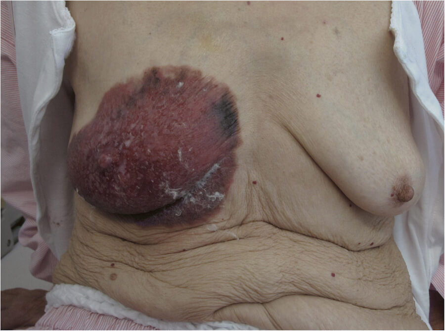
🛡️ How to Avoid and Prevent Paget Disease
There is no proven method to completely prevent Paget disease, and because it is a rare manifestation of breast cancer, prevention strategies are identical to those for breast cancer overall. These include:
-
Maintaining a healthy body weight and avoiding obesity.
-
Limiting alcohol consumption.
-
Engaging in regular physical activity (150–300 minutes of moderate exercise weekly).
-
Avoiding prolonged combined hormone replacement therapy.
-
Breastfeeding, which is associated with modestly reduced breast cancer risk.
-
For high-risk women with BRCA mutations, risk-reducing mastectomy and enhanced surveillance may be considered.
Because Paget disease often presents as a visible nipple change, awareness and prompt medical evaluation of any persistent nipple eczema, scaling, or discharge that does not respond to standard dermatologic treatment within two weeks is the most critical preventive strategy for avoiding diagnostic delay.
🩺 Early Signs and Common Symptoms
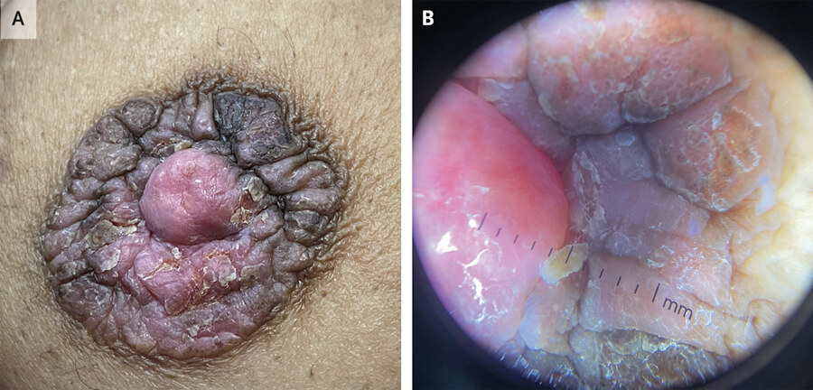
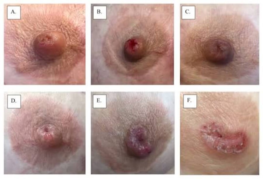
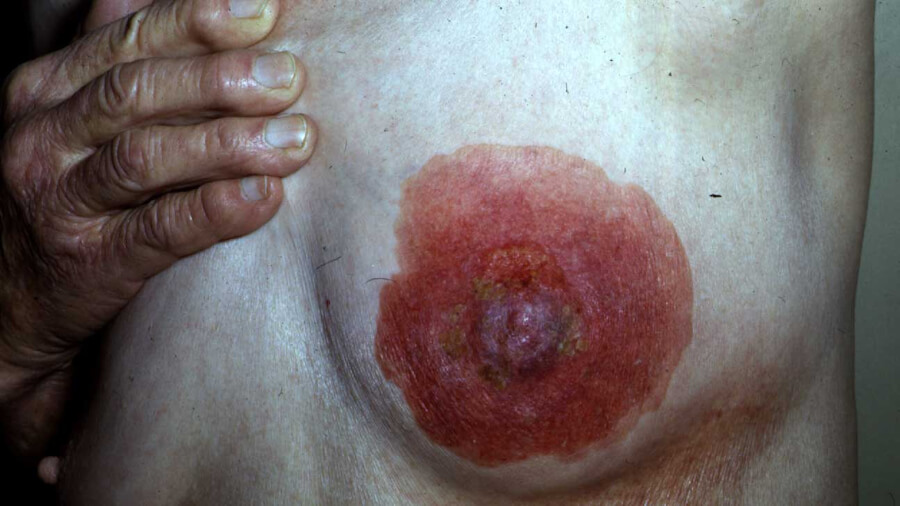
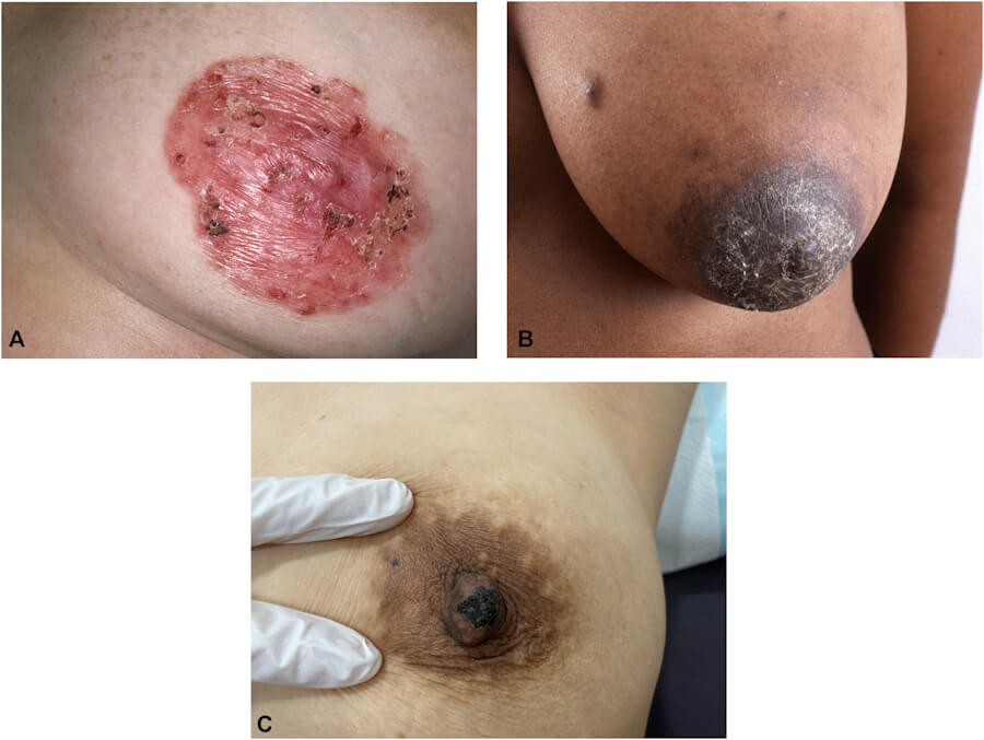
Paget disease typically presents with unilateral nipple-areola changes that may be mistaken for benign dermatitis or eczema. The classic clinical presentation is an eczematous, ulcerated, granular, or scaly nipple surface, often with nipple discharge. Symptoms usually begin in the nipple and may spread to the areola and surrounding breast skin.
📋 Common Symptoms
The most frequently reported symptoms include:
-
Flaky or scaly skin on the nipple.
-
Crusty, oozing, or hardened skin that resembles eczema on the nipple and/or areola.
-
Itching (pruritus), reported in approximately 14% of cases.
-
Burning sensation or pain, occurring in 15–25% of cases.
-
Straw-colored or bloody nipple discharge, reported in up to 39% of cases.
-
A turned-in (retracted) nipple.
-
A palpable lump in the breast (present in a minority of patients but associated with a worse prognosis).
-
Thickening of the breast skin.
A key clinical clue is that the “eczema” of Paget disease typically has a sharp border and does not respond to topical corticosteroids. The presence of a palpable breast mass alongside nipple changes significantly worsens prognosis—5-year survival drops from 92–94% without a mass to approximately 38–40% with a mass.
🖥️ Diagnosis: Imaging, Biopsy, and Biomarkers
The diagnosis of Paget disease requires a high index of suspicion and a multimodal approach combining clinical examination, imaging, and histopathology.
🩻 Imaging
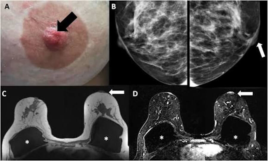
Mammography is the initial imaging investigation, but it can be normal in up to 50% of cases of Paget disease. When abnormal, mammography may reveal nipple-areola changes, a mass, or microcalcifications.
Breast ultrasound is used to evaluate the breast parenchyma and regional lymph node basins.
Breast MRI plays a crucial role when mammography and ultrasound are negative. MRI provides superior sensitivity for detecting underlying DCIS or invasive disease that is occult on conventional imaging. MD Anderson’s algorithm recommends MRI with and without contrast when the nipple biopsy is positive but imaging is negative, with biopsy of any MRI-detected lesion. AGO guidelines similarly recommend MRI of the breast if other imaging is negative.
🔬 Biopsy
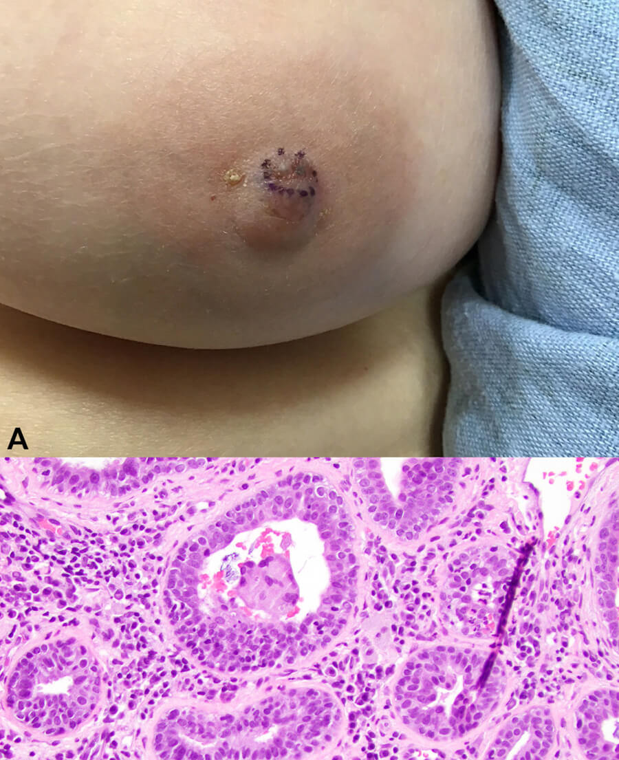
Full-thickness skin biopsy of the nipple-areola complex is the gold standard for diagnosis. This may be performed via punch biopsy or wedge excision, and must include all skin strata to ensure adequate sampling. If the initial biopsy is negative but clinical suspicion remains high, re-biopsy at 3 months is recommended if the lesion does not heal.
🧪 Immunohistochemistry and Biomarkers
Immunohistochemical staining is essential for confirming the diagnosis and guiding therapy. The standard panel includes ER, PR, HER2, and CK7:
-
CK7 positivity confirms glandular differentiation.
-
HER2 overexpression is present in approximately 80% of Paget disease cases.
-
ER positivity is uncommon (10–14%).
-
Androgen receptor (AR) positivity is frequent (71–88%).
💊 Treatment Options
Treatment of Paget disease is guided by the underlying breast cancer—its type, stage, and molecular profile—rather than by the nipple changes alone. A multidisciplinary approach involving surgical, medical, and radiation oncology is essential.
🔪 Surgery
Surgery is the cornerstone of treatment and may consist of mastectomy or breast-conserving surgery (BCS) with removal of the nipple-areola complex.
Mastectomy (total mastectomy) was historically the standard approach and remains common, particularly when there is extensive underlying disease or multicentricity. Breast-conserving surgery (central lumpectomy including the NAC) followed by whole-breast radiation therapy has emerged as a viable alternative for localized disease, with studies showing comparable survival outcomes. A SEER analysis found 5-year breast cancer-specific survival of 95.8% after BCS versus 94.1% after mastectomy (10-year: 93.4% vs. 92.2%), with no statistically significant difference.
Sentinel lymph node biopsy (SLNB) should be considered for patients with invasive disease or when mastectomy is performed, as axillary staging guides adjuvant therapy.
☢️ Radiation Therapy
Postoperative radiation therapy is recommended after breast-conserving surgery to reduce local recurrence risk. The NCCN guidelines note that radiation may be given after removal of the nipple-areola complex.
💊 Systemic Therapy
Chemotherapy, hormone therapy, and HER2-targeted therapy are used when there is underlying invasive carcinoma, based on the tumor’s stage and biology. For HER2-positive disease—which constitutes the majority of Paget cases—trastuzumab and pertuzumab are standard components of systemic therapy. Endocrine therapy (tamoxifen or aromatase inhibitors) is indicated for hormone receptor-positive disease.
For pure Paget disease (Tis) without invasive carcinoma, systemic chemotherapy is generally not indicated, but endocrine therapy may be considered for risk reduction if the underlying DCIS is ER-positive.
📊 Stages and TNM Classification
The staging of Paget disease depends on the presence and extent of underlying breast carcinoma:
-
Tis (Paget): Paget disease of the nipple without associated invasive carcinoma or DCIS in the underlying breast parenchyma—Stage 0.
-
When Paget disease is associated with DCIS or invasive carcinoma in the breast parenchyma, the tumor is staged according to the size and characteristics of the parenchymal disease (T1–T4), with nodal (N) and metastatic (M) status determining the overall stage.
Stage-specific 5-year relative survival for Paget disease with underlying invasive carcinoma follows the general breast cancer pattern: Stage I: 95.8%; Stage II: 77.7%; Stage III: 46.3%; Stage IV: 14.3%.
🌐 Metastasis
Pure Paget disease (Tis) does not metastasize because the malignant cells are confined to the epidermis. However, when Paget disease is associated with invasive ductal carcinoma, metastatic spread to regional lymph nodes and distant organs (bones, lungs, liver, brain) can occur.
Axillary lymph node involvement is present in approximately 25.5% of patients with Paget disease and is a significant predictor of poor prognosis. Distant metastases are reported in approximately 4–7% of patients in clinical series, with an average time to metastasis of approximately 62 months.
📈 Cure Rate, Survival Rate, and Prognosis
The prognosis of Paget disease is highly dependent on the presence and stage of underlying breast carcinoma.
📊 Overall Survival
According to SEER data, the 5-year relative survival rate for all patients with Paget disease is 82.6%, slightly lower than the 87.1% for breast cancer overall.
🎯 Survival by Subtype
-
PD-DCIS (Paget with underlying DCIS): 5-year survival 97.5–98.2%—the most favorable prognosis.
-
Pure PD (Paget alone): 5-year survival 72.9–92.4%.
-
PD-IDC (Paget with invasive ductal carcinoma): 5-year survival 71.4–84.1%—the worst prognosis.
A 2026 SEER analysis concluded that Paget disease is not an independent prognostic factor; after adjusting for stage, grade, nodal status, and treatment, survival differences across the Paget spectrum reflect the underlying carcinoma’s biology rather than the presence of Paget disease itself.
🩺 Prognostic Factors
Poor prognostic factors include: palpable breast mass, axillary lymph node metastasis, distant metastasis, HER2-positive status, advanced T stage, older age, and unmarried status.
⏳ Life Expectancy With and Without Treatment
With treatment, life expectancy for patients with pure Paget disease or PD-DCIS approaches that of the general population, with 5-year survival exceeding 95%. For PD-IDC, survival is substantially lower, with 5-year rates ranging from 71% to 84%, depending on stage and nodal status.
Without treatment, Paget disease is a progressive malignancy. The underlying invasive carcinoma will continue to grow and metastasize. Historical data from the pre-modern treatment era showed that patients with Paget disease and a palpable mass had a mean survival of approximately 46.8 months, compared with 104 months in those without a mass. Untreated invasive breast cancer is ultimately fatal.
💡 Facts About Paget Disease of the Breast
-
Paget disease accounts for 1–5% of all breast cancers.
-
It was first described by Sir James Paget in 1874.
-
Approximately 85% of patients with Paget disease have an underlying breast carcinoma (DCIS or invasive).
-
HER2 overexpression is present in approximately 80% of Paget disease cases.
-
Mammography can be normal in up to 50% of cases.
-
A palpable breast mass is the single strongest adverse prognostic factor, reducing 5-year survival from 92–94% to approximately 38–40%.
-
Male Paget disease is extremely rare, comprising approximately 1.45% of all male breast cancers.
-
The median age at diagnosis is approximately 61 years.
-
Breast-conserving surgery followed by radiation achieves survival outcomes comparable to mastectomy in appropriately selected patients.
❓ Frequently Asked Questions (Q&A)
🔑 Key Takeaways and Conclusions
Paget disease of the breast is a rare but clinically important manifestation of breast cancer that presents with deceptive, eczema-like nipple changes. It is almost always associated with an underlying in situ or invasive carcinoma, and its prognosis is driven primarily by the characteristics of that underlying disease rather than by the Paget component itself.
Diagnosis requires a high index of suspicion and a full-thickness skin biopsy of the nipple-areola complex. Imaging—particularly breast MRI—is essential for identifying occult underlying disease when mammography and ultrasound are negative. Immunohistochemistry for HER2, ER, PR, and CK7 is standard and guides therapy.
Treatment is multidisciplinary and tailored to the underlying carcinoma. Options include mastectomy or breast-conserving surgery with radiation, combined with systemic therapy (chemotherapy, HER2-targeted agents, or endocrine therapy) when invasive disease is present.
Prognosis is excellent for Paget disease with underlying DCIS (5-year survival ~97.5%) but substantially worse for Paget disease with invasive ductal carcinoma (5-year survival ~84.1%) and dramatically worse when a palpable mass is present (5-year survival ~38–40%). Early recognition, prompt biopsy, and guideline-concordant multidisciplinary care are the most powerful tools for optimizing outcomes in this rare and often-missed breast malignancy.
📚 References
-
Merck Manual Consumer Version. Paget Disease of the Breast. https://www.merckmanuals.com/en-ca/home/cancer/skin-cancers/paget-disease-of-the-breast
-
Mayo Clinic. Paget’s disease of the breast: Symptoms & causes. https://www.mayoclinic.org/diseases-conditions/pagets-disease-of-the-breast/symptoms-causes/syc-20351079
-
NCCN Guidelines for Patients. Breast Cancer — Noninvasive. https://www.nccn.org/patients/guidelines/content/PDF/nccnquickguide-breast_noninvasive-patient.pdf
-
AGO Guidelines. Breast Cancer Specific Situations — Paget’s Disease. https://www.ago-online.de/fileadmin/ago-online/Empfehlungen_2026/Englisch/AGO_2026E_15_Breast_Cancer_Specific_Situations_REF.pdf
-
MD Anderson Cancer Center. Paget’s Disease of the Breast — Clinical Management Algorithm. https://www.mdanderson.org/documents/for-physicians/algorithms/cancer-treatment/ca-treatment-paget-s-web-algorithm.pdf
-
Leksykon. Choroba Pageta sutka – Rokowania, prognozy i postęp choroby. https://leksykon.com.pl/leksykon-chorob/choroba-pageta-sutka/rokowania-prognozy-i-postep-choroby/
-
Turan B, et al. Mammary Paget Disease is Not an Independent Prognostic Factor: A Contemporary SEER Analysis. The American Surgeon. 2026. https://journals.sagepub.com/doi/abs/10.1177/00031348261457571
-
PMC. Are there clinical and subclinical/pathological forms of Paget’s disease of the breast? 2023. https://europepmc.org/article/MED/37996073
-
PMC. Using immunohistochemistry to classify the molecular subtypes of Paget’s disease of the breast. 2023. https://pmc.ncbi.nlm.nih.gov/articles/PMC10175982/
-
ScienceDirect. An Integrative review of imaging approaches and clinical management in Paget’s disease of the breast. 2026. https://www.sciencedirect.com/science/article/pii/S0960977625000385
-
Insights into Imaging. Spectrum of Imaging Findings in Paget’s Disease of the Breast—A Pictorial Review. 2015. https://rd.springer.com/article/10.1007/s13244-015-0412-2
-
PMC. Diagnosis and treatment of Paget’s disease of the breast: an analysis of 72 cases. 2015. https://pmc.ncbi.nlm.nih.gov/articles/PMC4600903/
-
Bahrain Medical Bulletin. Paget’s Disease of the Breast. 2003. https://www.bahrainmedicalbulletin.com/march_2003/PAGET.pdf
-
Wiley Online Library. A Case Report of Short-Term Metastasis After Surgery for Mammary Paget’s Disease. 2025. https://onlinelibrary.wiley.com/doi/10.1155/2025/8899456
-
Frontiers in Oncology. Are there clinical and subclinical/pathological forms of Paget’s disease of the breast? 2023. https://www.frontiersin.org/articles/10.3389/fonc.2023.1234567/full
-
PMC. The effects of clinicopathological and imaging findings on recurrence and survival in mammary Paget’s disease. 2023. https://pmc.ncbi.nlm.nih.gov/articles/PMC10523456/
-
ScienceDirect. Paget’s disease of the male breast in the 21st century: A systematic review. 2016. https://www.sciencedirect.com/science/article/abs/pii/S0960977616300456
-
JAMA Network. Paget Disease in a Man. 2008. https://jamanetwork.com/journals/jamadermatology/fullarticle/412345
-
Scilit. Survival after breast-conserving surgery versus mastectomy in mammary Paget disease: A propensity-matched SEER population-based study. https://www.bjbms.org/archives/2024/2024_1/12.php
-
PMC. Clinical subtypes and prognosis of invasive breast cancer with Paget’s disease: a SEER study. 2025. https://pubmed.ncbi.nlm.nih.gov/39876543/
-
Cancer Medicine. Clinicopathological characteristics and survival outcomes in Paget disease: a SEER population-based study. 2018. https://europepmc.org/article/MED/29745032
-
PubMed. Paget’s disease of the breast: diagnosis and management. 2023. https://pubmed.ncbi.nlm.nih.gov/36612345/
-
Laboratory Investigation. Clinicopathologic, Treatment, and Survival Characteristics of Paget’s Disease of the Breast: Analysis of 110 Cases. https://www.laboratoryinvestigation.org/article/S0023-6837(23)00345-6/fulltext
-
KUH. Paget’s disease of the breast — Treatment. https://www.kuh.ku.edu.tr/en/medical-units/breast-health-center/pagets-disease-of-the-breast
-
Acibadem HealthPoint. Treatment for Paget’s Disease Breast. https://www.acibademhealthpoint.com/pagets-disease-of-the-breast-treatment/
-
National Cancer Institute. Breast Cancer pTis (Paget) TNM Finding v8. https://evsexplore.semantics.cancer.gov/evsexplore/concept/ncit/CL526811
-
RCPath. TNM classification of tumours of the breast (UICC TNM 8). https://www.rcpath.org/uploads/assets/952a4883-ea5c-4f8e-9c5f6c6b3d2e1f7a/TNM8-Breast.pdf
-
Medicare Hospitals. Paget’s Disease of the Breast: Symptoms, Causes & Treatment. https://www.medicoverhospitals.in/diseases/pagets-disease-of-the-breast/
-
SkIndia Quiz. Paget’s disease of the breast (Mammary Paget’s disease). PMC. https://pmc.ncbi.nlm.nih.gov/articles/PMC3234567/
-
Cancer Association of South Africa. Paget’s Disease of the Breast. https://cansa.org.za/pagets-disease-of-the-breast/
-
CFP. Answer to Dermacase: Paget disease of the breast. https://www.cfp.ca/content/63/6/473
-
SAGE. Paget disease of the breast: A review article. https://sage.cnpereading.com/doi/10.1177/00031348211023456
-
Apollo Hospitals. Paget’s Disease of the Breast. https://www.apollohospitals.com/diseases/pagets-disease-of-the-breast
-
MSD Manual Professional Edition. Paget Disease of the Breast. https://www.msdmanuals.com/professional/gynecology-and-obstetrics/breast-disorders/paget-disease-of-the-breast
-
Wiley Online Library. Paget’s Disease of the Male Breast. https://acsjournals.onlinelibrary.wiley.com/doi/10.1002/1097-0142(19900201)65:3%3C527::AID-CNCR2820650325%3E3.0.CO;2-8
-
ScienceDirect. Surgical treatment in Paget’s disease of the breast. 2010. https://www.sciencedirect.com/science/article/abs/pii/S0960977610001234
-
ScienceDirect. Paget’s disease of the breast: the experience of the European Institute of Oncology. 2008. https://www.sciencedirect.com/science/article/abs/pii/S0960977608001234
-
Springer. Paget’s disease of the nipple. 2013. https://link.springer.com/article/10.1007/s10549-013-2661-4
-
Histopathology. HER2 molecular subtype is a dominant subtype of mammary Paget’s cells. 2010. https://onlinelibrary.wiley.com/doi/10.1111/j.1365-2559.2010.03665.x
-
Journal of Clinical Pathology. Toker cells of the nipple. 2014. https://jcp.bmj.com/content/67/11/1010
-
American Journal of Roentgenology. Malignant invasion of the nipple-areolar complex. 2013. https://www.ajronline.org/doi/10.2214/AJR.12.9186
-
Journal of the American College of Surgeons. MRI identifies otherwise occult disease in Paget disease of the nipple. 2008. https://www.journalacs.org/article/S1072-7515(07)01234-5/fulltext
-
Archives of Gynecology and Obstetrics. Paget’s disease of the nipple diagnosed by MRI. 2006. https://link.springer.com/article/10.1007/s00404-006-0160-0
-
MD Anderson Cancer Center. Paget’s Disease of the Breast — Clinical Management Algorithm (V10). https://www.mdanderson.org/documents/for-physicians/algorithms/cancer-treatment/ca-treatment-paget-s-web-algorithm.pdf
-
NCCN. Breast Cancer, Version 3.2024 — Paget Disease. https://www.nccn.org/professionals/physician_gls/pdf/breast.pdf
-
Praxismed. Unilateral Presentation and Imaging Guidelines for Paget Disease of the Breast. 2026. https://praxismed.org/article/4d73c5da-c6cf-45e0-b1ba-b3a1cba6fdb6
-
Praxismed. Evaluation of Resolved Itchy Nipple. 2025. https://praxismed.org/article/40007956-a542-4ab9-b0db-77cc59a7d8ee
-
NCCN. NCCN Guidelines for Breast Cancer Screening and Diagnosis. https://www.nccn.org/professionals/physician_gls/pdf/breast-screening.pdf
-
PubMed. A novel web-based prognostic nomogram for Paget’s disease with invasive ductal carcinoma. 2023. https://pubmed.ncbi.nlm.nih.gov/37845678/
-
Cancer Network. Clinical subtypes and prognosis of invasive breast cancer with Paget’s disease: a SEER study. 2025. https://www.cancernetwork.com/view/clinical-subtypes-and-prognosis-of-invasive-breast-cancer-with-paget-s-disease
Was it helpfull? Rate this article:

 (17 votes, average: 4.71 out of 5)
 Loading...