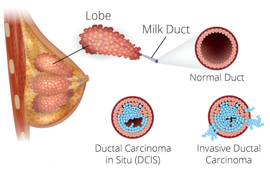
Ductal carcinoma in situ (DCIS) is a non-invasive neoplasm of the breast in which malignant epithelial cells proliferate within the lumen of the milk ducts but remain confined by the basement membrane and do not invade the surrounding stroma. Because the malignant cells have not breached the ductal wall, DCIS has no direct access to the lymphatic or haematogenous circulation, and pure DCIS therefore has essentially no metastatic potential. It is classified as stage 0 breast cancer in the TNM system and is widely regarded as a non-obligate precursor to invasive breast cancer: not every DCIS lesion will progress, but the presence of DCIS confers a substantially elevated risk of subsequent invasive disease. The widespread adoption of screening mammography has transformed the epidemiology of DCIS, converting what was once a largely palpable and often large lesion into a predominantly mammographically detected, non-palpable entity, and raising important questions about overdiagnosis and overtreatment.
📋 ICD-10 Classification
In the ICD-10-CM system, DCIS of the breast is coded under D05.1 — Intraductal carcinoma in situ of breast. The broader category D05 covers carcinoma in situ of the breast, and D05.1 is the specific code for the ductal (intraductal) form. In the ICD-10-GM (German modification) and many international ICD-10 versions, the equivalent code is D05.1 — Carcinoma in situ der Milchgänge (carcinoma in situ of the milk ducts). The ICD-O morphology code for DCIS is M8201/2 (carcinoma in situ, ductal). It is important to note that D05.1 is a non-billable, non-specific code in the US ICD-10-CM because it contains more specific subcodes; clinical documentation should specify the subtype and location where possible.
🔬 What Causes DCIS?
🧬 Molecular and Cellular Origins
DCIS arises from a clonal proliferation of epithelial cells originating in the terminal duct lobular unit (TDLU). The initiating event is the acquisition of genetic and epigenetic alterations in ductal epithelial cells that confer a growth advantage and allow the cells to proliferate within the ductal lumen while remaining constrained by the basement membrane. Unlike invasive carcinoma, DCIS cells have not acquired the capacity to degrade the basement membrane and invade the stroma.
The molecular landscape of DCIS is heterogeneous. Low-grade DCIS frequently harbours alterations in the PI3K/AKT pathway, whereas high-grade DCIS more often shows TP53 mutations, HER2 amplification, and genomic instability. The transition from DCIS to invasive ductal carcinoma (IDC) is associated with the accumulation of additional genetic and epigenetic changes, including alterations that promote epithelial–mesenchymal transition, angiogenesis, and immune evasion.
🎯 Risk Factors
The risk factors for DCIS largely overlap with those for invasive breast cancer, consistent with the view that DCIS lies on the same pathogenic continuum.
Established risk factors include:
-
Older age — DCIS incidence increases with age, and the majority of cases are diagnosed in women over 50.
-
Positive family history — a first-degree relative with breast cancer increases risk, and the risk is greater if the relative was diagnosed at a young age.
-
Personal history of breast cancer — a prior breast cancer in one breast increases the risk of a new primary in either breast.
-
Prior breast biopsy with atypical findings — including atypical ductal hyperplasia (ADH) and lobular carcinoma in situ (LCIS).
-
Reproductive and hormonal factors — early menarche (before 12), late menopause (after 55), nulliparity, and older age at first full-term pregnancy are associated with increased risk.
-
Dense breast tissue — high mammographic density increases risk and also reduces the sensitivity of mammography.
-
Alcohol consumption — even moderate intake is associated with increased risk.
-
Postmenopausal combined hormone therapy — long-term use (10 or more years) is associated with increased risk of DCIS.
-
Obesity — particularly postmenopausal obesity.
-
Smoking — associated with a modest increase in risk.
Chemoprevention with tamoxifen reduces the incidence of both invasive breast cancer and DCIS, whereas raloxifene reduces invasive breast cancer but not DCIS. This differential effect underscores the hormonal sensitivity of DCIS and the importance of estrogen receptor signalling in its pathogenesis.
🛡️ How to Avoid and Prevent DCIS
There is no guaranteed method to prevent DCIS, but risk-reduction strategies include:
-
Avoiding or limiting long-term combined hormone replacement therapy, particularly beyond 5–10 years of use.
-
Maintaining a healthy body weight and engaging in regular physical activity, which are associated with lower breast cancer risk.
-
Limiting alcohol consumption — even one drink per day may increase risk.
-
Breastfeeding, which has been associated with a reduced risk of both DCIS and LCIS.
-
Chemoprevention with tamoxifen (for both pre- and postmenopausal high-risk women) or aromatase inhibitors (for postmenopausal women) can reduce the incidence of DCIS and invasive breast cancer in high-risk individuals.
-
Risk-reducing mastectomy may be considered in women with very high genetic risk (e.g., BRCA1/2 or TP53 pathogenic variants).
-
Regular screening mammography does not prevent DCIS but enables detection at a non-invasive stage, allowing treatment before progression to invasive disease.
🧫 Types of DCIS
DCIS is a heterogeneous group of lesions classified by nuclear grade (low, intermediate, high) and architectural pattern. Nuclear grade is the most important prognostic feature, while architectural pattern provides additional descriptive information.
🔹 Comedo Type
The comedo pattern is characterised by high nuclear grade, numerous mitoses, and central luminal necrosis with associated microcalcifications. Comedo DCIS is associated with a higher risk of local recurrence and progression to invasive carcinoma than non-comedo subtypes.
🔹 Solid Type
In solid DCIS, the duct is filled with a solid sheet of malignant cells without significant luminal spaces. Solid DCIS is the predominant architectural pattern, seen in approximately 61% of cases in large series, and is particularly common in high-grade lesions.
🔹 Cribriform Type
Cribriform DCIS is characterised by punched-out, regular spaces within a proliferating cell population, creating a sieve-like appearance. It is the second most common pattern and is frequently seen in low- and intermediate-grade lesions.
🔹 Micropapillary and Papillary Types
Micropapillary DCIS shows small, bulbous projections of cells into the ductal lumen without fibrovascular cores, whereas papillary DCIS has true fibrovascular cores. These patterns are more commonly associated with low- and intermediate-grade lesions.
🔹 Other Patterns
Less common patterns include clinging, flat, and apocrine DCIS. Mixed patterns are common, and a single lesion may show more than one architectural type.
🔹 Grading
Nuclear grade — low, intermediate, or high — is determined by the degree of nuclear pleomorphism, mitotic activity, and necrosis. High-grade DCIS is associated with a higher risk of recurrence and progression, while low-grade DCIS has a more indolent course.
🩺 Symptoms and Early Signs
The majority of DCIS cases are asymptomatic and are detected on screening mammography.
When symptoms do occur, they may include:
-
A breast lump — uncommon in screen-detected DCIS but possible in larger or palpable lesions.
-
Nipple discharge — may be blood-stained.
-
A rash or eczema-like change on the nipple — may be red and scaly.
-
Nipple ulceration or itching — if the DCIS extends into the nipple-areolar complex.
-
Breast pain — DCIS does not typically cause pain, and pain is not a reliable indicator.
In men, DCIS may present with a bloody nipple discharge or a lump, as men do not undergo routine screening mammography.
🖥️ Diagnosis: Imaging, Biopsy, and Pathology
📸 Mammography
Screening mammography is the primary modality by which DCIS is detected. DCIS most commonly appears as microcalcifications — small clusters of irregular calcium deposits within the ducts. The calcifications of DCIS are often pleomorphic (varying in size and shape) and may be linear or branching in high-grade lesions. Architectural distortion without a mass may also be a sign of DCIS.
Digital breast tomosynthesis (3D mammography) improves the detection of DCIS compared with 2D digital mammography, particularly in dense breasts, by reducing tissue overlap.
🔊 Ultrasound
Ultrasound is used as a supplemental tool to evaluate mammographic abnormalities and to guide biopsy. DCIS may appear as a hypoechoic area, ductal extension, or clustered microcysts, but ultrasound is less sensitive than mammography for detecting microcalcifications.
🧲 Magnetic Resonance Imaging (MRI)
Breast MRI is the most sensitive imaging modality for DCIS but is not routinely used for screening average-risk women. It may be used in specific circumstances: to evaluate the extent of disease when breast-conserving surgery is being considered, to detect additional foci in dense breasts, or to screen high-risk women (e.g., BRCA carriers). DCIS on MRI typically appears as non-mass enhancement in a linear, segmental, or regional distribution.
🩻 Other Imaging
Contrast-enhanced mammography (CEM) and molecular breast imaging (MBI) are emerging modalities that may improve DCIS detection, but their roles are not yet fully established. PET/CT and bone scans are not indicated for the staging of DCIS because of its non-invasive nature and negligible metastatic potential.
🔪 Biopsy
Tissue diagnosis is mandatory for DCIS. Stereotactic core needle biopsy is the standard approach for mammographic calcifications, while ultrasound-guided biopsy may be used for sonographically visible lesions. Vacuum-assisted biopsy may be preferred to obtain sufficient tissue and reduce the risk of underestimating invasive disease.
Pathology confirms the diagnosis by demonstrating malignant cells confined within the ductal lumen, with an intact basement membrane. The pathology report should include nuclear grade, architectural pattern, presence of necrosis (especially comedo necrosis), margin status, and hormone receptor status (ER/PR). HER2 status may be assessed, particularly in high-grade or pleomorphic lesions, although it does not currently guide treatment in DCIS outside of clinical trials.
💊 Treatment Types
The treatment of DCIS is aimed at preventing progression to invasive breast cancer and reducing the risk of local recurrence. Because DCIS is non-invasive, chemotherapy is not indicated after surgical treatment.
🔪 Surgery
Breast-conserving surgery (lumpectomy) followed by whole-breast radiation therapy is the standard of care for most women with DCIS who are candidates for breast conservation. Mastectomy is reserved for extensive or multicentric disease, cases where clear margins cannot be achieved with breast conservation, or patient preference. Sentinel lymph node biopsy (SLNB) is generally not indicated for pure DCIS but may be considered in cases where invasive disease is suspected or when mastectomy is performed.
Margin status is a critical determinant of local recurrence. A negative margin (no ink on tumour) is associated with a lower risk of recurrence, although the optimal margin width for DCIS remains debated.
☢️ Radiation Therapy
Adjuvant whole-breast radiation therapy after lumpectomy reduces the risk of ipsilateral recurrence by approximately 50%, but it has not been shown to improve breast cancer-specific survival in most studies. The decision to add radiation should be individualised based on recurrence risk, patient age, and preferences. Some low-risk patients (e.g., small, low-grade DCIS with clear margins) may reasonably omit radiation.
💊 Endocrine Therapy
Adjuvant endocrine therapy with tamoxifen or an aromatase inhibitor is recommended for patients with hormone receptor–positive DCIS to reduce the risk of ipsilateral and contralateral breast events. Tamoxifen reduces the risk of subsequent breast cancer (both invasive and DCIS) by approximately 30–50% in women with ER-positive DCIS, but it is associated with side effects including hot flashes, vaginal dryness, and rare but serious risks of endometrial cancer and venous thromboembolism. Aromatase inhibitors (e.g., anastrozole, exemestane) are an alternative for postmenopausal women and may have a more favourable side-effect profile for some patients. Importantly, endocrine therapy reduces recurrence but has not been shown to improve overall survival in DCIS.
🧪 Active Surveillance (Emerging)
Clinical trials such as COMET, LORIS, and LORD are evaluating active surveillance (watchful waiting) as an alternative to immediate surgery for low-risk DCIS. Early results suggest that active monitoring may be feasible for carefully selected patients, but surgery remains the standard of care pending maturation of these trials. These trials represent an important step toward reducing overtreatment of indolent DCIS.
🎯 Targeted Therapy (Investigational)
HER2-targeted therapy (e.g., trastuzumab) has been investigated in HER2-positive DCIS, but data from the NSABP B-43 trial have not shown a statistically significant benefit, and this approach remains investigational.
📊 Stages
DCIS is classified as stage 0 in the AJCC TNM staging system (Tis, N0, M0). By definition:
-
T (tumour): Tis (DCIS) — carcinoma in situ.
-
N (node): N0 — no regional lymph node metastasis.
-
M (metastasis): M0 — no distant metastasis.
Because DCIS is confined to the duct and has not invaded the basement membrane, it cannot spread to lymph nodes or distant organs. When DCIS is found together with an invasive component (e.g., invasive ductal carcinoma), the stage is determined by the invasive component, not by the DCIS.
🌐 Metastasis
Pure DCIS does not metastasise. Distant metastasis following a diagnosis of pure DCIS is extremely rare, with an incidence of less than 0.5% in most series. When metastasis does occur in a patient with a history of DCIS, it is almost always associated with a preceding or synchronous invasive recurrence or with occult invasive disease that was not detected at the time of the original diagnosis. The rare cases of distant metastasis without documented invasive recurrence may reflect microinvasion or limitations in pathological sampling.
The risk of progression to invasive carcinoma in untreated DCIS is estimated at 25–60% over time, with high-grade and comedo-necrosis lesions carrying the highest risk.
📈 Cure Rate and Survival Rate
The prognosis for DCIS is excellent. Survival rates at 10 years are 98.9% and at 20 years are 96.7% in large cohort studies of women treated for DCIS. The 10-year breast cancer-specific survival rate exceeds 98%, and most people diagnosed with DCIS are successfully treated.
However, a diagnosis of DCIS is associated with a 3.36-fold increased risk of breast cancer mortality compared with the general population, and this elevated risk persists for more than 15 years after diagnosis. This increased mortality is largely attributable to recurrence as invasive disease or to occult invasive cancer present at the time of diagnosis. The 20-year actuarial risk of ipsilateral invasive recurrence is approximately 13.9%.
When DCIS recurs, approximately 50% of recurrences are invasive, and these invasive recurrences carry the prognosis of the corresponding stage of invasive breast cancer. When DCIS recurs as non-invasive disease, survival rates are the same as for the initial DCIS occurrence.
Factors associated with a higher risk of recurrence include: young age at diagnosis (under 40), high nuclear grade, comedo necrosis, larger lesion size, positive or close surgical margins, and symptomatic (rather than screen-detected) presentation.
⏱️ Life Expectancy With and Without Treatment
With treatment, the vast majority of patients with DCIS have a normal life expectancy. The risk of dying from breast cancer after a DCIS diagnosis is very low — approximately 3.3% at 20 years in large contemporary cohorts — and this risk is similar whether patients are treated with breast-conserving surgery plus radiation or with mastectomy.
Without treatment, DCIS is associated with a 25–60% risk of progression to invasive carcinoma over time. The natural history of untreated DCIS is incompletely understood because ethical constraints limit the study of observation alone, but historical series and contemporary active surveillance trials provide estimates. In the LORIS trial, the 5-year invasive disease-free rate was 87.5% in the active monitoring group compared with 94.2% in the surgery group, indicating that not all low-risk DCIS progresses rapidly and that some lesions may remain indolent for years. However, because progression to invasive cancer carries a risk of metastasis and death, treatment is recommended for the majority of patients with DCIS.
🕊️ Palliative Care
Palliative care is rarely required for DCIS itself because DCIS is a non-invasive, highly curable condition with an excellent prognosis. When palliative or supportive care is relevant, it is typically in the context of:
-
Symptom management during treatment (e.g., pain after surgery, skin reactions during radiation, side effects of endocrine therapy).
-
Psychosocial support for anxiety, fear of recurrence, and body image concerns, which can be significant even for non-invasive disease.
-
Survivorship care, including surveillance for recurrence, management of treatment-related side effects, and health promotion.
-
End-of-life care in the rare circumstance that a patient with a history of DCIS develops metastatic invasive breast cancer despite adequate local treatment. In this setting, palliative care follows the principles applicable to metastatic breast cancer, focusing on comfort, symptom control, and quality of life.
Supportive care is an umbrella term encompassing all services that help patients and families cope with the condition and its treatment, from diagnosis through survivorship or, in rare cases, advanced disease.
❓ Five Questions and Answers
Basics, Biology, & Molecular Characteristics
1. What is Ductal Carcinoma In Situ (DCIS)? DCIS is a non-invasive breast lesion in which abnormal cells grow inside milk ducts and have not broken through the duct wall.
2. What does “in situ” mean in cancer diagnosis? “In situ” means “in place”—abnormal cells are confined to their original tissue layer and have not invaded nearby tissue.
3. What is Stage 0 breast cancer? Stage 0 is non-invasive breast cancer/carcinoma in situ, including DCIS, with no spread beyond the duct.
4. Is DCIS considered real cancer, pre-cancer, or a non-invasive lesion? It is best understood as a non-invasive precursor lesion; it is called Stage 0 cancer but usually has excellent prognosis.
5. Where in the breast does DCIS originate (milk ducts)? DCIS originates in the epithelial lining of breast milk ducts, often near the terminal duct lobular unit.
6. What percentage of non-invasive breast cancers are DCIS (~80–85%)? About 80–85% of non-invasive breast cancers are DCIS.
7. How common is DCIS (accounting for ~20% of all newly diagnosed breast cancers)? DCIS accounts for about 20% of screen-detected newly diagnosed breast cancers.
8. What is the difference between DCIS and Invasive Ductal Carcinoma (IDC)? DCIS stays inside the duct; IDC has broken through the duct wall into surrounding breast tissue.
9. What is the basement membrane of the milk duct, and why is it critical in DCIS? It is a thin structural boundary around ducts; DCIS remains confined by it, which prevents direct spread.
10. What does it mean if cancer cells break through the basement membrane? It means the lesion is no longer pure DCIS and has become invasive cancer.
11. What is the difference between DCIS and LCIS (Lobular Carcinoma In Situ)? DCIS is a ductal non-invasive precursor; LCIS is a lobular marker of increased breast cancer risk rather than a direct precursor.
12. Why is LCIS classified as a risk factor, while DCIS is considered a non-invasive cancer precursor? LCIS usually does not progress directly to invasive cancer at the same site; DCIS is a more direct precursor to invasive ductal carcinoma.
13. How fast does DCIS grow or progress? DCIS growth is usually slow, often over years, but grade and biology vary.
14. Does all untreated DCIS turn into invasive breast cancer? No; many cases would never become invasive.
15. What percentage of DCIS cases would progress to invasive cancer if left untreated (~20–50%)? Estimates suggest about 20–50% may progress if untreated, depending on grade and follow-up.
16. How long does it take for DCIS to become invasive (often 5 to 10+ years, or never)? Progression, if it occurs, often takes years—commonly 5–10+ years—or may never happen.
17. Can doctors predict which individual DCIS tumors will become invasive and which will remain harmless? Not with certainty; grade, size, margins, necrosis, and genomic tests help estimate risk.
18. What is the “overdiagnosis” and “overtreatment” debate surrounding DCIS? It is the concern that some low-risk DCIS would never cause harm, yet surgery/radiation may be used.
19. What are the histological architecture patterns (subtypes) of DCIS? Common patterns include comedo, cribriform, papillary, micropapillary, and solid.
20. What is Comedo DCIS (comedocarcinoma)? Comedo DCIS is a pattern with high-grade cells and central ductal necrosis.
21. What is central necrosis (comedo necrosis) in DCIS? It is dead cell debris in the center of ducts, often with calcification.
22. Why is Comedo DCIS considered more aggressive than non-comedo types? It is often high grade and associated with higher recurrence/progression risk.
23. What is Cribriform DCIS? Cribriform DCIS has punched-out spaces within the duct, like a sieve.
24. What is Papillary DCIS? Papillary DCIS has finger-like projections growing inside the duct.
25. What is Micropapillary DCIS? Micropapillary DCIS has small bulbous projections without fibrovascular cores.
26. What is Solid DCIS? Solid DCIS fills the duct with a solid sheet of abnormal cells.
27. Can a single DCIS tumor contain multiple architectural patterns? Yes; mixed patterns are common.
28. What is Nuclear Grade in DCIS? Nuclear grade describes how abnormal the cell nuclei look and how fast the lesion may behave.
29. What is Low-Grade (Grade 1 / well-differentiated) DCIS? Low-grade DCIS has small, uniform nuclei and generally lower risk.
30. What is Intermediate-Grade (Grade 2 / moderately-differentiated) DCIS? Intermediate-grade DCIS has features between low and high grade.
31. What is High-Grade (Grade 3 / poorly-differentiated) DCIS? High-grade DCIS has markedly abnormal nuclei and generally higher recurrence/progression risk.
32. Why is nuclear grade the strongest predictor of DCIS recurrence and progression? Grade reflects cellular abnormality and growth behavior, strongly linked to recurrence risk.
33. How does cell nuclear size, pleomorphism, and chromatin structure determine grade? Larger, more variable nuclei and coarse chromatin indicate higher grade.
34. What is the Van Nuys Prognostic Index (VNPI) for DCIS? VNPI is a scoring system using grade, margin width, size, and age to estimate recurrence risk.
35. How does the Van Nuys index combine grade, margin width, size, and age to guide treatment? Higher scores suggest higher risk and may favor radiation or mastectomy; lower scores may support less treatment.
36. What are hormone receptors in DCIS? They are estrogen/progesterone receptors on cells that can drive growth and guide endocrine therapy.
37. What percentage of DCIS cases are Estrogen Receptor-positive (ER+)? About 75–80% of DCIS is ER-positive.
38. What percentage of DCIS cases are Progesterone Receptor-positive (PR+)? Many ER+ DCIS cases are also PR+, but rates vary.
39. What is HER2 status in DCIS? HER2 status describes whether DCIS cells overexpress HER2; it is common in high-grade DCIS.
40. Why is HER2 amplification common in DCIS (~30–50% of high-grade cases) compared to invasive cancer? HER2 positivity is often present in high-grade DCIS but may be lost as cancer becomes invasive.
41. Does HER2-positive status in DCIS mean the patient needs Herceptin / targeted therapy? Usually no; HER2 status in pure DCIS does not routinely change treatment.
42. Is Herceptin (trastuzumab) approved or recommended for non-invasive DCIS? It is not standard or routinely recommended for pure DCIS.
43. What is Triple-Negative DCIS (ER-, PR-, HER2-)? It is DCIS lacking ER, PR, and HER2 expression.
44. How common is triple-negative DCIS (~10%), and how does it behave? It is uncommon, about 10%, and may be higher grade with different biology.
45. What is the Ki-67 proliferation index in DCIS? Ki-67 estimates how many cells are dividing; it is not routinely used alone for DCIS decisions.
46. Can men get Ductal Carcinoma In Situ? Yes, men can develop DCIS, though it is rare.
47. What percentage of male breast cancers present as DCIS (<5%)? Less than 5% of male breast cancers present as DCIS.
48. What is the average age of diagnosis for DCIS (~50–60 years)? Average age is often around 50–60 years, especially with screening.
49. Has the incidence of DCIS increased since the introduction of routine screening mammography? Yes, largely because mammography detects calcified DCIS.
50. Why is DCIS often referred to as a “mammographic disease”? Most DCIS is found as mammographic microcalcifications before symptoms.
51. What is multifocal DCIS? Multifocal DCIS means separate DCIS foci in the same breast quadrant.
52. What is multicentric DCIS? Multicentric DCIS means DCIS in different quadrants of the same breast.
53. What is extensive DCIS (spanning large areas or multiple quadrants of the breast)? Extensive DCIS is widespread disease that may make breast-conserving surgery difficult.
54. What is microinvasive DCIS (DCIS with microinvasion / T1mic)? It is DCIS with tiny foci of invasion ≤1 mm.
55. What defines microinvasion (invasive cells measuring ≤1 millimeter)? Microinvasion is invasive cancer measuring ≤1 mm in greatest dimension.
56. How is DCIS with microinvasion managed differently from pure DCIS? It may require sentinel node evaluation and is staged/handled more like early invasive cancer.
57. What is Paget’s Disease of the Nipple? It is nipple/areola skin disease caused by underlying ductal cancer cells, often DCIS.
58. How is Paget’s disease of the nipple linked to underlying DCIS (~80–90% of cases)? About 80–90% of Paget cases have underlying DCIS or invasive cancer.
59. What gene mutations are commonly associated with DCIS (BRCA1, BRCA2, TP53, CHEK2)? BRCA1/2, TP53, CHEK2, PALB2, ATM and other genes may increase risk.
60. Is pure DCIS systemic or strictly localized? Pure DCIS is strictly localized to ducts.
61. Can pure DCIS metastasize to distant organs (lungs, liver, bones, brain)? No; pure DCIS cannot metastasize because it has not invaded vessels.
62. Why can pure DCIS NOT metastasize (because blood and lymphatic vessels are outside the basement membrane)? Metastasis requires invasion into vessels, which pure DCIS has not done.
63. If DCIS cannot metastasize, why do a tiny percentage of women with a DCIS history die of breast cancer (<1–2%)? Deaths are usually due to undetected invasive recurrence or a new invasive cancer.
64. Is breast cancer death after DCIS caused by undetected invasive microinvasion or secondary invasive recurrences? Yes, usually from invasive recurrence or missed invasive component.
65. What is the tumor microenvironment surrounding milk ducts in DCIS? It includes myoepithelial cells, stroma, immune cells, and blood vessels.
66. What role do myoepithelial cells play in holding DCIS cells inside the duct? They form an outer layer that helps contain DCIS.
67. What happens when myoepithelial cell layers break down or degrade? Loss of this layer is associated with progression to invasive cancer.
68. What role do stromal fibroblasts and immune cells play in DCIS progression? They can promote or restrain invasion and influence immune response.
69. Are tumor-infiltrating lymphocytes (TILs) present in high-grade DCIS? Yes, TILs are more common in high-grade DCIS.
70. What genomic alterations drive the transition from normal duct epithelium to DCIS? Alterations in growth, cell cycle, and DNA repair pathways contribute.
71. What genomic alterations drive the transition from DCIS to invasive ductal carcinoma? Additional mutations, microenvironment changes, and myoepithelial loss contribute.
72. Is DCIS a direct precursor to invasive ductal carcinoma, or an evolutionary parallel lesion? It is often a precursor, but not every DCIS follows that path.
73. What is Atypical Ductal Hyperplasia (ADH)? ADH is a benign high-risk ductal proliferation that falls short of DCIS.
74. How does ADH differ from low-grade DCIS under the microscope (based on size/extent: usually <2 mm)? ADH is usually focal/<2 mm; DCIS involves more extensive ductal involvement.
75. What is flat epithelial atypia (FEA)? FEA is a mild atypical ductal change and a risk marker.
76. Can FEA, ADH, and DCIS co-exist in the same breast tissue biopsy? Yes, they can coexist.
77. Is DCIS hereditary? Most DCIS is not hereditary, though inherited mutations increase risk.
78. What percentage of DCIS cases occur in women with a inherited BRCA gene mutation (~5–10%)? About 5–10% of breast cancers overall are hereditary; DCIS specifically is less commonly hereditary.
79. What is the cell of origin for ductal carcinoma in situ? It arises from ductal epithelial cells, often in the terminal duct lobular unit.
80. What is terminal duct lobular unit (TDLU) in breast anatomy? The TDLU is the milk-producing lobule and terminal duct where many breast cancers begin.
81. Why do calcifications form inside milk ducts containing DCIS cells? Necrotic debris and secretions can calcify within ducts.
82. What causes dystrophic calcification in comedo necrosis (dying cancer cell debris calcifying)? Dying cells release debris that calcifies.
83. What is the difference between microcalcifications and macrocalcifications? Microcalcifications are tiny and often suspicious; macrocalcifications are larger and usually benign.
84. Are macrocalcifications in the breast linked to DCIS (usually benign)? Usually they are benign, not typically DCIS.
85. Does DCIS cause systemic inflammation or abnormal blood markers? No; pure DCIS does not usually cause systemic inflammation or abnormal blood markers.
86. What is the impact of breast density on DCIS cellular biology? Density affects mammographic detection, not proven DCIS biology directly.
87. Is DCIS more aggressive in young women (<40 years old)? Young age is associated with higher recurrence risk, though DCIS is uncommon under 40.
88. What is the rate of high-grade nuclear features in premenopausal vs postmenopausal DCIS? High-grade features can occur in both; premenopausal DCIS may more often be high grade.
89. How is DCIS classified under the WHO Classification of Breast Tumors? WHO classifies DCIS as a non-invasive ductal carcinoma in situ lesion with subtypes/grades.
90. Can DCIS turn into lobular cancer? Pure DCIS does not usually “turn into” lobular cancer; invasive lobular cancer has different biology.
91. What is E-cadherin status in DCIS (positive membrane expression)? DCIS is typically E-cadherin positive.
92. Why is E-cadherin staining used to distinguish DCIS (positive) from LCIS (negative)? DCIS is positive; LCIS is usually negative, helping pathologists distinguish them.
93. What is p63 and calponin IHC staining used for in DCIS pathology (identifying myoepithelial cells)? They highlight myoepithelial cells to confirm the duct wall is intact.
94. How does cytokeratin CK5/6 and CK8/18 staining help analyze DCIS? They help characterize epithelial differentiation and distinguish benign from malignant patterns.
95. What is the role of oncogene amplification (e.g., CCND1, MYC) in DCIS? Amplifications can contribute to growth and progression but are not routine treatment targets in DCIS.
96. What is the clonal relationship between synchronous DCIS and adjacent invasive cancer? They are often clonally related, supporting DCIS as a precursor in many cases.
97. Can DCIS regress or disappear spontaneously without treatment? Spontaneous regression is possible but uncommon and not reliably predictable.
98. What is the medical definition of Stage 0 carcinoma in situ? Stage 0 is non-invasive cancer confined to the original tissue with no spread.
99. Where can patients find certified educational resources about DCIS? Trusted sources include NCCN, ACS, Breastcancer.org, Komen, and academic cancer centers.
100. What key biological features should every patient know about their DCIS pathology report? Grade, size, margins, necrosis, ER/PR status, and architecture are key.
Signs, Symptoms, & Screening
101. What are the early warning signs of DCIS? Most DCIS has no signs; screening microcalcifications are the most common finding.
102. Does DCIS usually cause any noticeable symptoms? Usually no.
103. What percentage of DCIS cases are completely asymptomatic at diagnosis (~85–90%)? About 85–90% are asymptomatic and screen-detected.
104. Can DCIS cause a palpable breast lump? It can, but this is uncommon.
105. How often does DCIS present as a distinct lump (<10% of cases)? Less than 10% present as a lump.
106. When DCIS does cause a lump, what does it feel like? It may feel like a firm or irregular area, but cannot be distinguished by feel alone.
107. What skin changes can occur with DCIS? Skin changes are uncommon unless Paget’s disease or invasive cancer is present.
108. Can DCIS cause nipple discharge? Yes, especially with duct involvement, but most DCIS does not.
109. What color is nipple discharge caused by DCIS (spontaneous, clear, or bloody)? It may be clear, bloody, or spontaneous.
110. Does single-duct or multi-duct nipple discharge indicate a higher concern for DCIS? Single-duct spontaneous/bloody discharge is more concerning and needs evaluation.
111. What nipple changes are associated with Paget’s disease and underlying DCIS? Scaling, crusting, redness, itching, and erosion of the nipple/areola.
112. Can DCIS cause redness, scaling, or crusting of the nipple/areola? Yes, if Paget’s disease is present.
113. Can DCIS cause nipple itching or burning sensations? It can with Paget’s disease, but most itching is benign.
114. Why is Paget’s disease of the nipple frequently misdiagnosed initially as eczema or dermatitis? It looks like a skin rash and often fails steroid treatment.
115. How long should “eczema” of the nipple be treated with topical steroids before requesting a biopsy? Persistent nipple rash should be evaluated promptly; biopsy is needed if it does not resolve.
116. Can DCIS cause breast pain or tenderness? Usually no; pain is not a typical DCIS symptom.
117. Is localized breast pain a typical symptom of DCIS? No.
118. Can DCIS cause skin dimpling, puckering, or nipple retraction? These suggest invasive cancer or architectural distortion and need urgent evaluation.
119. How is DCIS typically discovered (screening mammography)? Most often by screening mammography showing microcalcifications.
120. What is a screening mammogram vs a diagnostic mammogram? Screening is routine; diagnostic adds targeted views for a concern.
121. What does DCIS look like on a mammogram? Often clustered, linear, or segmental microcalcifications.
122. What are microcalcifications on a mammogram? Tiny calcium deposits in breast tissue.
123. What pattern of microcalcifications suggests DCIS (clustered, linear, segmental, or pleomorphic)? Clustered, linear, segmental, branching, or pleomorphic patterns are suspicious.
124. What does “clustered microcalcifications” mean? A group of tiny calcifications close together.
125. What does “segmental or linear branching calcifications” mean (calcifications filling a duct tree)? It means calcifications follow a duct’s branching pattern, suggesting DCIS.
126. What does “fine pleomorphic” or “fine linear” calcifications mean on a radiology report? They are irregular tiny calcifications with higher suspicion for malignancy.
127. Are all breast microcalcifications caused by DCIS? No; most are benign.
128. What percentage of mammographic microcalcifications turn out to be benign (~80%)? About 80% are benign.
129. What benign conditions cause microcalcifications (fibrocystic changes, fat necrosis, fibroadenoma, duct ectasia)? Fibrocystic change, fat necrosis, fibroadenoma, and duct ectasia can cause them.
130. What is 3D Mammography (Digital Breast Tomosynthesis / DBT)? DBT creates 3D mammographic slices for better tissue visualization.
131. Is 3D mammography better at detecting DCIS than standard 2D digital mammography? It improves cancer detection overall; microcalcification evaluation often uses both.
132. Does 3D tomosynthesis reduce false-positive call-backs for microcalcifications? It can reduce callbacks overall, though calcifications may still need magnification views.
133. Can dense breast tissue hide DCIS on a mammogram? Yes, density can hide lesions.
134. How does high breast density affect mammographic sensitivity for microcalcifications? Density lowers sensitivity, though calcifications may still be visible.
135. What is a breast ultrasound, and can it detect DCIS? Ultrasound uses sound waves; it is not ideal for calcifications but can evaluate masses/ducts.
136. Why is ultrasound usually poor at visualizing microcalcifications alone? Ultrasound is less sensitive to tiny calcifications than mammography.
137. When is ultrasound useful in evaluating DCIS (when evaluating a mass, dilated duct, or associated invasive component)? It helps evaluate masses, ducts, and possible invasion.
138. What is a breast MRI (Magnetic Resonance Imaging)? MRI uses contrast and magnets to image breast tissue.
139. Is breast MRI recommended for routine screening or staging of pure DCIS? Not routinely; it is selective.
140. What does DCIS look like on dynamic contrast-enhanced breast MRI (non-mass enhancement / NME)? Often as non-mass enhancement.
141. What pattern of NME on MRI suggests extensive DCIS (segmental or ductal distribution)? Segmental or ductal non-mass enhancement suggests extensive DCIS.
142. What is the sensitivity of breast MRI for high-grade vs low-grade DCIS? MRI is more sensitive for high-grade DCIS than low-grade.
143. Why is pre-operative breast MRI controversial in pure DCIS management? It may find more disease but can lead to wider surgery without proven survival benefit.
144. Does pre-operative breast MRI lower recurrence rates, or does it lead to unnecessary wider surgeries and mastectomies? Evidence does not clearly lower recurrence and may increase mastectomy rates.
145. What is Contrast-Enhanced Mammography (CEM) for evaluating DCIS? CEM combines mammography with contrast to show blood flow.
146. What is Molecular Breast Imaging (MBI) or Sestamibi nuclear scan? MBI uses a radioactive tracer to image metabolically active breast tissue.
147. What is BI-RADS (Breast Imaging-Reporting and Data System)? BI-RADS standardizes mammography reporting and risk categories.
148. What does BI-RADS Category 0 mean (incomplete, needs additional imaging)? Incomplete; more imaging is needed.
149. What does BI-RADS Category 1 mean (negative)? Negative/normal.
150. What does BI-RADS Category 2 mean (benign finding)? Benign finding.
151. What does BI-RADS Category 3 mean (probably benign, <2% risk, 6-month follow-up recommended)? Probably benign; short-interval follow-up is typical.
152. Should microcalcifications rated BI-RADS 3 be biopsied immediately or followed? Usually followed, unless other features warrant biopsy.
153. What does BI-RADS Category 4 mean (suspicious abnormality, biopsy recommended)? Suspicious; biopsy is recommended.
154. What is the difference between BI-RADS 4A (low concern 2-10%), 4B (moderate concern 10-50%), and 4C (high concern 50-95%)? They grade increasing suspicion from low to high.
155. What does BI-RADS Category 5 mean (highly suggestive of malignancy, >95% risk)? Highly suspicious; biopsy is required.
156. What does BI-RADS Category 6 mean (known biopsy-proven malignancy)? Known biopsy-proven cancer.
157. How do radiologists decide whether calcifications require a stereotactic biopsy? Suspicious morphology/distribution and BI-RADS 4/5 prompt stereotactic biopsy.
158. What is magnification view on a diagnostic mammogram? A magnified image to assess calcification shape and distribution.
159. Why are compression and magnification views necessary to evaluate calcification shape and distribution? They improve detail and help distinguish benign from suspicious calcifications.
160. Can physical activity, trauma, or deodorants cause microcalcifications on mammograms? Trauma can cause fat necrosis calcifications; deodorant does not cause true calcifications.
161. Why are women asked to avoid applying antiperspirant, powder, or lotion on the day of a mammogram? These can create artifacts mimicking calcifications.
162. Can metallic particles in aluminum antiperspirants mimic microcalcifications on imaging? Yes, they can cause artifacts.
163. Can DCIS develop in both breasts at the same time (bilateral DCIS)? Yes, but it is uncommon.
164. What is the rate of bilateral synchronous DCIS at screening (<2-5%)? About <2–5%.
165. How do screening guidelines differ for high-risk women (BRCA mutation, prior radiation)? High-risk women often start earlier and add MRI.
166. At what age should women with an average risk start annual screening mammograms (age 40)? Many guidelines support starting at age 40, with shared decision-making.
167. What are the current screening guidelines from major organizations (USPSTF, ACS, NCCN)? They differ: USPSTF/ACS/NCCN vary by age, risk, and interval; discuss with your clinician.
168. Can DCIS be felt during a clinical breast exam (CBE) or self-exam? Usually no; most DCIS is not palpable.
169. What physical signs indicate that DCIS may have microinvasion or invasive disease? A mass, skin changes, nipple retraction, or lymphadenopathy suggest possible invasion.
170. Can DCIS cause swollen axillary lymph nodes? Pure DCIS does not.
171. Why does pure DCIS NOT cause swollen lymph nodes? It cannot spread to lymph nodes without invasion.
172. If a patient with “DCIS” has a positive lymph node, what does that indicate (an unidentified invasive tumor component)? It suggests an invasive component was missed or is present.
173. What is nipple duct lavage or ductal cytology, and is it used to diagnose DCIS? It samples duct fluid/cells but is not standard for DCIS diagnosis.
174. What is galactography / ductography, and when is it performed for bloody nipple discharge? It images ducts and is used selectively for abnormal discharge.
175. How does breast reduction surgery affect DCIS detection on future mammograms? It changes breast architecture and can make mammograms harder to interpret.
176. Can post-surgical scar tissue or fat necrosis mimic DCIS calcifications years later? Yes, scar and fat necrosis can mimic calcifications.
177. How do radiologists distinguish dystrophic surgical calcifications from DCIS recurrence? By morphology, stability, and sometimes biopsy/MRI.
178. What screening schedule is recommended after completing treatment for DCIS? Usually annual diagnostic mammography, sometimes more frequent initially.
179. Why are annual diagnostic mammograms (or 6-month post-radiation mammograms) performed on survivors? To monitor for recurrence and post-treatment changes.
180. Can a normal mammogram miss DCIS? Yes, especially non-calcified or dense-tissue DCIS.
181. What is the false-negative rate of mammography for DCIS (~10-15%, especially in non-calcified or dense breasts)? About 10–15% in some settings.
182. How does artificial intelligence (AI) assist radiologists in finding DCIS microcalcifications? AI can flag suspicious calcifications and support detection.
183. Can thermal imaging (thermography) accurately screen for DCIS? No; it is not an accurate screening method.
184. Why does the FDA warn AGAINST using thermography instead of mammography for breast cancer screening? There is no evidence it reliably detects breast cancer.
185. Can blood tests or tumor markers detect early DCIS? No; no blood test reliably detects pure DCIS.
186. Why are liquid biopsies and ctDNA currently unable to reliably detect pure non-invasive DCIS? Pure DCIS usually does not shed enough tumor DNA into blood.
187. What screening steps are taken if a pregnant woman presents with suspicious microcalcifications or discharge? Ultrasound-first evaluation and biopsy if needed, with shielding and team coordination.
188. Is mammography safe during pregnancy with abdominal lead shielding? It can be performed when needed with shielding and medical justification.
189. How do doctors evaluate DCIS symptoms in elderly women (>75-80 years old)? They individualize based on life expectancy, comorbidities, and patient goals.
190. Is screening mammography still beneficial for women with limited life expectancy? Benefit is limited; decisions should be individualized.
191. What diagnostic steps should be taken if a man notices a breast lump or nipple discharge? Clinical exam, imaging, and biopsy if suspicious.
192. Why are men often diagnosed at later stages due to lack of routine screening? Men are not routinely screened, so symptoms are often the first sign.
193. How do doctors evaluate microcalcifications found accidentally during chest CT scans? They correlate with mammography and biopsy if suspicious.
194. What red flag symptoms require an immediate diagnostic workup? New lump, skin changes, bloody nipple discharge, or persistent nipple rash.
195. How quickly should a biopsy be scheduled after receiving a BI-RADS 4 or 5 mammogram result? Promptly—typically within days to a few weeks.
196. How can patients cope with anxiety during the callback and diagnostic imaging phase (“screen anxiety”)? Ask for clear explanations, bring support, and use relaxation/communication strategies.
197. What questions should you ask your radiologist about suspicious microcalcifications? Ask about pattern, BI-RADS, biopsy need, and alternatives.
198. Can breast implants obscure DCIS microcalcifications on standard mammographic views? Yes; displacement views help.
199. What are Eklund displacement views for women with breast implants? Special views that push implants aside to image breast tissue.
200. What is the most important message regarding early screening and DCIS detection? Screening finds most DCIS early; abnormal findings need timely evaluation.
Causes, Risk Factors, & Prevention
201. What causes Ductal Carcinoma In Situ? DCIS results from genetic and hormonal changes in ductal cells; no single cause.
202. What are the primary risk factors for developing DCIS? Age, genetics, hormones, density, benign atypia, and lifestyle factors.
203. How does age increase the risk of DCIS? Risk rises with age, especially after menopause.
204. Why is DCIS most commonly diagnosed in postmenopausal women over age 50? Screening and cumulative risk increase detection in this group.
205. How does a family history of breast cancer increase DCIS risk? Family history raises underlying genetic/risk susceptibility.
206. Having a first-degree relative (mother, sister, daughter) with breast cancer: how much does it raise DCIS risk (approx 2x)? About twofold, depending on other factors.
207. What genetic mutations are linked to DCIS? BRCA1/2, TP53, PALB2, CHEK2, ATM and others.
208. How does a BRCA1 gene mutation affect DCIS risk? It increases overall breast cancer risk; DCIS may be part of the spectrum.
209. How does a BRCA2 gene mutation affect DCIS risk? It increases risk, often with ER+ disease.
210. Why are BRCA2 mutation carriers more prone to developing high-grade ER+ DCIS? BRCA2 tumors are often hormone-receptor positive.
211. What other high- and moderate-penetrance genes raise DCIS risk (TP53, PALB2, CHEK2, ATM)? TP53, PALB2, CHEK2, ATM, and others can increase risk.
212. What is a prior history of benign breast disease, and how does it affect DCIS risk? Some benign diseases, especially atypia, raise risk.
213. How does Atypical Ductal Hyperplasia (ADH) increase the risk of DCIS and invasive cancer (4 to 5x risk)? ADH raises risk about 4–5-fold.
214. How does Atypical Lobular Hyperplasia (ALH) or LCIS raise overall breast cancer risk? They are risk markers for later breast cancer.
215. Does high breast tissue density increase the risk of developing DCIS? Yes, dense tissue is a risk factor and lowers mammographic sensitivity.
216. How many times higher is breast cancer risk in women with extremely dense breasts (category D) vs fatty breasts? Risk is substantially higher, often several-fold.
217. How does reproductive history influence DCIS risk? Early menarche, late menopause, nulliparity, and older first birth raise risk.
218. Does early menarche (starting period before age 12) increase DCIS risk? Yes, via longer estrogen exposure.
219. Does late menopause (after age 55) increase DCIS risk? Yes.
220. Does nulliparity (never having carried a pregnancy to term) raise DCIS risk? Yes.
221. Does age at first childbirth (>30 years old) impact DCIS risk? Older first birth is associated with higher risk.
222. Does breastfeeding lower the risk of developing DCIS? Breastfeeding is associated with modestly lower risk.
223. How does hormone replacement therapy (HRT) affect DCIS risk? Combined HRT can increase risk.
224. Does combined estrogen-progestin HRT raise DCIS risk more than estrogen-only HRT? Combined HRT is associated with higher risk.
225. Does stopping HRT reduce future DCIS risk? Risk declines after stopping, though not immediately to baseline.
226. Do oral contraceptives (birth control pills) slightly increase DCIS risk? There is a small increase during/after use that declines after stopping.
227. Does bioidentical hormone therapy carry the same risks as traditional HRT? Bioidentical hormones are not proven safer; risk depends on formulation and duration.
228. How does body weight and obesity affect DCIS risk? Postmenopausal obesity increases risk, especially ER+ disease.
229. Why does postmenopausal obesity increase ER+ DCIS risk (due to peripheral estrogen production in adipose tissue)? Fat tissue produces estrogen via aromatase.
230. Does abdominal fat/visceral obesity carry a higher risk than peripheral fat? Visceral obesity is linked to higher metabolic/hormonal risk.
231. Does physical inactivity increase DCIS risk? Inactivity is associated with higher risk.
232. How does regular exercise lower circulating estrogen and insulin levels to protect against DCIS? Exercise lowers estrogen/insulin and supports healthy weight.
233. Does alcohol consumption raise the risk of developing DCIS? Yes, alcohol raises breast cancer risk.
234. How many drinks per day significantly increase hormone-receptor positive DCIS risk? Risk rises with each daily drink; less is better.
235. Does cigarette smoking increase DCIS risk? Smoking is associated with higher risk and worse outcomes.
236. Is secondhand smoke linked to premenopausal DCIS development? Evidence is limited but avoiding smoke is advisable.
237. Does prior chest radiation therapy (e.g., for Hodgkin lymphoma in youth) increase DCIS risk? Yes, substantially.
238. How many years after chest radiation does the risk of radiation-induced DCIS peak (10 to 20+ years)? Risk rises years later, often 10–20+ years.
239. Does diet play a direct role in causing or preventing DCIS? Diet influences risk indirectly through weight, hormones, and metabolism.
240. Does high intake of red meat or processed meats increase DCIS risk? Higher intake is associated with modestly higher risk.
241. Does a Mediterranean-style diet lower breast cancer and DCIS incidence? It is associated with lower risk and better overall health.
242. Do phytoestrogens or soy foods increase or decrease DCIS risk? Whole soy foods are generally neutral or protective.
243. Why is dietary soy (isoflavones) safe and potentially protective when consumed in whole food forms? It binds estrogen receptors weakly and does not act like human estrogen.
244. Does dairy consumption raise DCIS risk? Evidence is mixed; no strong causal link.
245. Does high sugar or high glycemic index diet promote DCIS development? It may indirectly raise risk through obesity/insulin resistance.
246. Does Vitamin D deficiency correlate with higher DCIS risk? Low vitamin D is associated with higher breast cancer risk in some studies.
247. Can environmental toxins or endocrine-disrupting chemicals (EDCs) cause DCIS? Some EDCs are suspected risk contributors, but causality is not proven.
248. Does exposure to Bisphenol A (BPA), phthalates, or parabens raise hormone-driven DCIS risk? Concerns exist; evidence is not definitive.
249. Does night shift work or light exposure at night raise DCIS risk (melatonin suppression)? Long-term night shift work is associated with modestly higher risk.
250. Does chronic stress or grief cause cellular mutations leading to DCIS? Stress does not directly mutate cells, but may affect health behaviors.
251. Does physical breast trauma or injury cause DCIS? No; trauma does not cause DCIS.
252. Does wearing an underwire bra cause DCIS? No.
253. Does using aluminum-based antiperspirants cause DCIS? No proven link.
254. Do silicone or saline breast implants cause DCIS? No; implants do not cause DCIS.
255. Are women of certain racial or ethnic backgrounds at higher risk for DCIS? Risk varies by population and screening; disparities affect stage/outcomes.
256. Why is DCIS statistically diagnosed more frequently in Caucasian women (higher screening participation)? Higher screening uptake and detection explain much of the difference.
257. Why do Black women diagnosed with DCIS have higher rates of high-grade disease and recurrence? Disparities in access, biology, and treatment contribute.
258. Does Type 2 Diabetes or insulin resistance raise DCIS risk? It is associated with higher breast cancer risk.
259. Does high bone mineral density correlate with higher lifetime estrogen levels and DCIS risk? Yes, higher bone density is linked to higher estrogen exposure and risk.
260. Does diethylstilbestrol (DES) exposure in utero increase DCIS risk? DES exposure is associated with higher breast cancer risk.
261. Can breast reduction surgery lower overall DCIS risk (by reducing overall tissue volume)? It reduces tissue volume but is not a proven risk-reduction strategy.
262. Can genetic testing help identify women at high risk for DCIS? Yes, for inherited mutations in appropriate candidates.
263. Who should be referred for hereditary cancer genetic counseling? Strong family history, young onset, male breast cancer, triple-negative, or known mutation.
264. What risk assessment tools estimate a woman’s 5-year and lifetime risk of DCIS/breast cancer (Gail Model, Tyrer-Cuzick)? Gail and Tyrer-Cuzick are common risk models.
265. What score on the Tyrer-Cuzick model defines a “high-risk” woman (lifetime risk ≥20%)? Lifetime risk ≥20% is often considered high risk.
266. What chemoprevention (risk-reducing medication) options exist for high-risk women? Tamoxifen, raloxifene, and aromatase inhibitors.
267. What is Tamoxifen, and how is it used for breast cancer chemoprevention? A SERM used to reduce ER+ breast cancer risk.
268. How much does Tamoxifen reduce the risk of primary DCIS and invasive cancer in high-risk women (~50%)? About 50% reduction in ER+ breast cancer.
269. What is Raloxifene (Evista), and how does it compare to Tamoxifen for chemoprevention? A SERM with similar risk reduction but different side effects.
270. Are Aromatase Inhibitors (Anastrozole, Letrozole) used for breast cancer chemoprevention in postmenopausal women? Yes, in selected postmenopausal high-risk women.
271. What did the IBIS-II and MAP.3 trials show regarding Anastrozole and Exemestane in risk reduction? They showed meaningful reduction in breast cancer risk.
272. What are the side effects and risks of chemoprevention drugs? Hot flashes, joint pain, clots, uterine effects, and bone loss vary by drug.
273. What is low-dose Tamoxifen (“Baby Tam” – 5 mg daily)? A lower-dose tamoxifen strategy with fewer side effects.
274. Did the TAM-01 trial prove that 5 mg daily Tamoxifen retains 50% risk reduction with far fewer side effects? It showed meaningful risk reduction with better tolerability.
275. What is prophylactic (risk-reducing) mastectomy? Removal of healthy breasts to reduce cancer risk.
276. When is a bilateral prophylactic mastectomy considered for DCIS risk prevention (e.g., BRCA mutation carriers)? Mainly in very high-risk mutation carriers after counseling.
277. How much does prophylactic mastectomy reduce breast cancer risk (>90-95%)? It reduces risk by about 90–95%, not to zero.
278. Can lifestyle modifications effectively lower DCIS risk? Yes, modestly through weight, exercise, alcohol, and hormones.
279. What are modifiable vs non-modifiable risk factors for DCIS? Modifiable: weight, alcohol, activity; non-modifiable: age, genetics, reproductive history.
280. How does maintaining a healthy body mass index (BMI) post-menopause lower risk? It lowers estrogen exposure and metabolic risk.
281. How much exercise is recommended weekly for breast cancer prevention (150-300 mins moderate activity)? Aim for 150–300 minutes moderate activity plus strength training.
282. Does limiting alcohol intake to <3 drinks per week lower risk? Yes; less alcohol lowers risk.
283. Does stopping combined hormone replacement therapy reduce risk? Yes, risk declines after stopping.
284. Does breast cysts or fibroadenomas increase DCIS risk? Simple cysts/fibroadenomas do not substantially increase risk.
285. Does mastitis or breast inflammation raise DCIS risk? No clear causal link.
286. How does the gut and breast microbiome influence estrogen metabolism (the “estrobolome”)? Microbes can affect estrogen metabolism; clinical significance is still evolving.
287. What is Polygenic Risk Score (PRS) testing for breast cancer risk? It combines many small genetic variants to estimate risk.
288. Can high-risk screening protocols (annual MRI + mammogram) prevent DCIS from becoming invasive? They improve early detection, not true prevention.
289. What lifestyle changes should a woman make after being diagnosed with ADH or LCIS? Maintain healthy weight, limit alcohol, exercise, and follow screening.
290. Can Vitamin D3 supplementation reduce DCIS development risk? It may support overall health; not proven to prevent DCIS.
291. Are there ongoing clinical trials testing new chemoprevention agents (e.g., topical Tamoxifen gel)? Yes, including topical and low-dose agents.
292. How does topical transdermal Tamoxifen gel applied to the breast work without systemic side effects? It delivers drug locally with lower blood levels.
293. Does thyroid disease or hypothyroidism raise DCIS risk? No consistent strong link.
294. Can repeated low-dose radiation from annual screening mammograms cause DCIS? No; screening mammography radiation is very low and not a meaningful DCIS cause.
295. What public health policies aim to reduce environmental endocrine disruptors? Chemical regulation, labeling, and exposure-reduction policies.
296. What is the single most effective action a postmenopausal woman can take to reduce hormone-receptor positive DCIS risk? Maintain healthy weight and limit alcohol; discuss risk-reducing options with a clinician.
297. At what age do risk factors become most pronounced? Risk rises with age, especially after menopause.
298. Can DCIS be 100% prevented? No; risk can be reduced but not eliminated.
299. What key risk factor questions should you discuss with your primary doctor or gynecologist? Family history, genetics, density, hormones, alcohol, weight, and screening.
300. What is the scientific consensus on DCIS causes and prevention? DCIS is multifactorial; no single cause, and risk reduction is partial.
Diagnosis, Biopsy, & Pathology Reports
301. How is Ductal Carcinoma In Situ diagnosed? By imaging findings plus tissue biopsy.
302. What procedure is required to collect breast tissue for a DCIS diagnosis? Core needle biopsy or surgical biopsy.
303. What is a image-guided core needle biopsy? A biopsy guided by mammogram, ultrasound, or MRI.
304. What is a stereotactic core needle biopsy? A mammography-guided biopsy for calcifications.
305. Why is stereotactic biopsy the gold standard for sampling microcalcifications? It precisely targets calcifications.
306. How is a stereotactic core biopsy performed (using a specialized digital mammography table and vacuum-assisted device)? The breast is compressed, targeted, numbed, and sampled with a vacuum device.
307. Is a stereotactic breast biopsy painful? What local numbing agent is used? Local lidocaine numbing is used; pressure and noise are common, pain is usually mild.
308. What is a vacuum-assisted core biopsy (VACB) device? A device that uses suction to obtain multiple larger cores.
309. Why is a vacuum-assisted biopsy preferred for microcalcifications (allows multiple larger tissue cores through one insertion)? It obtains more tissue and improves calcification sampling.
310. What is a specimen radiograph (x-ray of biopsy core samples)? An x-ray of the biopsy specimens to confirm calcifications were obtained.
311. Why must the radiologist x-ray the tissue cores during surgery (to confirm calcifications were successfully harvested)? To verify the target was sampled.
312. What happens if the biopsy specimen radiograph shows no calcifications? The biopsy may need repeat or closer correlation.
313. What is a metallic biopsy marker clip, and why is it placed at the biopsy site? A small clip marks the site for future imaging/surgery.
314. Is the biopsy clip permanent, and does it cause pain or set off airport metal detectors? It is usually permanent, painless, and generally does not set off detectors.
315. What are the risks and side effects of a stereotactic core biopsy (bleeding, hematoma, infection, bruising)? Bruising, bleeding, hematoma, and rare infection.
316. What is an ultrasound-guided core needle biopsy, and when is it used for DCIS? Used for visible masses/ducts on ultrasound.
317. What is an MRI-guided core needle biopsy, and when is it necessary? Used for MRI-only lesions.
318. What is a surgical excisional biopsy (wire-localized biopsy)? Surgical removal of an abnormality, often wire-localized.
319. When is a surgical excisional biopsy performed instead of a core needle biopsy (if core biopsy is inconclusive or technically difficult)? When core biopsy is inconclusive or not feasible.
320. Can a Fine Needle Aspiration (FNA) biopsy diagnose DCIS? Usually no; FNA cannot show architecture.
321. Why is FNA insufficient for diagnosing DCIS (cannot distinguish in situ from invasive cancer due to lack of tissue architecture)? It samples cells, not tissue structure.
322. What happens to the tissue sample after it is removed (sent to pathology for formalin fixation and staining)? It is fixed, processed, stained, and examined.
323. How long does it take to get pathology results back after a breast biopsy (usually 3 to 7 business days)? Usually several business days.
324. What information is included on a DCIS Pathology Report? Diagnosis, grade, size, margins, necrosis, receptors, and architecture.
325. How do pathologists differentiate DCIS from Atypical Ductal Hyperplasia (ADH)? By extent/architecture and degree of atypia.
326. How do pathologists differentiate DCIS from Invasive Ductal Carcinoma (IDC)? By whether cells breach the duct/myoe epithelial layer.
327. What is the “underestimation rate” of core needle biopsy in DCIS? The chance that final surgery finds invasive cancer.
328. What does “underestimation” mean (when a core biopsy shows pure DCIS, but final surgical excision reveals invasive cancer)? It means the core missed an invasive component.
329. What percentage of core-biopsy DCIS cases are upgraded to invasive cancer after surgery (~15–20%)? About 15–20% in many series.
330. What factors increase the risk of upgrade to invasive cancer (high grade, mass lesion on imaging, large size, palpability)? High grade, mass, large size, and palpability.
331. How is DCIS nuclear grade reported (Grade 1 Low, Grade 2 Intermediate, Grade 3 High)? As low, intermediate, or high nuclear grade.
332. What architectural pattern features are noted on the path report (comedo, cribriform, micropapillary, solid)? The report notes the pattern(s) present.
333. What is Comedo Necrosis, and why is its presence explicitly stated on pathology reports? It is central duct necrosis linked to higher risk and is reported.
334. How is the physical size/extent of DCIS measured on biopsy and surgical specimens (in millimeters or centimeters)? By measuring extent on slides and correlating with imaging.
335. Why is measuring exact DCIS size difficult for pathologists (due to discontinuous or patchy duct distribution)? DCIS can be patchy and discontinuous.
336. How do pathologists evaluate Estrogen Receptor (ER) and Progesterone Receptor (PR) status? By immunohistochemistry.
337. What percentage of positive cells defines ER-positive status (≥1% nuclear staining)? ≥1% nuclear staining is generally ER-positive.
338. What does an Allred Score or H-score mean on a hormone receptor report? They quantify receptor expression intensity/proportion.
339. Is HER2 testing routinely performed on pure DCIS biopsy specimens? Not routinely.
340. Why do many guidelines recommend AGAINST routine HER2 testing for pure DCIS (since it does not alter systemic treatment)? HER2 does not usually change pure DCIS management.
341. What immunohistochemistry (IHC) markers confirm myoepithelial cell layer integrity (p63, smooth muscle myosin heavy chain, calponin)? p63, smooth muscle myosin heavy chain, and calponin.
342. What does negative p63 staining mean (loss of myoepithelial layer, indicating invasive cancer)? It suggests invasion.
343. What is genomic tumor profiling for DCIS? Tests that estimate recurrence risk and radiation benefit.
344. What is the Oncotype DX Breast DCIS Score test? A genomic test estimating 10-year DCIS recurrence risk.
345. How does the Oncotype DX DCIS score work (analyzing 12 genes to calculate 10-year local recurrence risk)? It analyzes 12 genes to estimate local recurrence risk.
346. What do DCIS Score results mean (Low Risk <39, Intermediate Risk 39–54, High Risk ≥55)? Low <39, intermediate 39–54, high ≥55.
347. How does the Oncotype DX DCIS score help women decide whether to add radiation therapy after lumpectomy? It helps estimate radiation benefit.
348. What is the DCISionRT genomic test (PreludeDx)? A test combining biomarkers and clinical features to estimate radiation benefit.
349. How does DCISionRT combine 7 protein biomarkers plus 4 clinical features to predict radiation benefit? It produces risk and radiation-benefit scores.
350. What is a “Residual Risk” score vs “Radiation Therapy Benefit” score on DCISionRT? Residual risk is recurrence risk; benefit score estimates radiation effect.
351. Are genomic tests for DCIS covered by Medicare and private insurance? Coverage varies; many plans cover in selected cases.
352. How do pathologists measure Surgical Margins after lumpectomy? By measuring distance from DCIS to inked margin.
353. What is a negative (clear) margin on lumpectomy pathology? No DCIS at the inked margin.
354. What is the consensus medical definition of an adequate clear margin for DCIS (≥2 mm)? ≥2 mm is generally considered adequate.
355. What is a close margin (<2 mm)? DCIS is present but <2 mm from the margin.
356. What is a positive margin (ink on DCIS cells)? DCIS touches the inked margin.
357. Why are wider margins (≥2 mm) required for DCIS compared to invasive cancer (“no ink on tumor”)? DCIS grows along ducts and is harder to clear.
358. What is margin re-excision surgery? Additional surgery to clear a close/positive margin.
359. How often do women require re-excision for close or positive margins after DCIS lumpectomy (~20–30%)? About 20–30%.
360. What is a second opinion on pathology slides? Review by another pathologist.
361. Why is obtaining a second pathology opinion from a dedicated breast pathologist recommended for DCIS? It improves diagnostic accuracy in borderline cases.
362. How often does a expert second opinion change a diagnosis between ADH, DCIS, or invasive cancer (~10–15%)? About 10–15%.
363. What is microinvasive DCIS (T1mic) on a path report? DCIS with invasive foci ≤1 mm.
364. How is microinvasive DCIS defined (invasive foci ≤1 mm in greatest dimension)? Invasion ≤1 mm.
365. Does microinvasive DCIS require sentinel lymph node biopsy? Often considered, depending on features.
366. What is Lymphovascular Invasion (LVI)? Cancer cells inside lymph/blood vessels.
367. Can LVI occur in pure DCIS (no, LVI only occurs in invasive disease)? No; LVI implies invasion.
368. What biopsy steps are taken if Paget’s disease of the nipple is suspected (full-thickness punch biopsy of nipple skin)? Nipple skin biopsy plus breast imaging.
369. What is a frozen section during surgery, and is it used to evaluate DCIS margins? Rapid intraoperative pathology; unreliable for DCIS margins.
370. Why are frozen sections unreliable for DCIS margins (due to tissue freezing artifact and scattered calcifications)? Freezing distorts tissue and DCIS is patchy.
371. What is MarginProbe device used during lumpectomy surgery? A device that assesses margins intraoperatively.
372. What is 3D specimen radiography / intraoperative specimen imaging? Imaging the surgical specimen to confirm target removal.
373. How do surgeons mark lumpectomy tissue orientations for the pathologist (using multi-colored inks and surgical sutures)? With inks and sutures/clips.
374. What does “multifocal DCIS” look like on a pathology specimen? Separate DCIS foci in the same quadrant.
375. What does “discontinuous DCIS” mean (skip lesions along the duct network)? DCIS with gaps along ducts.
376. How does pathologists’ ink color coding correspond to surgical margins (anterior, posterior, superior, inferior, medial, lateral)? Each ink color maps to a margin orientation.
377. What happens if DCIS extends to the deep fascial margin near the chest muscle? It may require re-excision or mastectomy.
378. What happens if DCIS extends to the skin margin? It may require re-excision or mastectomy.
379. How do pathologists report calcifications in relation to the tumor (luminal vs stromal calcifications)? They describe location and association with DCIS.
380. Why is it important that calcifications match between radiology and pathology reports? It confirms the targeted lesion was removed.
381. What is discordance between imaging and pathology? When imaging suspicion does not match pathology.
382. What steps are taken if mammography shows high-risk calcifications but biopsy shows benign tissue? Review, repeat biopsy, or excision.
383. What is a repeat biopsy or surgical excision for discordant findings? Additional sampling to resolve mismatch.
384. How long do pathology paraffin blocks and slide samples remain archived in hospitals (usually 10 to 20+ years)? Often 10–20+ years.
385. Can archived tissue blocks be retrieved years later for genomic testing or second opinions? Yes, usually with proper request.
386. What diagnostic tests are ordered if DCIS is diagnosed during pregnancy? Ultrasound, biopsy, and coordinated imaging/pathology.
387. What blood work is ordered before a DCIS biopsy (coagulation panel if taking blood thinners)? Usually none routine; coagulation tests if indicated.
388. Should aspirin or blood thinners (Eliquis, Plavix, Warfarin) be held before a core needle biopsy? Only per prescribing clinician; do not stop without advice.
389. How should patients care for their breast biopsy site at home (ice packs, supportive bra, avoiding heavy lifting for 24–48 hours)? Ice, supportive bra, and avoid heavy lifting briefly.
390. What signs of biopsy complications require emergency medical call (excessive bleeding, expanding hematoma, fever)? Heavy bleeding, expanding swelling, fever, or severe pain.
391. What is a Nurse Navigator, and how do they assist patients during the biopsy and diagnostic phase? They coordinate appointments, education, and support.
392. How does diagnostic delay affect DCIS outcomes? Modest delays are generally safe; prolonged delays should be avoided.
393. What questions should you ask your doctor when receiving your pathology report? Ask grade, size, margins, receptors, and recurrence risk.
394. What questions should you ask your surgeon about margin width? Ask if margins are ≥2 mm and whether re-excision is needed.
395. What questions should you ask about your Oncotype DX DCIS or DCISionRT score? Ask what the score means for radiation benefit.
396. Why is it helpful to request a printed copy of your full pathology report? It helps with second opinions and shared decisions.
397. How can medical terminology on a pathology report be translated into plain English for patients? Ask your clinician or use trusted patient guides.
398. What essential pathology details determine whether radiation therapy can be safely omitted? Low grade, small size, wide margins, ER+, and low genomic risk.
399. What is the ultimate diagnostic confirmation step for DCIS? Tissue pathology review.
400. Where can patients find trusted step-by-step pathology reading guides? NCCN, ACS, Breastcancer.org, and academic centers.
Stages, Risk Stratification, & Recurrence
401. What stage is Ductal Carcinoma In Situ? Stage 0.
402. Is DCIS always classified as Stage 0 breast cancer? Yes, pure DCIS is Stage 0.
403. What is the TNM staging classification for DCIS (Tis, N0, M0)? Tis N0 M0.
404. What does ‘Tis’ stand for in TNM staging (Carcinoma in situ)? Carcinoma in situ.
405. What does ‘Tis (DCIS)’ mean? DCIS as carcinoma in situ.
406. What does ‘Tis (Paget)’ mean (Paget’s disease of the nipple with or without underlying DCIS)? Paget disease without invasive cancer.
407. What does ‘N0’ status mean (no regional lymph node metastasis)? No lymph node metastasis.
408. What does ‘M0’ status mean (no distant metastasis)? No distant metastasis.
409. Can Stage 0 DCIS ever be classified as Stage 1, 2, 3, or 4 at initial diagnosis? Not if truly pure DCIS.
410. When does a “DCIS” diagnosis get re-staged to Stage 1 (if surgical pathology reveals invasive microinvasion >1 mm)? If invasion >1 mm is found.
411. What is Prognostic Risk Stratification in DCIS? Grouping by recurrence/progression risk.
412. Why do doctors stratify DCIS cases into Low-Risk, Intermediate-Risk, and High-Risk groups? To tailor treatment intensity.
413. What features define Low-Risk DCIS (small size <1–2 cm, low grade, ER+, non-comedo, wide clear margins, older age)? Small, low-grade, ER+, non-comedo, wide margins, older age.
414. What features define High-Risk DCIS (young age <40–50, high nuclear grade, comedo necrosis, large size >2–3 cm, close/positive margins)? Young age, high grade, comedo necrosis, large size, close margins.
415. What is the 10-year and 20-year overall survival rate for pure DCIS (>98–99%)? Excellent, generally >98–99%.
416. Why is overall survival for DCIS nearly 100% regardless of treatment type (lumpectomy vs mastectomy vs radiation)? Pure DCIS does not metastasize.
417. If overall survival is ~99%, what is the primary goal of DCIS treatment (preventing local recurrence and invasive progression)? Prevent local recurrence/invasive progression.
418. What is Local Recurrence in DCIS? Return of DCIS or cancer in the treated breast.
419. What is the baseline risk of local recurrence after lumpectomy alone without radiation (~25–30% at 10 years)? About 25–30% at 10 years.
420. How much does Radiation Therapy reduce local recurrence risk after lumpectomy (reduces risk by ~50%)? About 50%.
421. What is the local recurrence rate after lumpectomy plus radiation (~10–15% at 10 years)? About 10–15%.
422. What is the local recurrence rate after Mastectomy for DCIS (<1–2% at 10 years)? <1–2%.
423. When DCIS recurs in the breast, what form does the recurrence take? It can be DCIS or invasive cancer.
424. What percentage of DCIS recurrences come back as non-invasive DCIS (~50%)? About 50%.
425. What percentage of DCIS recurrences come back as Invasive Breast Cancer (~50%)? About 50%.
426. Why is an invasive recurrence dangerous (because invasive cancer carries distant metastatic risk)? Invasive cancer can metastasize.
427. What is In-Breast Tumor Recurrence (IBTR)? Recurrence in the treated breast.
428. What is True Recurrence (recurrence at or near the original surgical site) vs Second Primary Tumor (new cancer elsewhere in the breast)? True recurrence is near the original site; second primary is elsewhere.
429. What patient factors most strongly drive recurrence risk? Age, grade, margins, size, necrosis, and receptor status.
430. How does age at diagnosis impact recurrence risk (women under 40 have significantly higher recurrence rates)? Younger age is associated with higher recurrence risk.
431. How does nuclear grade impact recurrence risk (Grade 3 has 2 to 3x higher recurrence risk than Grade 1)? High grade raises recurrence risk several-fold.
432. How does surgical margin width impact recurrence risk? Wider clear margins lower recurrence risk.
433. What is the recurrence risk for margins <1 mm vs 2 mm vs >5 mm? Risk is higher with <1 mm; ≥2 mm is adequate; >5 mm may be slightly lower.
434. How does tumor size impact recurrence risk (tumors >2.5–3 cm have higher local recurrence rates)? Larger DCIS has higher recurrence risk.
435. How does comedo necrosis impact recurrence risk? It is associated with higher risk.
436. How does hormone receptor status impact recurrence risk (ER-negative DCIS has higher local recurrence rates)? ER-negative DCIS has higher recurrence risk.
437. What is the Van Nuys Prognostic Index (VNPI) scoring system for recurrence risk? A score using grade, margins, size, and age.
438. How does the VNPI score guide surgical decision-making (low score = lumpectomy alone; moderate = lumpectomy + radiation; high = mastectomy)? Higher scores favor more treatment.
439. What is the Oncotype DX DCIS Score for predicting 10-year local recurrence risk? A genomic recurrence-risk test.
440. How does Oncotype DX estimate risk of DCIS recurrence vs invasive recurrence separately? It estimates overall local recurrence and invasive recurrence risk.
441. What is the DCISionRT test for predicting 10-year recurrence risk with and without radiation? A test estimating recurrence and radiation benefit.
442. What is the risk of developing cancer in the opposite (contralateral) breast after DCIS (~0.5% per year / ~5-10% lifetime)? About 0.5% per year, ~5–10% lifetime.
443. Is the risk of contralateral breast cancer higher in BRCA mutation carriers? Yes.
444. What is the CTS5 (Cancer Treatment Stratification 5) score, and is it used for DCIS? CTS5 estimates late recurrence risk in invasive ER+ cancer; not standard for DCIS.
445. What is the role of Ki-67 proliferation index in predicting recurrence? It may add prognostic information but is not standard alone.
446. How does post-lumpectomy endocrine therapy (Tamoxifen or Anastrozole) lower recurrence risk? It reduces ER+ recurrence and new primary risk.
447. How much does Tamoxifen reduce local recurrence after lumpectomy + radiation (~30–50% reduction)? About 30–50%.
448. Does Tamoxifen reduce recurrence in both the treated breast and the opposite breast? Yes.
449. Does Tamoxifen benefit women with ER-negative DCIS (no, endocrine therapy only works for ER+ DCIS)? No.
450. What is the long-term risk of dying from breast cancer after a DCIS diagnosis (~1–2% at 20 years)? About 1–2% at 20 years.
451. What landmark research by Dr. Steven Narod analyzed breast cancer mortality in over 100,000 DCIS patients? A large population study of DCIS outcomes.
452. What did the Narod study reveal about radiation therapy (radiation prevents local recurrence but does NOT change overall 20-year survival)? Radiation reduces local recurrence but not overall survival in that analysis.
453. Why does preventing local recurrence NOT always change overall survival (because survival is already nearly 100%)? Because pure DCIS survival is already excellent.
454. Which sub-groups of DCIS patients have a higher statistical risk of breast cancer mortality (women diagnosed under age 35, Black women)? Younger women and Black women have higher mortality risk.
455. What is the impact of obesity on DCIS recurrence risk? Obesity is associated with higher recurrence risk.
456. Does smoking increase local recurrence risk after radiation therapy? Smoking is associated with worse outcomes.
457. Does continuing alcohol consumption increase recurrence risk? Higher alcohol intake is associated with higher risk.
458. How do doctors monitor DCIS survivors for recurrence? Physical exams and annual diagnostic mammography.
459. What follow-up imaging schedule is recommended after DCIS treatment (annual diagnostic mammogram, or every 6 months initially)? Usually annual diagnostic mammogram; sometimes 6-month initial follow-up.
460. Is routine PET-CT or CT body scanning recommended for DCIS follow-up? No.
461. Why are body scans NOT recommended for DCIS (because pure DCIS does not spread systemically)? Pure DCIS does not metastasize.
462. What signs or symptoms should prompt a DCIS survivor to request an immediate evaluation (new lump, skin changes, nipple discharge)? New lump, skin changes, or bloody discharge.
463. How is a local DCIS recurrence treated if it occurs after lumpectomy + radiation? Usually mastectomy; sometimes local excision if feasible.
464. Can a patient receive radiation twice to the same breast (usually no; mastectomy is standard for recurrence after prior radiation)? Usually no; mastectomy is standard.
465. What is Partial Breast Re-Irradiation (re-irradiation), and is it ever an option? It is selective re-treatment in special cases.
466. How is an invasive local recurrence treated after prior DCIS (mastectomy or wide excision + axillary evaluation + systemic therapy based on tumor features)? Treated as invasive breast cancer.
467. Does a local recurrence after DCIS automatically mean the cancer has metastasized? No.
468. What is the 5-year and 10-year survival rate after an invasive local recurrence is treated? Generally good but depends on stage/biology.
469. What is the role of circulating tumor cells (CTCs) or ctDNA in monitoring DCIS recurrence? Research only; not standard.
470. Are liquid biopsies approved for routine DCIS surveillance (not currently)? No.
471. How do doctors calculate individual recurrence risk when discussing treatment trade-offs? Using clinical features, genomic tests, and nomograms.
472. What online tools estimate DCIS recurrence risk (e.g., Memorial Sloan Kettering DCIS Risk Nomogram)? MSKCC nomogram and similar tools.
473. How does the MSKCC DCIS Nomogram combine 10 clinical variables to predict 5- and 10-year recurrence rates? It combines age, grade, margins, size, necrosis, and treatment factors.
474. Why is risk communication challenging when explaining DCIS to patients (“cancer” label vs low mortality)? The word cancer causes fear despite excellent prognosis.
475. How does the term “carcinoma” impact patient perception of risk and anxiety? It often increases anxiety and aggressive choices.
476. Should low-risk DCIS be renamed to “DUCA” (Ductal Lesion of Uncertain Malignant Potential) or “IDLE” (Inert Lesion of Epithelial Origin)? Some experts propose renaming to reduce overtreatment.
477. How would changing the name of low-grade DCIS impact patient treatment choices and overtreatment anxiety? It might reduce anxiety and overtreatment.
478. What clinical trials are evaluating active surveillance for low-risk DCIS (COMET, LORIS, LORD, PASSPORT trials)? COMET, LORIS, LORD, and PASSPORT.
479. What is the COMET trial (Comparing Operative to Medical Endocrine Therapy for Low-Risk DCIS)? A US trial comparing surgery vs active surveillance for low-risk DCIS.
480. What is the LORIS trial in the UK? A UK trial of active monitoring vs surgery for low-risk DCIS.
481. What is the LORD trial in Europe? A European trial of active surveillance for low-risk DCIS.
482. What patient criteria allow entry into active surveillance trials (low-grade or intermediate-grade, ER+, no mass lesion)? Low/intermediate grade, ER+, no mass.
483. What is the recurrence rate in active surveillance trials compared to surgery? Early data show feasibility; long-term rates pending.
484. How does patient compliance with active surveillance imaging (mammogram every 6 months) affect safety? Good compliance is essential for safety.
485. Can a patient on active surveillance decide to have surgery later if imaging shows changes? Yes.
486. How does tumor grade alter the safety of active surveillance? Higher grade is less suitable.
487. Why is active surveillance NEVER recommended for High-Grade (Grade 3) DCIS? High-grade DCIS has higher progression risk.
488. Why is active surveillance NEVER recommended for DCIS with comedo necrosis or microinvasion? These features imply higher risk.
489. What percentage of women choose active surveillance when offered a clinical trial option? A minority, often about 15–30%.
490. How does age influence treatment choice and risk management? Older women may choose less treatment; younger women often face higher recurrence risk.
491. What is the prognosis for elderly women (>75-80) with low-grade ER+ DCIS treated with Tamoxifen alone or observation? Excellent, especially with competing health risks.
492. How do comorbidities affect 10-year risk-benefit calculations for aggressive treatment? Comorbidities may reduce the benefit of aggressive treatment.
493. What essential questions should you ask your oncologist about your personal recurrence risk? Ask absolute risk, invasive risk, and treatment benefit.
494. How does obtaining clear margins (>2 mm) reduce recurrence risk more than any single drug? Clear margins strongly reduce local recurrence.
495. What role does patient preference play in choosing between lumpectomy + radiation vs mastectomy vs active surveillance? Patient values are central.
496. Why do two women with the exact same DCIS pathology make completely different surgical choices? Personal values, anxiety, and lifestyle differ.
497. How can decision-aid tools help patients weigh recurrence risk against side effects? They clarify trade-offs and values.
498. What is the ultimate goal of risk stratification in DCIS? Match treatment intensity to risk and preference.
499. Where can patients find validated risk calculators for DCIS? MSKCC nomogram, clinician tools, and academic centers.
500. What is the most reassuring fact about DCIS prognosis and survival? Pure DCIS has excellent long-term survival.
Surgery & Margin Management
501. What surgical treatments are used for Ductal Carcinoma In Situ? Lumpectomy or mastectomy.
502. What is a Lumpectomy (Breast-Conserving Surgery / Partial Mastectomy / Wide Local Excision)? Removal of DCIS plus clear margins while preserving the breast.
503. What is a Mastectomy (Total / Simple Mastectomy)? Removal of the entire breast.
504. Lumpectomy vs Mastectomy: How do you choose the right surgery for DCIS? Based on extent, margins, genetics, radiation options, and preference.
505. Is long-term overall survival identical between Lumpectomy + Radiation and Mastectomy for DCIS? Yes, generally equivalent.
506. If survival is equal, why would a woman choose a Mastectomy for DCIS? To avoid radiation, reduce recurrence anxiety, or due to extent/genetics.
507. What clinical factors make Mastectomy necessary for DCIS (large tumor size relative to breast, widespread calcifications, multifocal/multicentric disease, positive margins after re-excision, contraindications to radiation)? Extensive disease, positive margins, multicentricity, or radiation contraindication.
508. What patient factors lead to choosing a Lumpectomy (desire to preserve natural breast, smaller localized tumor, willingness to undergo radiation and follow-up imaging)? Desire for breast preservation and willingness to follow radiation/surveillance.
509. What is a Surgical Margin in DCIS surgery? The tissue edge around the removed DCIS.
510. Why is margin status the most critical technical factor in DCIS surgery? It strongly predicts local recurrence.
511. What is a negative (clear) margin for DCIS? No DCIS at the inked margin.
512. What is the SSO-ASTRO-ASCO consensus guideline margin width for DCIS (≥2 mm)? ≥2 mm.
513. What happens if surgical margins are positive (ink on tumor cells)? Re-excision or mastectomy is usually recommended.
514. What happens if surgical margins are close (<2 mm)? Re-excision may be considered.
515. Why does DCIS require a 2 mm clear margin, whereas invasive cancer only requires “no ink on tumor” (0 mm)? DCIS spreads along ducts and is harder to clear.
516. What is a Re-Excision surgery (re-excision lumpectomy)? Additional surgery to clear margins.
517. How is a re-excision surgery performed (removing additional tissue around the specific positive or close margin site)? The involved margin cavity is re-excised.
518. How many times can a surgeon attempt a re-excision before recommending a mastectomy (usually 1 to 2 times)? Usually 1–2 attempts.
519. What percentage of women undergoing lumpectomy for DCIS require a re-excision for margins (~20–30%)? About 20–30%.
520. Why is achieving clear margins in DCIS harder than in invasive cancer (because DCIS grows unpredictably along milk ducts without a solid mass)? DCIS is patchy and duct-based.
521. What is Pre-Operative Localization before DCIS lumpectomy? Marking the DCIS site before surgery.
522. Why is pre-operative localization required for DCIS (because DCIS microcalcifications cannot be seen or felt during surgery)? DCIS is often non-palpable.
523. What is Wire Localization (needle/wire localization)? A wire guides the surgeon to the site.
524. How is wire localization performed on the morning of surgery under mammographic or ultrasound guidance? Imaging guides wire placement.
525. What are wireless localization technologies (Savi Scout radar, Magseed magnetic marker, LOCALIZER RFID tags)? Wireless markers for surgical guidance.
526. How do wireless seed reflectors improve surgical precision and patient comfort compared to traditional wires? They avoid protruding wires and can be placed earlier.
527. Can wireless seeds be placed days or weeks before surgery? Yes, often.
528. What is Oncoplastic Breast Surgery (Oncoplastic Lumpectomy) for DCIS? Cancer removal with plastic-surgery reshaping.
529. How do oncoplastic techniques combine cancer removal with plastic surgery tissue rearrangement? They rearrange remaining tissue for better shape.
530. How does oncoplastic surgery allow larger tissue resections with wider margins while preventing breast deformity? It uses glandular rearrangement.
531. What is a contralateral balancing procedure (breast reduction or lift on the opposite breast for symmetry)? Surgery on the other breast for symmetry.
532. What is a Total (Simple) Mastectomy for DCIS? Removal of the whole breast.
533. What tissues are removed during a total simple mastectomy (entire breast tissue, nipple-areola complex, skin, fascia over chest muscle)? Breast tissue, nipple-areola, skin, and fascia.
534. What is a Skin-Sparing Mastectomy? Mastectomy preserving most skin.
535. What is a Nipple-Sparing Mastectomy (NSM)? Mastectomy preserving nipple-areola.
536. Is nipple-sparing mastectomy safe for DCIS? Yes, in selected cases.
537. What criteria allow nipple-sparing mastectomy in DCIS (DCIS located >1–2 cm from the nipple, negative sub-areolar biopsy)? DCIS away from nipple and negative subareolar biopsy.
538. What is a Bilateral (Double) Mastectomy for unilateral DCIS? Removal of both breasts.
539. Why do many women diagnosed with DCIS in one breast request removal of both breasts? Anxiety reduction and symmetry.
540. Does choosing a double mastectomy for unilateral DCIS improve overall survival (no, overall survival remains ~99%)? No.
541. How does double mastectomy impact peace of mind and eliminate the need for future screening mammograms? It may reduce anxiety but does not eliminate all risk.
542. What is an Aesthetic Flat Closure (“going flat”) after mastectomy? A flat chest contour without reconstruction.
543. What is a Goldilocks Mastectomy / closure? A tailored flat closure using remaining tissue.
544. What is Sentinel Lymph Node Biopsy (SLNB)? Removal of the first draining lymph node(s).
545. Is Sentinel Lymph Node Biopsy necessary for pure DCIS? No, not for pure DCIS.
546. Why is SLNB routine for Mastectomy in DCIS, but NOT required for standard Lumpectomy? Mastectomy can make later SLNB harder if invasion is found.
547. Why is SLNB performed during a Mastectomy for DCIS (because if final pathology unexpectedly reveals invasive cancer, SLNB cannot be performed later once breast lymphatic pathways are disrupted)? To preserve the option if invasion is found.
548. What is the risk of finding invasive cancer (upgrade) on final pathology after mastectomy for DCIS (~5–10%)? About 5–10%.
549. What is the risk of finding a positive sentinel lymph node in pure DCIS (<1–5%, usually isolated tumor cells or microinvasion)? <1–5%.
550. What is an Axillary Lymph Node Dissection (ALND), and is it ever performed for DCIS (no, ALND is never indicated for pure DCIS)? No, not for pure DCIS.
551. What is Breast Reconstruction after mastectomy for DCIS? Rebuilding breast shape after mastectomy.
552. What is Immediate Reconstruction vs Delayed Reconstruction? Immediate at mastectomy; delayed later.
553. Why is immediate reconstruction popular for DCIS (because radiation is rarely needed after mastectomy for DCIS)? Radiation is usually not needed.
554. What are tissue expanders, and how do they work? Temporary implants stretched over time.
555. What is implant-based breast reconstruction (silicone vs saline implants)? Reconstruction using implants.
556. What is direct-to-implant (one-stage) reconstruction? Immediate permanent implant placement.
557. What is prepectoral vs subpectoral implant placement? Above vs below chest muscle.
558. What is Autologous (Flap) Reconstruction using the patient’s own tissue? Reconstruction using the patient’s tissue.
559. What is a DIEP Flap (Deep Inferior Epigastric Perforator flap) reconstruction? Abdominal tissue flap preserving muscle.
560. How does a DIEP flap differ from a TRAM flap (spares abdominal wall muscles)? DIEP spares rectus muscle.
561. What is a Latissimus Dorsi flap? Back muscle/tissue flap.
562. What is a PAP or GAP flap? Flaps from thigh/gluteal tissue.
563. What is Acellular Dermal Matrix (ADM) used for in implant reconstruction? Support material for implants.
564. What is autologous fat grafting / fat transfer to refine breast contours after reconstruction? Fat injection for contour refinement.
565. What is nipple and areola reconstruction and 3D nipple tattooing? Recreating nipple appearance.
566. How long is the physical recovery period after a Lumpectomy for DCIS (1 to 2 weeks)? About 1–2 weeks.
567. How long is the recovery period after a Mastectomy without reconstruction (3 to 4 weeks)? About 3–4 weeks.
568. How long is the recovery period after Mastectomy with flap reconstruction (6 to 8+ weeks)? About 6–8+ weeks.
569. What are surgical drains (Jackson-Pratt / JP drains), and how are they cared for at home? Drains remove fluid and are emptied/measured.
570. How long do surgical drains stay in place after mastectomy (usually 1 to 3 weeks)? Usually 1–3 weeks.
571. What are common post-surgical complications (seroma, hematoma, wound infection, skin flap necrosis)? Seroma, hematoma, infection, and flap necrosis.
572. What is a seroma, and how is it drained in clinic if necessary? Fluid collection drained with a needle.
573. What is Post-Mastectomy Pain Syndrome (PMPS) or phantom breast pain? Persistent pain after mastectomy.
574. What nerve blocks (PECS block, Paravertebral block) minimize pain during surgery? Regional nerve blocks.
575. What is the risk of Lymphedema after SLNB (~5%) vs Lumpectomy alone (0%)? About 5% after SLNB; minimal after lumpectomy alone.
576. How soon after DCIS surgery can you drive a car? When off narcotics and comfortable, often within days to weeks.
577. How soon after DCIS surgery can you resume heavy lifting (>10 lbs) or exercise? Usually after several weeks; follow surgeon advice.
578. What post-surgical bra or compression garment should be worn after lumpectomy or reconstruction? Supportive, non-underwire bra per surgeon.
579. How long after diagnosis should DCIS surgery take place (typically within 4 to 8 weeks)? Usually within weeks; not an emergency.
580. Does waiting 8–12 weeks for surgery increase the risk of DCIS progressing to invasive cancer (studies show no adverse impact for modest delays)? Modest delays do not appear harmful.
581. Can a pregnant woman undergo surgery for DCIS (yes, surgery can be safely performed during any trimester)? Yes, with coordination.
582. How do surgeons minimize cosmetic defect or indentation after lumpectomy? Oncoplastic techniques and tissue rearrangement.
583. What is hidden scar technique in breast surgery? Incisions placed in less visible areas.
584. What is the Women’s Health and Cancer Rights Act (WHCRA) regarding insurance coverage for reconstruction and symmetry procedures? It requires coverage for reconstruction and symmetry.
585. What questions should you ask your breast surgeon before lumpectomy or mastectomy? Ask margins, re-excision risk, nodes, radiation, and reconstruction.
586. What questions should you ask your plastic surgeon during reconstruction consultation? Ask options, timing, recovery, risks, and symmetry.
587. How does hospital and surgeon volume affect positive margin rates and cosmetic outcomes? Higher-volume surgeons often have better outcomes.
588. Why is choosing a dedicated fellowship-trained breast surgical oncologist recommended? Specialized expertise improves care.
589. What items should you pack in your surgical bag for mastectomy or lumpectomy recovery? Front-button shirts, phone charger, toiletries, pillow, drain holder.
590. How do you prepare your home for recovery after breast surgery (front-button shirts, extra pillows, drain holders)? Set up comfortable clothing, pillows, and drain supplies.
591. Can DCIS recur in the chest wall or scar after a total mastectomy? Rarely.
592. What is the local recurrence rate after mastectomy for DCIS (<1–2%)? <1–2%.
593. How is a chest wall recurrence after mastectomy evaluated and treated (biopsy, surgical excision, systemic therapy/radiation)? Biopsy and individualized treatment.
594. What is capsular contracture around breast implants, and how is it treated? Scar tightening around implant; may need surgery.
595. What is Breast Implant Illness (BII), and how is it evaluated? A controversial symptom cluster; evaluate with clinicians.
596. What is BIA-ALCL (Anaplastic Large Cell Lymphoma) linked to textured breast implants? A rare lymphoma linked to textured implants.
597. How does scar tissue from lumpectomy look on future screening mammograms? As stable architectural distortion/scar.
598. What is fat necrosis after lumpectomy or flap reconstruction, and how does it feel (benign hard lump)? Benign firm fat lump.
599. How do doctors distinguish fat necrosis from DCIS recurrence on imaging (ultrasound, MRI, or core biopsy)? Imaging and biopsy if needed.
600. What is the most important advice for women making surgical decisions for DCIS? Choose based on extent, risk, and personal values—not fear alone.
Radiation Therapy & Omission Strategies
601. What is Radiation Therapy for Ductal Carcinoma In Situ? Targeted x-rays to reduce local recurrence after lumpectomy.
602. Why is radiation therapy offered after lumpectomy for DCIS? To lower local recurrence risk.
603. How does radiation therapy work (using high-energy x-rays to destroy any remaining microscopic DCIS cells in the breast)? It damages remaining abnormal cells.
604. Does radiation therapy improve overall survival in DCIS? Generally no clear overall survival benefit.
605. If radiation does NOT improve overall survival, why is it routinely recommended? To reduce local recurrence and invasive recurrence.
606. How much does radiation therapy reduce the risk of local recurrence after lumpectomy (by approximately 50%)? About 50%.
607. What is the baseline 10-year local recurrence risk after lumpectomy alone vs lumpectomy plus radiation (~25-30% reduced to ~10-15%)? About 25–30% down to 10–15%.
608. Does radiation therapy reduce the risk of both DCIS recurrences and invasive recurrences? Yes.
609. What is Whole Breast Irradiation (WBI)? Radiation to the whole breast.
610. What is standard fractionated whole breast radiation (daily treatments for 4 to 6 weeks)? Daily treatment over 4–6 weeks.
611. What is Hypofractionated Whole Breast Irradiation (HF-WBI)? Shorter course with larger daily doses.
612. Why is hypofractionated radiation now the standard protocol for DCIS (delivering higher daily doses over 3 to 4 weeks with equal safety and efficacy)? It is shorter and equally effective.
613. What is the FAST-Forward radiation protocol (delivering whole breast radiation in 5 daily fractions over 1 week)? A 1-week whole-breast regimen.
614. Is 1-week ultra-hypofractionated radiation approved for DCIS? It is used in some settings; follow local guidelines.
615. What is a Radiation Boost (tumor bed boost)? Extra radiation to the surgical cavity.
616. What is Accelerated Partial Breast Irradiation (APBI)? Radiation to the cavity plus margin only.
617. How does APBI differ from Whole Breast Irradiation (targeting only the surgical cavity plus a 1–2 cm margin instead of the whole breast)? It targets less tissue.
618. Who is an eligible candidate for APBI in DCIS (women ≥50 years, low/intermediate grade, screen-detected, size ≤2–3 cm, negative margins ≥2 mm)? Selected low-risk, older women.
619. What delivery techniques are used for Partial Breast Irradiation (APBI)? External beam, brachytherapy, or intraoperative.
620. What is 3D Conformal External Beam Partial Breast Radiation (3D-CRT)? External radiation to the cavity.
621. What is Intensity-Modulated Radiation Therapy (IMRT)? Radiation shaped to target tissue.
622. What is Intraoperative Radiation Therapy (IORT)? Radiation during surgery.
623. How is IORT delivered (single high dose of radiation applied directly into the lumpectomy cavity in the operating room during surgery)? One dose to the cavity.
624. What is Brachytherapy (balloon catheter or multi-catheter interstitial radiation, e.g., SAVI, Contura)? Internal radiation to the cavity.
625. What are the advantages of APBI (completed in 1 to 5 days, less exposure to healthy lung/heart tissue, lower fatigue)? Shorter treatment and less normal-tissue exposure.
626. What are the potential drawbacks of APBI (slightly higher local skin/seroma changes in some techniques, strict selection criteria)? Strict eligibility and some local effects.
627. Can Radiation Therapy be safely omitted (skipped) after lumpectomy for low-risk DCIS? Yes, in selected low-risk patients.
628. What clinical trial evidence supports omitting radiation in low-risk DCIS (RTOG 9804, EORTC 10801, NRG CC005)? Trials show low recurrence in highly selected low-risk DCIS.
629. What did the RTOG 9804 trial show regarding low-risk DCIS (10-year local recurrence rate without radiation was ~11% vs ~3% with radiation)? About 11% vs 3%.
630. What criteria define a patient eligible for omitting radiation (age ≥50–60, low/intermediate nuclear grade, small size ≤1–2 cm, wide clear margins ≥2–5 mm, ER+ on endocrine therapy)? Low-risk features and clear margins.
631. How do genomic tests (Oncotype DX DCIS Score, DCISionRT) identify patients who derive minimal benefit from radiation? They estimate low recurrence/benefit.
632. What is a “Low” Oncotype DX DCIS score (<39), and what is the 10-year recurrence risk without radiation (~8–10%)? Low score, about 8–10% recurrence without radiation.
633. What is a “Low Risk / Low Benefit” score on DCISionRT (identifying women whose 10-year recurrence risk changes by <1–2% with radiation)? It suggests minimal radiation benefit.
634. Why do many older women (age ≥65–70) with low-risk DCIS choose to omit radiation after lumpectomy? Low absolute benefit and competing health risks.
635. Is radiation therapy EVER recommended after a total Mastectomy for DCIS? Rarely; not for pure DCIS.
636. Why is post-mastectomy radiation therapy (PMRT) NEVER indicated for pure DCIS (because mastectomy removes all breast tissue and recurrence risk is <1–2%)? Recurrence risk is very low.
637. When would radiation be considered after mastectomy initially planned for DCIS (only if final surgical path reveals unexpected extensive invasive cancer or positive deep margins)? If unexpected invasive cancer or positive margins.
638. How is radiation therapy planned (simulation scan)? A planning CT maps treatment fields.
639. What is a radiation simulation appointment (CT scan used to map exact treatment fields and mark skin tattoo dots or ink aligners)? Planning scan and skin marks.
640. What is Prone Positioning radiation (lying face down on a special table so the breast falls away from the heart and lungs)? Face-down positioning to spare organs.
641. What is Deep Inspiration Breath Hold (DIBH) technique? Holding breath to move heart away.
642. How does DIBH protect the heart and lungs during left-breast radiation (taking a deep breath pushes the heart away from the chest wall)? It increases distance from the heart.
643. What are the acute (short-term) side effects of radiation therapy for DCIS? Skin redness, fatigue, breast swelling.
644. What skin changes occur during breast radiation (radiation dermatitis, redness, tanning, peeling, dryness, itching)? Redness, dryness, tanning, peeling.
645. What skin care products are safe to use during radiation therapy (Aquaphor, Miaderm, Calendula cream, plain aloe vera)? Gentle moisturizers per radiation team.
646. What skin care products should be avoided before daily radiation sessions (heavy oils, metallic creams, scented lotions)? Heavy, metallic, or scented products.
647. Does radiation therapy cause breast pain, swelling, or heaviness during treatment? Yes, temporary swelling/heaviness can occur.
648. Does radiation therapy cause fatigue? Yes, commonly.
649. How long does radiation-induced fatigue last after treatment ends (usually resolves within 2 to 8 weeks)? Usually weeks.
650. Does radiation therapy cause hair loss on the head (no, radiation only affects hair in the direct treatment path on the chest/armpit)? No head hair loss.
651. What are the chronic (long-term or delayed) side effects of breast radiation? Fibrosis, skin changes, rare lung/heart effects.
652. What is radiation-induced breast fibrosis (firmness, hyper-pigmentation, or mild shrinkage of the treated breast)? Firmness and skin changes.
653. What is telangiectasia (tiny visible blood vessels on the skin surface years after radiation)? Tiny skin vessels.
654. What is the risk of radiation pneumonitis (mild inflammation of lung tissue under the treated area, <1% risk)? <1%.
655. What is the risk of long-term cardiac toxicity or coronary disease from left-breast radiation (<1% with modern DIBH and IMRT)? Very low with modern techniques.
656. What is the risk of rib fracture or chest wall pain after radiation (<1% risk)? <1%.
657. What is the risk of secondary radiation-induced cancers (e.g., angiosarcoma, lung cancer) decades later (extremely low, ~0.1%)? Extremely low.
658. Does radiation therapy increase the risk of lymphedema after lumpectomy (minimal risk unless axillary nodes were radiated)? Minimal unless nodes treated.
659. How does radiation therapy affect future breast reconstruction options if a recurrence happens later? It can complicate reconstruction.
660. Can a woman who receives radiation after lumpectomy have a DIEP flap or implant reconstruction if she needs a mastectomy later? Yes, with planning.
661. How long after lumpectomy surgery should radiation therapy begin (typically within 4 to 12 weeks)? Usually within 4–12 weeks.
662. What happens if radiation therapy is delayed past 12 weeks (studies show modest delays up to 12 weeks do not compromise control)? Modest delays appear safe.
663. How long does a daily radiation treatment session take (typically 10 to 15 minutes total, with actual beam-on time of 1 to 3 minutes)? About 10–15 minutes.
664. Is external beam radiation therapy painful while being delivered (no, invisible and painless like a standard x-ray)? No.
665. Can you drive yourself to and from daily radiation therapy appointments? Usually yes.
666. Can you continue working a job while undergoing 3 to 4 weeks of daily radiation? Many people do, with fatigue adjustments.
667. What clothing should you wear to daily radiation appointments (comfortable, soft, front-opening cotton tops)? Soft, comfortable clothing.
668. How do radiation oncologists monitor skin reactions weekly during treatment? Weekly skin checks.
669. What is moist desquamation (peeling skin in skin folds under the breast), and how is it managed with specialized dressings? Peeling skin managed with dressings.
670. Is it safe to swim or take hot baths during radiation therapy (no, pool chlorine and soaking should be avoided until skin heals)? Avoid until skin heals.
671. Is sun exposure safe on irradiated breast skin (sunscreen and covering skin recommended permanently for irradiated areas)? Protect irradiated skin from sun.
672. What dietary modifications or supplements are recommended during radiation (avoiding mega-dose antioxidant pills that could protect cancer cells)? Avoid mega-dose antioxidants unless approved.
673. How does age influence the decision to add or omit radiation therapy? Older age may favor omission in low-risk disease.
674. Why do guidelines strongly recommend radiation for young women (<40-50 years) with DCIS (due to higher baseline recurrence risk)? Younger age carries higher recurrence risk.
675. What questions should you ask your radiation oncologist at your initial consultation? Ask recurrence benefit, side effects, and technique.
676. What questions should you ask about hypofractionated vs Partial Breast Radiation options? Ask eligibility and evidence.
677. What questions should you ask about DIBH heart protection for left-breast DCIS? Ask if DIBH is used and heart dose.
678. How do financial logistics, daily transportation, and time off work affect a patient’s choice regarding radiation? Practical burdens can influence choice.
679. What transportation assistance programs exist for patients undergoing daily radiation? American Cancer Society and local programs.
680. What is the success rate of radiation therapy in preventing local DCIS recurrence (~85–90% disease-free at 10 years)? About 85–90% disease-free.
681. Can you receive radiation therapy if you have an underlying autoimmune disease (e.g., scleroderma, active lupus)? Sometimes, with caution.
682. Why are collagen vascular diseases relative contraindications to breast radiation (due to severe skin fibrosis and tissue necrosis)? They increase toxicity risk.
683. Can a pregnant woman receive radiation therapy (no, radiation is strictly contraindicated during pregnancy due to fetal risk)? No.
684. How is DCIS managed during pregnancy if lumpectomy is performed (radiation is delayed until after childbirth)? Radiation is delayed until after delivery.
685. How does breast size affect radiation delivery and side effect risk (larger, pendulous breasts have higher skin reaction rates)? Larger breasts may have more skin reaction.
686. How do radiation therapists align skin markings daily using lasers? Lasers align patient position.
687. What digital imaging checks (Cone-Beam CT / OBI) verify patient positioning before turning on the radiation beam? Imaging verifies setup.
688. What happens after the final day of radiation therapy (“ringing the bell”)? Treatment ends; skin care and follow-up continue.
689. How long after finishing radiation does skin take to fully heal (usually 2 to 4 weeks)? Usually weeks.
690. When should follow-up diagnostic imaging be scheduled after completing radiation therapy (usually 6 months post-radiation)? Often around 6 months.
691. How does post-radiation scar tissue look on follow-up mammograms (skin thickening, trabecular edema)? Skin thickening and edema can occur.
692. What clinical trials are comparing radiation vs active surveillance in low-risk DCIS? Several active-surveillance and omission trials.
693. What is the future outlook for personalized radiation therapy in DCIS? More genomic/imaging-based selection.
694. How will AI and radiomics better select candidates for radiation omission? By predicting recurrence/benefit more precisely.
695. What non-invasive biomarker tests will replace traditional clinical criteria for radiation decisions? Genomic and biomarker assays may refine decisions.
696. How do patient decision-aids help women balance fear of recurrence against 3–4 weeks of daily treatment? They clarify trade-offs.
697. What is the single most important realization about radiation therapy for DCIS? It reduces local recurrence but is not life-saving for pure DCIS.
698. Radiation is an option to reduce local recurrence risk, NOT a life-saving mandatory treatment for Stage 0 DCIS. Correct.
699. Where can patients view video tutorials explaining breast radiation simulation and treatment? Cancer center websites and trusted patient organizations.
700. What resources assist DCIS survivors in caring for post-radiation skin long-term? Radiation oncology teams and survivorship resources.
Endocrine (Hormone) Therapy & Prevention
701. What is Endocrine (Hormone) Therapy for Ductal Carcinoma In Situ? Medication that blocks estrogen-driven growth in ER+ DCIS.
702. Why is endocrine therapy offered after surgery (and radiation) for DCIS? To reduce recurrence and new primary risk.
703. How does endocrine therapy work in DCIS (blocking estrogen from binding to ER+ ductal cells or stopping estrogen production)? It blocks estrogen signaling or production.
704. Does endocrine therapy improve overall survival in Stage 0 DCIS? No clear overall survival benefit.
705. If endocrine therapy does NOT improve overall survival, what is its primary benefit? Reducing local recurrence and contralateral cancer.
706. How much does endocrine therapy reduce the risk of local DCIS recurrence (~30–50% reduction)? About 30–50%.
707. Does endocrine therapy reduce the risk of developing a NEW cancer in the opposite (contralateral) breast (~50% reduction)? Yes, about 50%.
708. Is endocrine therapy effective for ER-negative DCIS (no, endocrine therapy ONLY benefits ER-positive DCIS)? No.
709. What percentage of DCIS cases are ER-positive (~75–80%)? About 75–80%.
710. What medications are used for endocrine therapy in DCIS? Tamoxifen and aromatase inhibitors.
711. What is Tamoxifen? A SERM that blocks estrogen receptors.
712. How does Tamoxifen work (Selective Estrogen Receptor Modulator / SERM that blocks estrogen receptors in breast tissue)? It blocks estrogen in breast tissue.
713. Is Tamoxifen prescribed for both premenopausal and postmenopausal women with DCIS? Yes.
714. What is the standard daily dose of Tamoxifen for DCIS (20 mg orally once daily)? 20 mg daily.
715. What is “Baby Tam” (Low-Dose Tamoxifen – 5 mg daily)? Low-dose tamoxifen.
716. What did the TAM-01 clinical trial prove about 5 mg daily Tamoxifen for DCIS? It reduced recurrence with fewer side effects.
717. How did 5 mg Tamoxifen compare to 20 mg Tamoxifen in the TAM-01 trial (halved local recurrence risk with side effect rates equal to placebo)? It retained benefit with better tolerability.
718. Why is low-dose Tamoxifen (5 mg daily for 3 years) becoming a popular option for DCIS patients? Better tolerability.
719. What are Aromatase Inhibitors (AIs)? Drugs that block estrogen production.
720. How do Aromatase Inhibitors work (blocking the aromatase enzyme that converts androgens into estrogen in fat and muscle tissue)? They block aromatase.
721. Can Aromatase Inhibitors be given to premenopausal women without ovarian suppression (no, AIs ONLY work in postmenopausal women)? No.
722. What are the three approved Aromatase Inhibitors (Anastrozole / Arimidex, Letrozole / Femara, Exemestane / Aromasin)? Anastrozole, letrozole, exemestane.
723. What did the NSABP B-35 and IBIS-II DCIS clinical trials show comparing Anastrozole vs Tamoxifen in postmenopausal women? AIs are effective, with different side effects.
724. Did Anastrozole show superior recurrence reduction compared to Tamoxifen in postmenopausal women under age 60? Some data suggest modest advantage.
725. How long is endocrine therapy typically taken for DCIS (standard duration is 5 years; or 3 years for low-dose Tamoxifen)? Usually 5 years; low-dose may be 3 years.
726. Is extending endocrine therapy beyond 5 years ever recommended for DCIS (no, 5 years is the maximum standard for Stage 0 DCIS)? No.
727. Can endocrine therapy be safely omitted (skipped) for DCIS? Yes, it is optional.
728. Why do up to 30–50% of women offered endocrine therapy for DCIS choose to decline or stop taking it early? Side effects and modest absolute benefit.
729. How do doctors and patients weigh the modest absolute risk reduction (e.g., 3-5% drop in 10-year recurrence) against 5 years of side effects? Shared decision-making.
730. What are the common side effects of Tamoxifen? Hot flashes, vaginal discharge, cramps, mood changes.
731. Does Tamoxifen cause hot flashes and night sweats? Yes.
732. Does Tamoxifen cause vaginal discharge, dryness, or irritation? Yes.
733. Does Tamoxifen cause leg cramps or joint stiffness? Yes.
734. Does Tamoxifen cause mood shifts, fatigue, or brain fog? Can occur.
735. Does Tamoxifen cause weight gain? Some report it, but evidence is mixed.
736. What are the rare, serious risks of Tamoxifen? Clots, endometrial cancer, cataract.
737. What is the risk of blood clots (Deep Vein Thrombosis / DVT, Pulmonary Embolism / PE) with Tamoxifen (<1% risk)? <1%.
738. What is the risk of uterine (endometrial) cancer or uterine sarcoma with Tamoxifen (<1% risk, primarily in postmenopausal women)? <1%.
739. What is the risk of cataracts with Tamoxifen? Small increased risk.
740. What are the common side effects of Aromatase Inhibitors (Anastrozole, Letrozole, Exemestane)? Joint pain, bone loss, hot flashes.
741. Why do Aromatase Inhibitors cause joint pain and stiffness (AI-induced arthralgia)? Estrogen suppression affects joints.
742. Do Aromatase Inhibitors cause bone loss (osteopenia and osteoporosis) and fracture risk? Yes.
743. How is bone mineral density monitored during AI therapy (baseline DEXA scan, repeated every 2 years)? DEXA at baseline and periodically.
744. What bone-protective medications are prescribed if bone loss occurs (Calcium + Vitamin D3, Bisphosphonates like Fosamax/Zometa, or Prolia)? Calcium/vitamin D and bone drugs.
745. Do Aromatase Inhibitors cause high cholesterol or cardiovascular risk? They can worsen lipids modestly.
746. Do Aromatase Inhibitors cause severe vaginal dryness and dyspareunia (painful sex)? Yes, commonly.
747. How do the side effect profiles of Tamoxifen vs Anastrozole compare in postmenopausal women? Tamoxifen: clots/uterine; AI: joints/bones.
748. Why do younger postmenopausal women often tolerate Anastrozole better (no blood clot or uterine cancer risk), while older women tolerate Tamoxifen better (less joint/bone pain)? Side-effect trade-offs vary with age.
749. How can hot flashes from endocrine therapy be managed without systemic estrogen? Non-hormonal medications and behavioral strategies.
750. What non-hormonal prescription medications treat hot flashes (Fezolinetant / Veozah, Venlafaxine / Effexor, Gabapentin, Oxybutynin)? Fezolinetant, venlafaxine, gabapentin, oxybutynin.
751. How does Fezolinetant (Veozah) work (NK3 receptor antagonist blocking brain temperature control signals)? It blocks NK3 receptors in the brain.
752. Are SSRI/SNRI antidepressants safe to take with Tamoxifen? Some are; avoid strong CYP2D6 inhibitors.
753. Which antidepressants interfere with Tamoxifen liver metabolism (CYP2D6 enzyme inhibitors like Prozac/Fluoxetine and Paxil/Paroxetine)? Fluoxetine and paroxetine.
754. Which antidepressants are safe to take with Tamoxifen (Effexor/Venlafaxine, Lexapro/Escitalopram, Wellbutrin/Bupropion)? Venlafaxine, escitalopram, bupropion are often preferred.
755. What non-drug therapies relieve hot flashes (acupuncture, cognitive behavioral therapy, cooling vests)? Acupuncture, CBT, cooling strategies.
756. How can AI-induced joint pain be managed (regular aerobic exercise, resistance training, switching AI brands, duloxetine)? Exercise, switching drugs, duloxetine.
757. How can vaginal dryness and sexual discomfort be safely managed during endocrine therapy? Moisturizers, lubricants, and non-hormonal options.
758. Are non-hormonal vaginal moisturizers (hyaluronic acid gels) and lubricants (silicone-based) safe and effective? Yes.
759. Are low-dose vaginal estrogen tablets/creams safe for DCIS survivors taking Tamoxifen or AIs? Often considered acceptable after discussion.
760. What is the consensus on vaginal estrogen for DCIS (considered acceptable for severe symptoms after non-hormonal options fail, especially with Tamoxifen)? Acceptable in selected cases after discussion.
761. Can you take herbal supplements like Black Cohosh or St. John’s Wort during endocrine therapy? Discuss first; some interact.
762. Why should St. John’s Wort be avoided with Tamoxifen (induces liver enzymes that reduce Tamoxifen effectiveness)? It can lower tamoxifen levels.
763. How can hair thinning caused by endocrine therapy be treated (topical Minoxidil / Rogaine, low-level laser light, spironolactone)? Minoxidil, laser, and dermatology care.
764. What should you do if side effects from endocrine therapy become intolerable? Discuss dose/switch/stop with oncologist.
765. How does switching from one AI to another AI (e.g., Anastrozole to Exemestane) restore drug tolerance in ~50% of women? Some tolerate a different AI better.
766. How does switching from an AI to Tamoxifen (or lowering Tamoxifen dose to 5 mg) help retain risk reduction with fewer side effects? It can improve tolerability while retaining some benefit.
767. What happens if a patient completely stops taking endocrine therapy early? Recurrence risk reduction is reduced.
768. Is taking endocrine therapy for 1 to 2 years better than not taking it at all (yes, partial duration provides proportional risk reduction)? Yes.
769. Does endocrine therapy cause permanent chemical menopause in premenopausal women (no, Tamoxifen does not stop ovary function)? No.
770. Can a young woman get pregnant while taking Tamoxifen? No—contraception is required.
771. Why is strict non-hormonal contraception (barriers, IUD) mandatory while taking Tamoxifen (Tamoxifen is teratogenic and causes birth defects)? Tamoxifen can harm a fetus.
772. How long must a woman wait after stopping Tamoxifen before trying to conceive (at least 2 months for drug washout)? At least about 2 months.
773. Can a young survivor pause Tamoxifen for pregnancy and resume it later (POSITIVE trial protocol)? Yes, under specialist guidance.
774. What blood tests or monitoring are required while taking Tamoxifen (routine gynecological exams to report abnormal bleeding)? Report abnormal bleeding; routine GYN care.
775. What abnormal symptom must be reported immediately on Tamoxifen (any postmenopausal vaginal bleeding or spotting)? Postmenopausal bleeding.
776. Why is postmenopausal bleeding on Tamoxifen evaluated with a pelvic ultrasound and endometrial biopsy? To rule out endometrial pathology.
777. What abnormal symptoms must be reported on Aromatase Inhibitors (severe bone pain, unexpected fractures)? Severe bone pain or fracture.
778. How does compliance with daily pill-taking impact DCIS outcomes? Better compliance improves benefit.
779. What digital health apps help patients track daily endocrine therapy pill compliance? Medication reminder apps.
780. Does drinking alcohol interact with Tamoxifen or Aromatase Inhibitors? No major interaction, but alcohol raises recurrence risk.
781. Are there specific foods that interfere with endocrine therapy (grapefruit juice should be consumed in moderation)? Grapefruit may interact with some drugs; moderation advised.
782. Is soy consumption safe while taking Tamoxifen? Yes, whole soy foods are generally safe.
783. Why is dietary soy safe to eat alongside Tamoxifen (competitive binding without blocking Tamoxifen efficacy)? It does not block tamoxifen’s benefit.
784. How do doctors determine if a DCIS patient is a “poor metabolizer” of Tamoxifen (CYP2D6 gene testing)? CYP2D6 testing exists but is not routine.
785. Is CYP2D6 genetic testing routinely recommended before starting Tamoxifen (guidelines currently do not require routine testing)? No.
786. What is Raloxifene (Evista), and is it approved for DCIS treatment (approved for risk reduction, but Tamoxifen/AIs are preferred post-lumpectomy)? Used for risk reduction, not standard DCIS treatment.
787. What novel oral SERDs (Selective Estrogen Receptor Degraders) are in clinical trials for DCIS? Several SERDs are under study.
788. What are transdermal topical Tamoxifen gels applied directly to the breast? Experimental local tamoxifen.
789. How do topical Tamoxifen gels achieve high breast tissue drug levels with minimal systemic blood levels in trials? They deliver drug locally.
790. What questions should you ask your oncologist before starting endocrine therapy for DCIS? Ask absolute benefit, side effects, and alternatives.
791. What questions should you ask about low-dose “Baby Tam” (5 mg) availability? Ask if low-dose is appropriate/available.
792. How does endocrine therapy interact with other common medications (statins, blood pressure drugs, thyroid meds)? Few major interactions; review all meds.
793. What financial assistance programs help un-insured or under-insured patients afford endocrine medications? Manufacturer and nonprofit assistance programs.
794. Are generic versions of Anastrozole, Letrozole, Exemestane, and Tamoxifen equally effective as brand-name versions? Yes, generics are equivalent.
795. How do patient support groups help women navigate hormone therapy side effects? Peer support and practical strategies.
796. What percentage of overall DCIS recurrence reduction is achieved by combining surgery + radiation + endocrine therapy (>80–90% reduction)? Often >80–90%.
797. Why is endocrine therapy an individual CHOICE for Stage 0 DCIS, rather than a mandatory life-or-death requirement? Benefit is risk reduction, not survival.
798. How can a 3-month trial of endocrine therapy help a patient decide if side effects are manageable for them? It provides personal tolerability experience.
799. Where can patients access validated side-effect management tools for breast cancer hormone therapy? Oncology teams and trusted survivorship resources.
800. What is the single most empowering message regarding DCIS endocrine therapy? It is optional; decide based on benefit, side effects, and values.
Active Surveillance & Overtreatment Debates
801. What is Active Surveillance (Watchful Waiting / Monitoring) for DCIS? Close imaging follow-up without immediate surgery.
802. Why is Active Surveillance being investigated as an alternative to immediate surgery for low-risk DCIS? To avoid overtreatment of indolent lesions.
803. What is the core rationale behind active surveillance (preventing unnecessary surgery and radiation for low-risk lesions that may never harm the patient)? Many low-risk DCIS lesions never cause harm.
804. What is the definition of “Overtreatment” in DCIS? Treatment that provides little or no benefit and causes side effects.
805. Why do many leading breast cancer researchers argue that low-grade DCIS is currently overtreated? Because many low-grade lesions never progress.
806. What percentage of low-grade DCIS cases will NEVER progress to invasive cancer during a woman’s lifetime (~50–80%)? About 50–80%.
807. If a low-grade DCIS lesion never progresses, how does treating it with surgery/radiation affect the patient (exposes them to surgical risks and side effects without clinical benefit)? It exposes them to unnecessary risks.
808. What is the definition of “Overdiagnosis”? Detecting a lesion that would never cause symptoms or harm.
809. How does screening mammography cause overdiagnosis (detecting microscopic, slow-growing in situ lesions that would never cause physical symptoms)? It finds indolent lesions.
810. Is it possible to know with 100% certainty at biopsy whether an individual woman’s DCIS will remain indolent or turn invasive? No.
811. Why is the inability to distinguish indolent from aggressive DCIS the central dilemma in breast oncology today? It drives overtreatment and uncertainty.
812. What international randomized clinical trials are testing Active Surveillance vs Standard Surgery for DCIS? COMET, LORIS, LORD, PASSPORT.
813. What is the COMET Trial (Comparing Operative to Medical Endocrine Therapy for Low-Risk DCIS) in the United States? A US active-surveillance trial.
814. What are the inclusion criteria for the COMET trial (age ≥40, low-grade or intermediate-grade DCIS, ER-positive, HER2-negative, no mass lesion on imaging)? Low/intermediate grade, ER+, no mass.
815. What protocol do women in the COMET active surveillance arm follow (diagnostic mammogram + physical exam every 6 months, optional endocrine therapy)? 6-month imaging/exams with optional endocrine therapy.
816. What is the LORIS Trial (Low Risk DCIS Trial) in the United Kingdom? A UK active-monitoring trial.
817. What is the LORD Trial (Management of Low-Risk DCIS) in Europe? A European active-surveillance trial.
818. What is the PASSPORT Trial in Japan? A Japanese active-surveillance trial.
819. What primary outcomes are these clinical trials measuring (rate of invasive cancer development, overall survival, quality of life, surgical rates)? Invasive progression, survival, QOL, surgery rates.
820. What have initial data and feasibility studies from these trials shown (active surveillance is feasible, accepted by selected patients, and short-term safety is high)? Feasible with selected patients.
821. What happens if a woman on active surveillance shows changes on her 6-month mammogram? She is evaluated and may proceed to surgery.
822. Does detecting progression during active surveillance allow prompt, successful surgical treatment without compromising overall survival? Prompt treatment is usually effective.
823. What percentage of women in active surveillance protocols eventually decide to have surgery (~15–30% over 5 years, due to imaging changes or anxiety)? About 15–30%.
824. Is Active Surveillance safe for High-Grade (Grade 3) DCIS? No.
825. Why is Active Surveillance STRICTLY CONTRAINDICATED for High-Grade DCIS (high-grade DCIS has a high rate of rapid invasive progression)? High-grade DCIS has higher progression risk.
826. Is Active Surveillance safe for DCIS with Comedo Necrosis? No, generally not.
827. Why is Active Surveillance contraindicated for comedo DCIS? It is higher risk.
828. Is Active Surveillance safe for young women (<40–50 years old)? Generally no; younger age has higher risk.
829. Is Active Surveillance safe for DCIS presenting as a palpable breast mass? No.
830. Is Active Surveillance safe for ER-negative DCIS? Usually no.
831. What role does Endocrine Therapy play in Active Surveillance protocols? It may reduce progression risk.
832. Can taking Tamoxifen or Anastrozole alone without surgery control low-risk DCIS during active surveillance? It may help, but is still investigational.
833. What is “Medical Management” of DCIS (using hormone therapy to suppress ductal cells while monitoring with imaging)? Endocrine therapy plus imaging surveillance.
834. What did the VA Physician Health Study and autopsy studies reveal about silent, undiagnosed DCIS in women? Many silent DCIS lesions are found at autopsy.
835. What percentage of women who die of unrelated causes in old age have undetected DCIS on detailed autopsy (~10–15%)? About 10–15%.
836. What do autopsy studies prove about DCIS (that millions of women live their entire lives with harmless DCIS that never causes symptoms or harm)? Some DCIS never causes harm.
837. How has the “cancer” label impacted patient decision-making and fear? It increases fear and aggressive treatment.
838. How does hearing the word “carcinoma” affect a patient’s emotional response and push them toward aggressive choices like double mastectomy? It can push toward more aggressive choices.
839. What medical organizations have proposed re-naming low-grade DCIS (e.g., National Cancer Institute / NCI working groups)? NCI and expert groups have discussed renaming.
840. What alternative names have been proposed for low-grade DCIS? IDLE, DUCA, and similar terms.
841. What does IDLE stand for (Indolent Lesion of Epithelial Origin)? Indolent Lesion of Epithelial Origin.
842. What does DUCA stand for (Ductal Lesion of Uncertain Malignant Potential)? Ductal Lesion of Uncertain Malignant Potential.
843. How would removing the word “carcinoma” from Stage 0 DCIS alter patient anxiety and overtreatment rates? It might reduce anxiety/overtreatment.
844. How do professional guidelines currently position Active Surveillance outside of clinical trials? Surgery remains standard; trials are encouraged.
845. Do NCCN or ASCO guidelines currently recommend Active Surveillance as standard care outside a clinical trial (guidelines still consider surgery the standard of care, but support trial enrollment)? No, not standard outside trials.
846. Why are medical guidelines cautious about adopting active surveillance universally (waiting for long-term 10-year randomized trial data)? Long-term data are pending.
847. How do doctors handle medicolegal concerns regarding active surveillance? Through informed consent and trial protocols.
848. How do patients manage anxiety while living with untreated low-risk DCIS in their breast (“living with a ticking time bomb” perception)? Counseling, support, and scheduled imaging.
849. What psychological traits make a woman a good candidate for active surveillance (comfort with uncertainty, low health anxiety, high compliance with imaging)? Comfort with uncertainty and good compliance.
850. What psychological traits suggest a woman should choose surgery instead (high cancer anxiety, desire for definitive physical removal)? High anxiety or desire for removal.
851. How do genomic markers (e.g., DCISionRT, Oncotype DX) assist in selecting candidates for active surveillance? They help identify very low-risk lesions.
852. Can a artificial intelligence (AI) model analyzing baseline mammograms predict which DCIS lesions will remain stable? Research is ongoing.
853. What is the role of breast MRI in monitoring women on active surveillance? It may be used selectively.
854. Does annual breast MRI increase the safety of active surveillance by catching subtle invasive changes early? It may help in selected cases.
855. What is the cost difference between active surveillance vs immediate lumpectomy + radiation? Active surveillance shifts costs to long-term imaging.
856. How does active surveillance affect long-term healthcare resource utilization (shifting costs to long-term high-resolution imaging)? More imaging over time.
857. What percentage of women offered active surveillance in clinical trials decline and request surgery? A substantial minority.
858. How does age influence the appropriateness of active surveillance? Older women with low-risk DCIS may be good candidates.
859. Why is active surveillance particularly attractive for frail elderly women (>75-80) with significant heart or lung disease? Less treatment burden.
860. What is the risk of dying from competing medical causes vs low-risk DCIS in an 80-year-old woman? Competing causes often dominate.
861. How does patient-doctor shared decision making operate in low-risk DCIS care? Through discussion of risks, benefits, and values.
862. What is a “Decision Aid” for DCIS treatment, and how does it help patients choose between options? A tool that clarifies trade-offs.
863. How do second opinions from academic comprehensive cancer centers help clarify active surveillance eligibility? They provide expert review.
864. What happens if a patient on active surveillance develops microinvasion (prompt surgery is performed, with high cure rates)? Prompt surgery is usually curative.
865. Does delaying surgery during active surveillance compromise 10-year overall survival if invasive progression occurs (trial data suggests no loss of overall survival benefit)? Current data do not show compromise.
866. What is the role of dietary and lifestyle interventions during active surveillance (exercise, weight management, alcohol reduction)? Support overall health and risk reduction.
867. Can lifestyle modifications actively suppress low-grade DCIS growth during surveillance? Not proven to suppress DCIS directly.
868. What questions should you ask your oncologist if you are interested in Active Surveillance? Ask eligibility, monitoring, and conversion-to-surgery criteria.
869. Is there an active COMET or active surveillance clinical trial open near your location? Check clinicaltrials.gov and cancer centers.
870. What is the monitoring schedule if you choose active surveillance outside a clinical trial? Often every 6 months initially.
871. How often must diagnostic mammograms and clinical exams be performed during surveillance (every 6 months for the first 2–5 years)? About every 6 months initially.
872. What specific imaging changes trigger a recommendation to convert from surveillance to surgery (new calcifications, microcalcification density increase, mass appearance, architectural distortion)? New mass, distortion, or increased calcifications.
873. How does vacuum core biopsy sampling itself impact small DCIS lesions (sometimes completely removing tiny calcifications during biopsy)? It can remove some calcifications.
874. What happens if a follow-up mammogram shows all calcifications were removed by the initial biopsy needle? Surveillance may continue, but surgery is often still standard.
875. Does complete radiologic excision by biopsy needle mean the patient is cured without surgery? No.
876. Why is surgical excision still standard even if calcifications disappear on post-biopsy mammograms (microscopic DCIS cells often remain in surrounding duct walls)? Microscopic disease may remain.
877. How are pathologists improving diagnostic consistency to avoid misclassifying ADH as DCIS? Using criteria, consensus review, and IHC.
878. How does digital spatial profiling enhance our understanding of indolent vs aggressive DCIS ductal microenvironments? It maps molecular features in tissue.
879. What ethical responsibilities do doctors have when informing patients about overtreatment risks? Full informed consent and shared decisions.
880. How do patient advocacy groups view the active surveillance movement in DCIS? Views vary; many support research and choice.
881. What support networks exist for women participating in DCIS active surveillance trials? Trial navigators and patient communities.
882. How does active surveillance for DCIS compare to active surveillance for low-risk prostate cancer (Gleason 6)? Both aim to avoid overtreatment of low-risk disease.
883. Why did active surveillance become the standard of care for low-risk prostate cancer, and will DCIS follow the same path? Prostate cancer has long-term data; DCIS data are still maturing.
884. What key milestones must be met before active surveillance becomes a standard guideline option for DCIS? Long-term randomized safety data.
885. What are the expected publication dates for full 10-year data from the COMET, LORIS, and LORD trials? Over the coming years; exact dates vary.
886. How can patients stay updated on published clinical trial results for DCIS management? ClinicalTrials.gov, oncology meetings, and trusted organizations.
887. What is the most important message for a woman considering active surveillance for DCIS? It requires strict follow-up and willingness to convert to surgery if needed.
888. Active surveillance requires rigorous compliance with 6-month diagnostic imaging and a commitment to prompt surgery if changes occur. Correct.
889. What choice is right for a woman who cannot sleep at night knowing DCIS is in her breast (surgery is the right choice for her peace of mind)? Surgery may be the right choice for peace of mind.
890. Why is there no “one-size-fits-all” correct answer in DCIS management? Risk and values vary.
891. How does personal value system (prioritizing avoiding surgery vs prioritizing removing all cancer risk) dictate the best choice? Values strongly shape the best choice.
892. What role do multi-disciplinary tumor boards play in evaluating borderline low-risk DCIS cases? They provide consensus review.
893. Can a woman start active surveillance and change her mind to have surgery 6 months or 2 years later? Yes.
894. Does choosing lumpectomy eliminate the need for active imaging follow-up (no, annual diagnostic imaging is still required)? No; follow-up is still needed.
895. What digital decision tools help patients map their personal values against DCIS treatment choices? Decision aids and shared-decision tools.
896. Where can patients read verified experiences from women enrolled in the COMET trial? Through trial sites and patient communities.
897. What is the ultimate balance in modern DCIS care (avoiding overtreatment while ensuring safety)? Personalized, risk-adapted care.
898. How is precision medicine transforming Stage 0 breast cancer management? Through genomic risk and tailored treatment.
899. What summary takeaway empowers women facing a new DCIS diagnosis today? DCIS is highly treatable; take time to make a values-based choice.
900. DCIS is a non-emergency, highly treatable condition—you have time to gather facts, seek second opinions, and make the choice that aligns with your personal values. Correct.
Survivorship, Lifestyle, Myths, & Resources
901. What is life like after surviving Ductal Carcinoma In Situ? Most survivors live full, healthy lives with follow-up.
902. Am I considered a “breast cancer survivor” if I had Stage 0 DCIS? Yes, many people consider themselves survivors.
903. Why do many women with DCIS experience invalidation or “survivor guilt” (“you only had Stage 0, it wasn’t real cancer”)? Because DCIS is called Stage 0, others may minimize it.
904. How should DCIS patients cope with emotional dismissal from friends or family? Seek validation, support groups, and counseling.
905. Is the psychological impact and anxiety of a DCIS diagnosis equal to that of invasive breast cancer? It can be substantial and should be respected.
906. How often do you need follow-up diagnostic mammograms after finishing DCIS treatment? Usually annually.
907. What is the post-treatment imaging schedule (annual diagnostic mammogram, or alternating mammogram/MRI for high-risk patients)? Annual diagnostic mammogram; MRI for high-risk.
908. Why is a diagnostic mammogram (with targeted views) performed instead of a screening mammogram for DCIS survivors? To evaluate the treated area closely.
909. What is “Scanxiety” (fear and anxiety preceding routine follow-up mammograms)? Anxiety before follow-up scans.
910. How can DCIS survivors manage scanxiety before annual imaging appointments? Prepare, use relaxation, and bring support.
911. What dietary changes are recommended for DCIS survivors? Balanced diet, healthy weight, limited alcohol.
912. Does eating a Mediterranean diet lower the risk of breast cancer recurrence? It is associated with better overall outcomes.
913. Should DCIS survivors avoid sugar? No need to avoid all sugar; limit excess added sugar.
914. Fact vs Myth: Does dietary sugar cause DCIS to recur? Myth; sugar does not directly cause DCIS recurrence.
915. Fact: Sugar does not directly feed cancer cells; maintaining healthy blood glucose and insulin levels through balanced nutrition supports overall health. Correct.
916. Is a Ketogenic (Keto) diet safe or necessary for DCIS survivors? Not necessary; discuss with clinician.
917. Is Intermittent Fasting beneficial for breast cancer survivors? Possible metabolic benefits, but not proven for DCIS recurrence.
918. How does overnight fasting (>13 hours) correlate with lower breast cancer recurrence risk in epidemiological studies? Some studies suggest association, not proof.
919. Is soy consumption safe for ER-positive DCIS survivors? Yes, whole soy foods are safe.
920. Fact vs Myth: Does eating tofu, edamame, or soy milk cause DCIS recurrence? Myth; whole soy is safe.
921. Fact: Whole soy foods contain phytoestrogens (isoflavones) that bind to ER-beta receptors and do NOT increase recurrence risk. Correct.
922. Should DCIS survivors avoid processed meats and high-nitrate foods? Limit processed meats for overall health.
923. Should DCIS survivors consume organic foods to avoid pesticide exposure? Not required; wash produce and eat a healthy diet.
924. Are green tea, cruciferous vegetables (broccoli, kale, Brussels sprouts), and flaxseeds beneficial for estrogen metabolism? Healthy foods; not proven DCIS cures.
925. What dietary supplements are recommended for DCIS survivors? Only as needed; avoid high-dose supplements without discussion.
926. Why should high-dose antioxidant supplements be discussed with your oncologist during active treatment? They may interfere with treatment.
927. Is Vitamin D3 supplementation recommended for DCIS survivors (maintaining blood levels >40–50 ng/mL)? Correct deficiency; target sufficiency with clinician guidance.
928. Is Exercise safe and recommended after recovering from DCIS treatment? Yes.
929. How much exercise should a DCIS survivor aim for weekly (150–300 minutes of moderate aerobic activity plus 2 strength sessions)? 150–300 minutes plus strength.
930. How does regular physical activity reduce breast cancer recurrence risk by up to 30–40%? Through hormones, weight, and inflammation.
931. What exercises are safe post-lumpectomy or post-mastectomy? Walking, stretching, and gradual strength training.
932. How do you safely rebuild upper body and chest strength after breast surgery? Start gently and progress with guidance.
933. What is Physical Therapy / Rehabilitation for breast cancer survivors? Therapy for mobility, strength, and function.
934. How can a physical therapist help treat post-surgical shoulder tightness, loss of motion, or cording? Stretching and manual therapy.
935. Can you swim, run, or do yoga with breast implants or flap reconstruction? Usually yes after healing.
936. How does body mass index (BMI) impact recurrence risk in postmenopausal DCIS survivors? Higher BMI raises risk.
937. How can postmenopausal women safely lose weight after DCIS treatment? Diet, exercise, and clinician support.
938. Is alcohol consumption safe for DCIS survivors? Less is better; limit or avoid.
939. How many alcoholic drinks per week is considered safe (<3 drinks per week to minimize ER+ recurrence risk)? <3 drinks/week is a reasonable low-risk goal.
940. Is smoking cessation mandatory for DCIS survivors? Strongly recommended.
941. How does smoking impact surgical wound healing, flap survival, and long-term overall health? It worsens healing and health.
942. How does DCIS treatment affect intimacy and sexual health? Body image, dryness, and fatigue can affect intimacy.
943. How do you manage vaginal dryness and painful intercourse caused by low estrogen or hormone therapy? Moisturizers, lubricants, and clinician advice.
944. What non-hormonal lubricants (silicone-based) and vaginal moisturizers (hyaluronic acid) are recommended? Silicone lubricants and hyaluronic moisturizers.
945. Is pelvic floor physical therapy helpful for post-treatment sexual dysfunction? Yes, for some.
946. How do couples talk openly about body image adjustments, mastectomy scars, or reconstructed breasts? Open communication and counseling.
947. What specialized lingerie, prosthetics, or swimsuits exist for women who choose aesthetic flat closure (“going flat”)? Specialty bras, prosthetics, and swimwear.
948. How do you find a certified mastectomy fitter for post-surgical bras and breast prostheses? Through oncology centers and certified fitter directories.
949. Does health insurance cover post-mastectomy prostheses and custom fitting under the WHCRA? Often yes.
950. How does DCIS treatment affect fertility in young women? Surgery/radiation generally do not cause infertility; endocrine therapy delays pregnancy.
951. Does Tamoxifen cause temporary or permanent infertility (Tamoxifen does not cause permanent ovarian failure, but pregnancy must be avoided while taking it)? Generally temporary delay; pregnancy must be avoided.
952. How long must a young survivor delay pregnancy after a DCIS diagnosis (usually until surgery/radiation is complete and Tamoxifen is paused)? Until treatment complete and tamoxifen paused.
953. Can a survivor safely breastfeed after lumpectomy and radiation (the untreated breast functions normally; the irradiated breast produces little to no milk)? Yes; irradiated breast may produce less milk.
954. How do you talk to young children or teenagers about a Stage 0 DCIS diagnosis? Use simple, honest, age-appropriate language.
955. How can family members and friends best support a loved one recovering from DCIS? Listen, help practically, and respect choices.
956. How do you handle unsolicited medical advice or toxic positivity from well-meaning acquaintances? Set boundaries and redirect to your care team.
957. What workplace rights protect DCIS survivors (Americans with Disabilities Act / ADA, FMLA)? ADA and FMLA may provide protections.
958. How do you request workplace accommodations during radiation or surgical recovery? Ask HR for reasonable accommodations.
959. What financial assistance programs help manage medical co-pays, high deductibles, or time off work (e.g., Pink Fund, Triage Cancer)? Pink Fund, Triage Cancer, and others.
960. Myth: Does eating baking soda / alkaline water cure DCIS? Myth; it does not cure DCIS.
961. Fact: Alkaline water or baking soda does not change cellular pH or cure DCIS; relying on unproven alternative remedies allows disease progression. Correct.
962. Myth: Does wearing an underwire bra cause DCIS? Myth.
963. Fact: Underwire bras do not restrict lymphatic flow or cause cellular mutations; bra wear has zero connection to DCIS. Correct.
964. Myth: Does using aluminum antiperspirants or deodorants cause DCIS? Myth.
965. Fact: Decades of epidemiological research show no causal link between antiperspirants and breast cancer. Correct.
966. Myth: Can squeezing or pressing the breast during a mammogram cause DCIS cells to burst and spread? Myth.
967. Fact: Mammographic compression cannot burst milk ducts or spread cancer cells. Correct.
968. Myth: Does a needle biopsy cause DCIS cells to “seed” along the needle track and spread? Myth.
969. Fact: Core needle biopsies do not increase the risk of cancer spread; modern biopsy needles use protective coaxial cannulas. Correct.
970. Myth: Can exposure to room air during surgery cause DCIS to spread rapidly? Myth.
971. Fact: Air exposure during surgery has no effect on cancer biology or cellular spread. Correct.
972. Myth: Is finding a lump the only way to catch breast cancer early? Myth.
973. Fact: Over 85–90% of DCIS cases present as non-palpable microcalcifications on routine mammograms with no lump. Correct.
974. Myth: If you have no family history of breast cancer, are you safe from DCIS? Myth.
975. Fact: Over 80–85% of women diagnosed with DCIS have no family history or inherited genetic mutation. Correct.
976. Myth: Does sugar “feed” DCIS and cause it to turn invasive overnight? Myth.
977. Fact: Simple dietary sugar restriction does not shrink tumors; healthy cell metabolism requires balanced nutrition. Correct.
978. Myth: If you undergo a total mastectomy for DCIS, is your future breast cancer risk 0%? Myth.
979. Fact: Mastectomy removes >95–98% of breast tissue, reducing recurrence risk to <1–2%, but absolute zero risk does not exist in biological systems. Correct.
980. Myth: Is Stage 0 DCIS a medical emergency requiring immediate surgery within 24 hours? Myth.
981. Fact: DCIS is a slow-growing, non-emergency condition; taking 4 to 8 weeks to gather facts, seek second opinions, and plan care is entirely safe. Correct.
982. What major advocacy and educational organizations support DCIS patients (Breastcancer.org, Susan G. Komen, American Cancer Society, Young Survival Coalition)? Breastcancer.org, Komen, ACS, YSC, and others.
983. What online patient communities connect DCIS survivors (e.g., Breastcancer.org DCIS discussion boards, ACS Cancer Survivors Network)? Breastcancer.org and ACS networks.
984. What is the role of Integrative Oncology in survivorship (acupuncture, massage, mindfulness meditation, yoga)? It supports symptoms and quality of life.
985. How does mindfulness meditation reduce anxiety and fear of recurrence in cancer survivors? It reduces stress and improves coping.
986. What is a written Survivorship Care Plan, and what details should it contain? A plan with treatment summary and follow-up schedule.
987. How do primary care physicians collaborate with surgical and medical oncologists during long-term DCIS follow-up? Through shared care and survivorship plans.
988. What digital health apps help manage long-term survivorship, daily pill tracking, and appointment logs? Medication and survivorship tracking apps.
989. How can DCIS survivors get involved in peer mentoring or patient advocacy (e.g., Guiding Eyes, Komen Advocates in Science)? Through advocacy organizations and mentorship programs.
990. How does participating in clinical research registries advance future DCIS care for the next generation? It improves evidence and personalized care.
991. What is the most important message for a woman transitioning from active DCIS treatment to long-term survivorship? Follow-up and healthy habits matter; prognosis is excellent.
992. You have survived Stage 0 breast cancer—your long-term prognosis is excellent, and you can look forward to a full, healthy life. Correct.
993. Always maintain your annual diagnostic follow-up mammograms. Correct.
994. Focus on healthy lifestyle habits: regular exercise, balanced nutrition, maintaining a healthy BMI, and limiting alcohol. Correct.
995. Partner with an oncology care team that listens to your concerns and respects your personal values. Correct.
996. Recognize that feeling anxious or conflicted after a Stage 0 diagnosis is completely normal—seek professional support if needed. Correct.
997. Remember that medical science is making rapid strides in personalizing DCIS care and eliminating overtreatment. Correct.
998. What key questions should you ask at your annual survivorship check-up? Ask about recurrence risk, imaging, symptoms, and lifestyle.
999. Where can you find immediate, free, certified patient navigation assistance today? Cancer centers, ACS, Komen, and patient navigator programs.
1000. What is the final takeaway? Knowledge is power: understand your pathology, know your options, and take charge of your health with confidence.
💡 Facts
-
DCIS accounts for approximately 20% of all breast cancers detected by screening mammography.
-
The incidence of DCIS rose from 1.87 per 100,000 in 1973–1975 to 32.5 per 100,000 in 2005, largely due to the widespread use of screening mammography.
-
Most DCIS cases are asymptomatic and detected only on mammography as microcalcifications.
-
Chemotherapy is never indicated for pure DCIS.
-
Pure DCIS does not metastasise, and the risk of distant metastasis after a DCIS diagnosis is less than 0.5%.
-
The 10-year survival rate for DCIS exceeds 98%, and the 20-year survival rate is approximately 96.7%.
-
Approximately 50% of DCIS recurrences are invasive, which is why long-term surveillance is important.
-
Tamoxifen reduces the risk of subsequent breast cancer (both invasive and DCIS) in women with ER-positive DCIS, but it has not been shown to improve overall survival.
-
Active surveillance trials (COMET, LORIS, LORD) are testing whether some low-risk DCIS can be safely observed without immediate surgery.
-
Men can develop DCIS, although it is rare; it may present with bloody nipple discharge or a lump.
🎯 Key Takeaways and Conclusions
Ductal carcinoma in situ is a non-invasive, stage 0 breast neoplasm with an excellent prognosis when treated. It is defined by malignant cells confined within the milk ducts and has no intrinsic metastatic potential. The widespread use of screening mammography has led to a dramatic increase in DCIS detection, and most cases are now found as asymptomatic microcalcifications.
Treatment is aimed at preventing progression to invasive breast cancer and includes breast-conserving surgery with or without radiation, mastectomy for extensive disease, and adjuvant endocrine therapy for hormone receptor–positive lesions. Chemotherapy is not indicated. Survival rates are greater than 98% at 10 years, and most patients have a normal life expectancy.
However, DCIS is not entirely benign: untreated DCIS carries a 25–60% risk of progression to invasive carcinoma, and even treated DCIS is associated with a small but persistent increase in breast cancer mortality, largely driven by invasive recurrences. Active surveillance is an emerging option for low-risk DCIS and may reduce overtreatment, but it remains investigational.
Patients should be counselled about the excellent prognosis, the rationale for local therapy, the role of endocrine therapy in reducing recurrence, and the importance of long-term surveillance. Shared decision-making should incorporate tumour grade, size, margin status, age, and patient preferences.
📚 References
-
ICD10Data.com. 2026 ICD-10-CM Diagnosis Code D05.1: Intraductal carcinoma in situ of breast. https://www.icd10data.com/ICD10CM/Codes/C00-D49/D00-D09/D05-/D05.1
-
Leitlinienprogramm Onkologie. Duktales Carcinoma in situ (DCIS). https://www.leitlinienprogramm-onkologie.de
-
Gesund.Bund.de. ICD-10-Code: Carcinoma in situ der Milchgänge (D05.1). https://gesund.bund.de
-
Virnig BA, et al. Ductal carcinoma in situ: risk factors and impact of screening. J Natl Cancer Inst Monogr. 2010;2010(41):113-116. https://pubmed.ncbi.nlm.nih.gov/20956813/
-
UPMC. Ductal Carcinoma in Situ Causes, Symptoms, and Treatments. https://www.upmc.com
-
PMC. DCIS and LCIS: Are the Risk Factors for Developing In Situ Breast Cancer Different? 2023. https://pmc.ncbi.nlm.nih.gov/articles/PMC10486634/
-
Jaffer S, et al. Histologic classification of ductal carcinoma in situ. Microsc Res Tech. 2002;59(2):92-101. https://pubmed.ncbi.nlm.nih.gov/12373719/
-
AJR Online. Ductal Carcinoma in Situ: The Mammographer’s Perspective. https://www.ajronline.org
-
PMC. Pathological features of 11,337 patients with primary ductal carcinoma in situ (DCIS). UK Sloane Project. 2020. https://pmc.ncbi.nlm.nih.gov/articles/PMC7674964/
-
Penn Medicine. Ductal carcinoma in situ (DCIS). https://www.pennmedicine.org/conditions/ductal-carcinoma-in-situ
-
Johns Hopkins Medicine. Ductal Carcinoma in Situ (DCIS). https://www.hopkinsmedicine.org/health/conditions-and-diseases/breast-cancer/ductal-carcinoma-in-situ
-
Cancer Research UK. Ductal carcinoma in situ (DCIS) symptoms. https://www.cancerresearchuk.org
-
Mayo Clinic. Ductal carcinoma in situ (DCIS) — Symptoms and causes. https://www.mayoclinic.org/diseases-conditions/dcis/symptoms-causes/syc-20371889
-
BMJ Best Practice. Breast cancer in situ — Symptoms, diagnosis and treatment. https://bestpractice.bmj.com/topics/en-us/717
-
Mayo Clinic. Ductal carcinoma in situ (DCIS) — Diagnosis and treatment. https://www.mayoclinic.org/diseases-conditions/dcis/diagnosis-treatment/drc-20371895
-
ScienceDirect. Current approaches and emerging strategies in the treatment of ductal carcinoma in situ and premalignant breast lesions. Cancer Treat Rev. 2026;150:103220. https://www.sciencedirect.com/science/article/abs/pii/S0305737226001349
-
RadioGraphics. Advancements in Detection and Management of Ductal Carcinoma in Situ. 2025. https://pubs.rsna.org/radiology/doi/full/10.1148/rg.240174
-
Mayo Clinic. Ductal carcinoma in situ (DCIS) — Survival rates. https://www.mayoclinic.org/diseases-conditions/dcis/survival-rates/gnc-20604667
-
StatPearls. Ductal Carcinoma In Situ (DCIS) of the Breast. 2023. https://www.statpearls.com/articlelibrary/viewarticle/35923
-
ScienceDirect. Metastatic Invasive Breast Cancer Recurrence Following Curative-Intent Therapy for Ductal Carcinoma In Situ. 2011. https://www.sciencedirect.com
-
Taylor & Francis. Case Report: Liver Metastasis from Ductal Carcinoma In Situ of the Breast. 2026. https://www.tandfonline.com
-
NHS Right Decisions. Breast supportive care. https://www.rightdecisions.scot.nhs.uk
-
ScienceDirect. Leveraging endocrine sensitivity in ER-positive DCIS: Neoadjuvant therapy and nonoperative management. 2026. https://www.sciencedirect.com
-
PMC. Active surveillance trials for DCIS (COMET, LORIS, LORD). https://pmc.ncbi.nlm.nih.gov/articles/PMC11807286/
-
Medscape. Surveillance or Surgery for Low-Risk DCIS. 2026. https://www.medscape.com
-
ASCO Publications. Temporal trends in active surveillance and treatment strategies for low-risk, hormone receptor-positive ductal carcinoma in situ in the US. JCO Oncol Pract. 2025. https://ascopubs.org
-
MSKCC. What Is Ductal Carcinoma In Situ (DCIS), and How Do I Decide on the Right Treatment? https://www.mskcc.org
-
Breastcancer.org. DCIS (Ductal Carcinoma In Situ). https://www.breastcancer.org
-
Dana-Farber. Ductal Carcinoma in Situ (DCIS). https://youngandstrong.dana-farber.org
-
SingHealth. Ductal Carcinoma in Situ (DCIS) — Symptoms. https://www.singhealth.com.sg
-
HealthEd.govt.nz. Breast Conditions: Ductal Carcinoma In Situ. https://healthed.govt.nz
-
ScienceDirect. Ductal carcinoma in situ: a proposal for a new classification. Semin Diagn Pathol. 1994. https://pubmed.ncbi.nlm.nih.gov/7831528/
-
Wiley Online Library. Ten-years overall survival was reported by two studies with indirect comparisons. https://onlinelibrary.wiley.com
-
PCORI. DCIS active surveillance outcomes. https://www.pcori.org
-
KCE. Health states and utilities in DCIS. https://kce.fgov.be
-
Verywell Health. Is Stage 0 Breast Cancer Technically Cancer? https://www.verywellhealth.com
-
Sunnybrook. Ductal Carcinoma In Situ (DCIS) and the Treatment Options. https://sunnybrook.ca
-
PMC. Should low-risk DCIS lose the cancer label? An evidence review. 2023. https://pmc.ncbi.nlm.nih.gov/articles/PMC10127244/
-
AGO Online. Diagnostik und Therapie früher und fortgeschrittener Mammakarzinome. https://www.ago-online.de
-
J Med Assoc Thai. Treatment Outcomes of Patients with Ductal Carcinoma In Situ. https://www.jmatonline.com
-
MD Anderson. When DCIS recurs and remains noninvasive, survival rates are the same. https://openworks.mdanderson.org
-
CMAJ. Risk of invasive cancer after biopsy alone for DCIS. https://www.cmaj.ca
-
Acibadem Health Point. The Ductal Carcinoma In Situ Survival Rates Explored. https://www.acibademhealthpoint.com
-
Academic.oup.com. Active surveillance and de-escalation in DCIS. https://academic.oup.com
-
Nature. Breast Cancer Research — DCIS biology and progression. https://www.nature.com
-
RSNA Radiology. DCIS: State-of-the-Art Review. https://pubs.rsna.org
-
ASCO. Pathologic complete response and DCIS. https://ascopubs.org
-
NCI. Breast Cancer Treatment (PDQ®) — DCIS. https://www.cancer.gov
-
NCCN. NCCN Guidelines for Breast Cancer. https://www.nccn.org
-
ACS. Ductal Carcinoma in Situ (DCIS). https://www.cancer.org
Was it helpfull? Rate this article:

 (11 votes, average: 4.73 out of 5)
 Loading...