Invasive lobular carcinoma (ILC) is the second most common histological type of breast cancer, accounting for approximately 10–15% of all invasive breast cancers. It arises from the epithelial cells of the terminal duct lobular units and is defined by a unique discohesive growth pattern driven by the loss of the cell adhesion molecule E-cadherin. Unlike invasive ductal carcinoma (IDC), which typically forms a palpable mass, ILC cells often infiltrate the breast stroma in single-file lines, making detection more challenging on physical examination and conventional imaging.
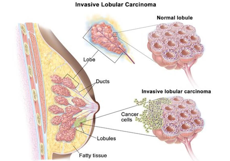
📋 ICD-10 Classification
In the International Classification of Diseases, Tenth Revision (ICD-10), invasive lobular carcinoma is classified under C50.9 — Malignant neoplasm of breast, unspecified site when the specific subsite is not documented, or under site-specific codes within the C50 category (e.g., C50.0–C50.9) depending on the quadrant involved. The corresponding ICD-O morphology code is 8520/3.
🔬 What Causes ILC?
The defining molecular event in ILC is the loss of functional E-cadherin, a protein encoded by the CDH1 gene located on chromosome 16q22. E-cadherin is essential for cell-to-cell adhesion; its absence allows cancer cells to detach and infiltrate surrounding tissues in a discohesive, single-file pattern. In more than 90% of ILC cases, E-cadherin loss results from somatic mutations, deletions, or promoter methylation of CDH1. Germline CDH1 mutations cause hereditary diffuse gastric cancer (HDGC) syndrome, which confers a high lifetime risk of both ILC and diffuse gastric cancer.
🧬 Genetic and Hormonal Risk Factors
Beyond CDH1, pathogenic variants in ATM, BRCA2, CHEK2, and PALB2 are associated with an increased risk of ILC, whereas BRCA1 mutations are not. ILC is more strongly associated with lifetime exposure to female hormones than IDC; risk factors include early menarche, late menopause, late age at first birth, and use of postmenopausal hormone therapy.
🛡️ How to Avoid and Prevent ILC
There is no guaranteed method to prevent ILC, but risk-reduction strategies include:
-
Limiting postmenopausal hormone therapy, particularly combined estrogen-progestin regimens, which have been linked to increased ILC risk.
-
Maintaining a healthy body weight and lifestyle — regular physical activity, a varied diet, moderate alcohol consumption, and smoking cessation reduce the risk of all breast cancers.
-
High-risk genetic carriers (e.g., CDH1 mutation carriers) should undergo intensive surveillance with annual breast MRI and mammography starting at age 30, and may consider risk-reducing mastectomy.
🧫 Types of ILC
ILC is not a single entity; histological subtyping recognizes several variants with differing biological behavior:
🔹 Classic ILC
The most common form, characterized by small, uniform cells infiltrating in single-file strands. Classic ILC typically has a favorable prognosis and is strongly hormone receptor–positive.
🔹 Pleomorphic ILC
A more aggressive variant (approximately 5% of ILC cases) with larger, pleomorphic cells, higher histological grade, and a greater likelihood of HER2 positivity and worse outcomes compared with classic ILC.
🔹 Other Variants
Additional patterns include solid, alveolar, tubulolobular, apocrine, signet-ring, histiocytoid, and mixed subtypes. The alveolar and tubulolobular variants tend to have a better prognosis, while solid and pleomorphic variants are associated with worse outcomes.
🩺 Symptoms and Early Signs
ILC often does not produce a discrete lump, and many cases are asymptomatic until detected on imaging. When symptoms occur, they may include:
-
An area of thickening, swelling, or fullness in the breast
-
Skin changes such as dimpling, puckering, or thickening (peau d’orange)
-
Nipple changes, including inversion or discharge
-
Breast pain or warmth
-
A lump near the armpit (axillary lymph node involvement)
Because these signs are subtle, ILC is often larger at diagnosis than IDC.
🖥️ Diagnosis: Imaging and Biopsy
Accurate diagnosis of ILC requires a multimodality approach because conventional mammography has limited sensitivity, particularly in dense breast tissue.
📸 Mammography
Mammography sensitivity for ILC is significantly lower than for IDC, dropping to as low as 11% in dense breasts in some studies. The infiltrative, non–mass-forming growth pattern of ILC contributes to this limitation.
🔊 Ultrasound
Breast ultrasound improves detection compared with mammography and is useful for evaluating palpable abnormalities and axillary lymph nodes, though it may underestimate tumor size.
🧲 Magnetic Resonance Imaging (MRI)
MRI is the most sensitive imaging modality for ILC, with detection rates approaching 100% for index lesions and superior accuracy for multifocal and multicentric disease. It is particularly recommended for dense breasts and for surgical planning.
🩻 Computed Tomography (CT) and PET/CT
Contrast-enhanced CT can delineate ILC margins with greater accuracy than conventional modalities. FDG-PET/CT is used for systemic staging, although ILC metastases are often hypometabolic and may be difficult to detect. The NCCN Guidelines now recommend fluoroestradiol (FES) PET for systemic staging in certain patients with recurrent or metastatic lobular breast cancer.
🔪 Biopsy
Core needle biopsy is the definitive diagnostic procedure. Histopathological confirmation requires demonstration of the characteristic single-file infiltrative pattern and, ideally, absence of E-cadherin expression on immunohistochemistry.
💊 Treatment Types
Treatment for ILC is multidisciplinary and stage-dependent, generally following the same principles as other invasive breast cancers, with important subtype-specific considerations.
🔪 Surgery
Breast-conserving surgery (lumpectomy) or mastectomy is the primary local treatment. Sentinel lymph node biopsy (SLNB) is the standard for axillary staging in clinically node-negative disease. ILC’s infiltrative growth pattern can make margin assessment challenging, and multifocality is common, sometimes necessitating mastectomy.
☢️ Radiation Therapy
Postoperative radiotherapy is indicated after breast-conserving surgery and for high-risk mastectomy cases, following standard breast cancer guidelines.
💊 Endocrine Therapy
Because approximately 90% of ILC tumors are hormone receptor–positive, endocrine therapy (tamoxifen or aromatase inhibitors) is the cornerstone of systemic treatment, typically continued for 5–10 years. ILC responds well to endocrine therapy, and this is often the optimal systemic choice.
🧪 Chemotherapy
ILC is less sensitive to neoadjuvant chemotherapy than IDC, with lower pathologic complete response (pCR) rates. In ER-positive/HER2-negative disease, chemotherapy-based regimens have not consistently improved response compared with endocrine-based therapy.
🎯 Targeted Therapy
CDK4/6 inhibitors (e.g., palbociclib, ribociclib, abemaciclib) combined with endocrine therapy improve outcomes in hormone receptor–positive, HER2-negative metastatic disease and are being incorporated into high-risk early-stage treatment. Anti-HER2 therapy is reserved for the minority (<10%) of ILC tumors that are HER2-positive.
🔬 Emerging Approaches
Investigational strategies include therapies targeting ILC-enriched mutations (e.g., PIK3CA, ERBB2) and synthetic lethality approaches such as ROS1 inhibition in CDH1-deficient tumors.
📊 Staging
ILC is staged using the AJCC TNM system, identical to other breast cancers. Staging incorporates tumor size (T), nodal involvement (N), distant metastasis (M), histological grade, hormone receptor status, and HER2 status.
-
Stage I: Tumor ≤2 cm, node-negative, no distant spread.
-
Stage II: Tumor 2–5 cm with limited nodal involvement, or >5 cm without nodal spread.
-
Stage III: Locally advanced disease with extensive nodal involvement or chest wall/skin involvement.
-
Stage IV: Distant metastasis.
Prognostic staging (clinical and pathological) incorporates biomarker status and is increasingly used to guide systemic therapy decisions.
🌐 Metastasis
ILC exhibits a distinctive metastatic pattern compared with IDC. While both subtypes metastasize to bone, liver, lungs, and brain, ILC has a greater tendency to spread to the peritoneum, retroperitoneum, and gastrointestinal tract. ILC metastases are frequently infiltrative and hypometabolic, making them difficult to detect on conventional and metabolic imaging; they may present late with complications such as bowel obstruction, hydronephrosis, or liver failure.
Other characteristic features include:
-
Bone metastases are common and may be mixed osteosclerotic/lytic.
-
Skip metastases (spread across discontinuous nodal levels or distant metastases without ipsilateral nodal involvement) are overrepresented in ILC (23.5% vs 6.7% in NST).
-
Late relapse can occur more than 10–20 years after initial diagnosis, necessitating long-term surveillance.
📈 Cure Rate and Survival Rate
Survival outcomes for ILC are generally comparable to IDC in early-stage disease, but important differences emerge over time and by stage.
📉 Five-Year Relative Survival
The overall 5-year relative survival rate for ILC is approximately 93%, slightly higher than the 90% observed for IDC. Stage-specific 5-year disease-specific survival for ILC has been reported as 90% (vs 88% for IDC) in stage-matched comparisons.
📊 Ten-Year Survival and Stage-Specific Differences
Survival for ILC is slightly higher than for IDC in the first seven years after diagnosis, but lower for regional and distant disease at 10 years. For distant (metastatic) disease, 10-year survival is 12.1% for ILC versus 19.6% for IDC. For regional-stage disease, 10-year survival is 76.4% for ILC versus 78.2% for IDC.
⏳ Metastatic ILC Survival
In metastatic disease, median overall survival (OS) varies by study and line of therapy:
-
30.2 months for metastatic ILC versus 33.7 months for metastatic IDC in the German OPAL real-world registry.
-
36 months median OS for ILC with bone-only metastases versus 42 months for IDC in a stage IV comparison.
-
49 months median OS for de novo stage IV ILC versus 69 months for IDC in another cohort.
⏱️ Life Expectancy With and Without Treatment
Life expectancy in ILC is highly stage-dependent. With guideline-concordant treatment, early-stage ILC is potentially curable, and the majority of patients achieve long-term survival. Metastatic ILC remains incurable, but systemic therapy—particularly endocrine therapy with CDK4/6 inhibition—can extend survival to a median of approximately 2.5–4 years in modern series.
Without treatment, ILC is a progressive malignancy that can metastasize and become life-limiting, although the slow-growing nature of classic ILC means that the trajectory can span years. Palliative care is appropriate at any stage to manage symptoms and improve quality of life.
🕊️ Palliative Care
Palliative care is an integral component of comprehensive ILC management, particularly for metastatic disease. Its goals are to relieve pain, manage symptoms, and support quality of life for patients and families. Modalities include:
-
Palliative radiotherapy for bone metastases, skull base metastases with cranial nerve involvement, and ocular metastases, which can improve quality of life even in terminal-stage disease.
-
Palliative systemic therapy (endocrine therapy, chemotherapy, or targeted agents) to control disease and alleviate symptoms.
-
Interventional procedures such as stent placement for gastric outlet obstruction caused by gastrointestinal metastases.
-
Hospice care for end-of-life support when disease-directed therapy is no longer beneficial.
❓ Questions and Answers
Basics, Biology, & Molecular Characteristics
-
What is invasive lobular carcinoma (ILC)?
ILC is a type of breast cancer that arises from the epithelial cells of the terminal duct lobular units and invades surrounding breast tissue. Its defining biological feature is the functional loss of E-cadherin, a protein that mediates cell-to-cell adhesion. -
Where does invasive lobular carcinoma start in the breast?
ILC begins in the lobules (the milk-producing glands) and the terminal duct lobular units (TDLUs) of the breast. -
What is the lobule in breast anatomy?
A lobule is a cluster of milk-producing alveoli within the breast, connected to a duct that carries milk toward the nipple. The terminal duct lobular unit (TDLU) is the functional milk-producing structure of the breast and the site of origin for lobular neoplasia and ILC. -
How common is invasive lobular carcinoma?
ILC accounts for 10–15% of all invasive breast cancers. -
What percentage of breast cancers are invasive lobular carcinoma (~10–15%)?
Approximately 10–15% of all breast cancers are ILC. -
Is ILC the second most common type of breast cancer?
Yes. ILC is the second most common histological type of breast cancer after invasive ductal carcinoma of no special type (IDC/NST). -
What is E-cadherin (CDH1), and why is it critical in ILC?
E-cadherin is a transmembrane glycoprotein encoded by the CDH1 gene on chromosome 16q22. It is essential for cell-to-cell adhesion in epithelial tissues. In ILC, loss of functional E-cadherin removes the “glue” that holds cells together, allowing them to detach and invade in single-file lines. -
What happens when E-cadherin protein expression is lost in breast tissue?
Loss of E-cadherin leads to a discohesive growth pattern, in which tumor cells infiltrate the stroma as individual cells or single-file strands rather than forming cohesive glandular structures or a solid mass. -
How does loss of E-cadherin cause ILC cells to grow in single-file lines?
Without E-cadherin-mediated adhesion, ILC cells cannot stick together to form a cohesive tumor mass. Instead, they migrate individually through the extracellular matrix, often aligning in “Indian file” strands along collagen bundles and tissue planes. -
What is the “Indian file” or single-file pattern in ILC pathology?
The “Indian file” pattern refers to the histological appearance of ILC cells arranged in single-file linear cords infiltrating the breast stroma. This pattern is a hallmark diagnostic feature of classic ILC. -
Why does ILC usually form a sheet or web rather than a distinct hard lump?
Because ILC cells lack E-cadherin and do not provoke a prominent desmoplastic (fibrotic) reaction, they spread diffusely through the breast tissue without forming a well-circumscribed mass. The result is a subtle thickening or web-like infiltration rather than a discrete lump. -
What is the difference between ILC and IDC (Invasive Ductal Carcinoma)?
IDC (now called invasive breast cancer of no special type, NST) arises from the milk ducts and typically forms a palpable, distinct mass with a desmoplastic reaction. ILC arises from the lobules, lacks E-cadherin, and grows in a diffuse, single-file pattern, often without a discrete lump. ILC also has a higher rate of multifocality and a distinctive metastatic pattern involving the peritoneum and gastrointestinal tract. -
What is the difference between LCIS (Lobular Carcinoma In Situ) and ILC?
LCIS is a non-invasive lesion in which abnormal cells fill and distend the lobules but do not break through the basement membrane. ILC is invasive cancer in which cells have breached the basement membrane and infiltrated the surrounding breast stroma. -
Is LCIS considered true cancer or a risk factor for ILC?
LCIS is not considered true invasive cancer. It is best regarded as a risk factor and a non-obligate precursor for subsequent invasive carcinoma (either ductal or lobular). It confers an approximately 8–10-fold increased relative risk of developing breast cancer. -
What is Pleomorphic LCIS (PLCIS)?
Pleomorphic LCIS is a variant of LCIS with larger, more atypical cells and a higher risk of progression to invasive carcinoma, particularly pleomorphic ILC. It often requires more aggressive management than classic LCIS. -
What are the histological variants of ILC?
Recognized variants include classic, pleomorphic, solid, alveolar, tubulolobular, signet-ring, apocrine, histiocytoid, and mixed subtypes. -
What is Classical Invasive Lobular Carcinoma?
Classic ILC is the most common variant, characterized by small, uniform cells infiltrating in single-file strands. It is typically hormone receptor–positive, HER2-negative, low-grade, and has a relatively favorable prognosis. -
What is Pleomorphic Invasive Lobular Carcinoma (PILC)?
PILC is a more aggressive variant (approximately 5% of ILC cases) with larger, pleomorphic cells, higher histological grade, and a greater likelihood of HER2 positivity and hormone receptor loss compared with classic ILC. -
Why is pleomorphic ILC considered more aggressive than classical ILC?
PILC is associated with higher histological grade, larger tumor size, more frequent HER2 amplification, and lower overall survival. The pleomorphic subtype occurs statistically significantly less often as the Luminal A subtype compared to others. -
What is Solid Variant Invasive Lobular Carcinoma?
The solid variant is characterized by cohesive sheets or nests of lobular cells with little intervening stroma, rather than single-file infiltration. It can be mistaken for ductal carcinoma and often has a higher grade than classic ILC. -
What is Alveolar Variant Invasive Lobular Carcinoma?
In the alveolar variant, tumor cells are arranged in small, rounded nests (alveoli) separated by thin fibrous septa. The ER-positivity rate is highest for the alveolar variant among the ILC subtypes. -
What is Tubulolobular Carcinoma?
Tubulolobular carcinoma shows features of both lobular and tubular carcinoma, with small tubules and single-file cords. It typically has a favorable prognosis. -
What is Mixed Invasive Ductal and Lobular Carcinoma (IDC/ILC)?
Mixed carcinomas show both ductal and lobular components and are classified as mixed when the lobular component constitutes a significant proportion of the tumor. -
What is Signet-Ring Cell Lobular Carcinoma?
Signet-ring cell carcinoma is a rare variant in which tumor cells contain a large cytoplasmic mucin vacuole that pushes the nucleus to the periphery, creating a signet-ring appearance. -
Is ILC considered slow-growing compared to IDC?
Classic ILC generally has a lower proliferation rate (Ki-67) than IDC, but its diffuse growth pattern and tendency for late recurrence mean it is not uniformly “slow-growing” in clinical behavior. -
What is the typical proliferation rate (Ki-67 score) in classical ILC?
Ki-67 is typically low (<15–20%) in classic ILC, consistent with its classification as a luminal A–type tumor. -
Are most ILC tumors hormone receptor-positive (ER+ and PR+)?
Yes. More than 90% of primary ILCs express estrogen receptor (ER) and progesterone receptor (PR). -
What percentage of ILC tumors are Estrogen Receptor-positive (ER+)?
Approximately 90–95% of ILC tumors are ER-positive. -
What percentage of ILC tumors are Progesterone Receptor-positive (PR+)?
Approximately 70–80% of ILC tumors are PR-positive. -
Is HER2-positive ILC rare?
Yes. HER2 amplification or overexpression is present in only 3–13% of ILC cases, and it is particularly rare in classic, non-pleomorphic ILC. -
What percentage of classical ILC tumors are HER2-negative (~90–95%)?
Approximately 90–97% of classic ILC tumors are HER2-negative. -
What is Triple-Negative Invasive Lobular Carcinoma (TN-ILC)?
TN-ILC lacks ER, PR, and HER2 expression. It is rare, accounting for approximately 2% of ILC cases, and is more common in pleomorphic variants. -
How common is triple-negative ILC?
Approximately 2% of all ILC cases are triple-negative. -
What intrinsic molecular subtype is classical ILC usually classified as (Luminal A)?
Classic ILC is most commonly classified as the Luminal A intrinsic subtype, characterized by high ER expression, low proliferation, and favorable prognosis. -
What intrinsic molecular subtype is pleomorphic ILC often classified as (Luminal B or HER2-enriched)?
Pleomorphic ILC is more frequently classified as Luminal B or HER2-enriched, reflecting its more aggressive biology. -
Can men get invasive lobular carcinoma?
Yes, men can develop ILC, but it is extremely rare. Male breast cancer accounts for <1% of all breast cancers, and ILC represents a small minority of male cases. -
Why is male ILC extremely rare compared to male IDC?
Male breast tissue contains far fewer lobules and less glandular tissue than female breast tissue, reducing the opportunity for lobular carcinogenesis. -
What is the average age of diagnosis for invasive lobular carcinoma (~60–65 years)?
The median age at diagnosis is approximately 60–65 years, with 67% of cases diagnosed in women over 60. -
Is ILC diagnosed at a later average age than IDC?
Yes. ILC is more common in postmenopausal women, and its incidence peaks later than that of IDC. -
Can young women under 40 get invasive lobular carcinoma?
Yes, although it is uncommon. Approximately 2% of ILC cases occur in women aged 20–39. -
What is multifocal ILC?
Multifocal ILC refers to two or more separate tumor foci within the same quadrant of the breast. -
What is multicentric ILC?
Multicentric ILC refers to tumor foci in different quadrants of the same breast. -
Why does ILC have a higher rate of multifocality and multicentricity than IDC?
The diffuse, infiltrative growth pattern of ILC makes it more likely to involve multiple areas of the breast simultaneously, and it is often underestimated by conventional imaging. -
What is bilateral ILC?
Bilateral ILC refers to synchronous or metachronous tumors in both breasts. -
What is the rate of synchronous or metachronous bilateral breast cancer in ILC (~10–15%)?
The rate of bilateral breast cancer in ILC is approximately 10–15%, higher than in IDC. -
Why is the opposite (contralateral) breast at higher risk in ILC patients?
The same hormonal and genetic risk factors that predispose to ILC in one breast also predispose the contralateral breast, and lobular neoplasia is often bilateral. -
What is extracellular matrix remodelling in ILC?
ILC cells interact with and remodel the extracellular matrix to facilitate their single-file invasion through tissue planes, a process that is distinct from the desmoplastic reaction typical of IDC. -
What role do catenins (p120-catenin, beta-catenin) play in ILC cell adhesion loss?
Catenins link E-cadherin to the actin cytoskeleton. When E-cadherin is lost, p120-catenin translocates to the cytoplasm, and this pattern can be detected by immunohistochemistry to support a diagnosis of ILC. -
What is p120-catenin cytoplasmic translocation on IHC staining?
In normal epithelial cells, p120-catenin is membrane-bound. In ILC with E-cadherin loss, p120-catenin shows diffuse cytoplasmic staining, which is a diagnostic hallmark. -
Why is p120-catenin IHC used to confirm a diagnosis of ILC when E-cadherin is ambiguous?
Cytoplasmic p120-catenin staining provides additional evidence of E-cadherin dysfunction when E-cadherin staining is equivocal or technically suboptimal. -
What is the CDH1 gene mutation, and how does it relate to ILC?
CDH1 encodes E-cadherin. Somatic CDH1 mutations are found in a large proportion of ILCs, and germline CDH1 mutations cause hereditary diffuse gastric cancer (HDGC) syndrome, which carries a high lifetime risk of ILC. -
What is Hereditary Diffuse Gastric Cancer (HDGC) syndrome?
HDGC is an autosomal dominant syndrome caused by germline CDH1 mutations, characterized by a high lifetime risk of diffuse gastric cancer and invasive lobular breast cancer in women. -
How is HDGC syndrome linked to invasive lobular carcinoma?
Women with germline CDH1 mutations have a lifetime risk of ILC estimated at approximately 37–60% in various studies. -
What is the lifetime risk of ILC for a female with a germline CDH1 mutation (~30–50%)?
Estimates range from approximately 37% to 60%, depending on the study population. -
What other gene mutations are commonly found in ILC tumors (PIK3CA, PTEN, AKT1, FOXA1)?
Recurrent somatic alterations in ILC include PIK3CA, PTEN, AKT1, FOXA1, ERBB2, and TP53 (the latter more common in pleomorphic ILC). -
Why are PIK3CA mutations particularly frequent in classical ILC (~40–50%)?
PIK3CA mutations activate the PI3K/AKT/mTOR pathway, promoting cell survival and proliferation. They are particularly common in hormone receptor–positive, luminal-type tumors such as classic ILC. -
What is ESR1 mutation in ILC, and when does it develop?
ESR1 mutations emerge in metastatic, endocrine-resistant ER-positive breast cancer, including ILC, and confer resistance to aromatase inhibitors by producing a constitutively active estrogen receptor. -
What is tumor stroma interaction in lobular carcinoma?
ILC cells interact with the surrounding stroma to facilitate their diffuse, single-file invasion, and the stroma in ILC is biologically distinct from the desmoplastic stroma of IDC. -
Does ILC trigger a prominent desmoplastic (fibrotic) tissue reaction like IDC does?
No. ILC typically provokes little to no desmoplastic reaction, which is one reason it does not form a hard, palpable lump and is difficult to detect by physical examination and mammography. -
Why does lack of desmoplastic reaction make ILC difficult to feel on physical exam?
Without a fibrotic reaction, ILC does not create a firm, distinct mass. Instead, it produces a subtle thickening or ill-defined fullness that is easily missed on palpation. -
What is the tumor microenvironment in ILC?
The ILC tumor microenvironment is characterized by low tumor-infiltrating lymphocytes (TILs) and a stroma that supports diffuse invasion rather than a robust immune response. -
Are tumor-infiltrating lymphocytes (TILs) usually low in classical ILC?
Yes. Classic ILC typically has low TIL levels, which may contribute to its relative resistance to immunotherapy. -
What is microinvasive lobular carcinoma (T1mic)?
Microinvasion refers to a focus of invasive carcinoma measuring ≤1 mm in greatest dimension. It is uncommon in lobular carcinoma but can occur. -
Is ILC considered a primary breast cancer?
Yes. ILC is a primary breast malignancy, not a metastasis from another site. -
Can ILC develop in ectopic or accessory breast tissue (e.g., armpit)?
Yes, breast cancer including ILC can arise in accessory breast tissue along the milk line, including the axilla. -
How long can invasive lobular carcinoma remain undetected in the breast?
Because of its subtle presentation, ILC can remain undetected for months to years and is often larger at diagnosis than IDC. -
What is tumor heterogeneity in mixed ductal-lobular carcinomas?
Mixed ductal-lobular carcinomas contain distinct ductal and lobular components, each with potentially different biological features and treatment responses. -
Is ILC more common in women who used combined Hormone Replacement Therapy (HRT)?
Yes. Combined estrogen-progestin HRT is associated with an increased risk of ILC, with relative risks exceeding 2 in some studies. -
Why did ILC incidence rise significantly during the peak era of HRT use in the 1990s?
The widespread use of combined HRT during the 1990s contributed to the rising incidence of ILC, which is more strongly associated with hormonal exposure than IDC. -
Did ILC incidence drop after the Women’s Health Initiative (WHI) report reduced HRT use?
Incidence trends have fluctuated, but the decline in combined HRT use after the WHI report was associated with a leveling or decline in ILC incidence in some populations. -
Is ILC more common in dense breast tissue?
Yes. High mammographic density is associated with an increased risk of ILC, and density also reduces the sensitivity of mammography for detecting ILC. -
How does high breast density complicate early detection of ILC?
Dense fibroglandular tissue can mask the subtle architectural distortion caused by ILC, leading to false-negative mammograms and delayed diagnosis. -
What is the tumor doubling time for classical ILC?
Classic ILC generally has a longer doubling time than high-grade IDC, consistent with its lower proliferation index. -
Is ILC a fast-metastasizing cancer in its early stages?
Classic ILC is generally not rapidly metastasizing in its early stages, but its diffuse local spread and potential for late distant recurrence mean it requires careful long-term surveillance. -
What is the cell of origin in lobular neoplasia?
Lobular neoplasia is believed to arise from luminal epithelial cells of the terminal duct lobular unit that acquire CDH1 alterations. -
What is the difference between Atypical Lobular Hyperplasia (ALH) and LCIS?
ALH and LCIS lie on a spectrum of lobular neoplasia. The distinction is quantitative: if more than 50% of the TDLU is involved, the lesion is classified as LCIS; lesser involvement is ALH. -
What is the combined term “Lobular Neoplasia”?
“Lobular neoplasia” (LN) encompasses both ALH and LCIS, recognizing them as a spectrum of non-obligate precursor lesions. -
Does ALH increase the risk of developing ILC in either breast?
Yes. ALH is associated with an approximately 4–5-fold increased relative risk of subsequent breast cancer, and the risk is bilateral, though it predominates in the ipsilateral breast. -
How many times does ALH raise lifetime breast cancer risk (approx 4–5x)?
Approximately 4–5 times the relative risk. -
How many times does LCIS raise lifetime breast cancer risk (approx 8–10x)?
Approximately 8–10 times the relative risk. -
Is ILC always aggressive?
No. Classic ILC is generally indolent and hormone-sensitive, but pleomorphic and other high-grade variants can be aggressive. The clinical behavior depends on the histological variant, grade, stage, and molecular features. -
What makes classical ILC behave differently from aggressive subtypes?
Classic ILC is typically low-grade, ER-positive, HER2-negative, and has a low proliferation rate, whereas pleomorphic and solid variants are more likely to be high-grade, HER2-positive, or triple-negative. -
Why is ILC sometimes referred to as a “stealth” breast cancer?
Because it often does not form a palpable lump, is frequently occult on mammography, and can spread to unusual sites, ILC is sometimes described as a “stealth” cancer. -
How does ILC invade surrounding healthy breast tissue without destroying overall architecture initially?
ILC cells infiltrate along existing tissue planes and collagen bundles in single-file cords, preserving the overall architectural framework of the breast to some extent, which contributes to its subtle presentation. -
What is concentric targetoid growth pattern around ducts in ILC?
In some ILC variants, tumor cells form a target-like arrangement around ducts, reflecting their infiltrative growth pattern. -
What is the difference between primary ILC and recurrent ILC?
Primary ILC is the initial tumor. Recurrent ILC is disease that returns after initial treatment, either locally (in the breast or chest wall), regionally (lymph nodes), or distantly (metastatic). -
Can ILC change its hormone receptor or HER2 status upon recurrence?
Yes. Biomarker conversion between primary and metastatic tumors occurs in a proportion of cases, which is why re-biopsy of metastatic lesions is recommended when feasible. -
What is HER2-low status in invasive lobular carcinoma?
HER2-low refers to tumors with HER2 immunohistochemistry 1+ or 2+ without gene amplification, a category that has become therapeutically relevant with HER2-directed antibody-drug conjugates. -
Are HER2-low tumors common in ILC?
HER2-low status is relatively common in ILC, particularly in hormone receptor–positive cases. -
What is androgen receptor (AR) expression level in classical ILC?
AR is expressed in a substantial proportion of ER-positive ILCs, and AR expression may influence endocrine responsiveness. -
What is FGFR1 amplification in lobular breast cancer?
FGFR1 amplification is a recurrent genomic alteration in some ILCs and is being explored as a therapeutic target. -
How does loss of E-cadherin alter growth factor signaling pathways (EGFR, IGFR)?
E-cadherin loss can release catenins and alter signaling through EGFR, IGFR, and other receptor tyrosine kinase pathways, contributing to ILC cell survival and invasion. -
What is the role of lipid metabolism genes in lobular cancer cell survival?
ILC cells may rely on altered lipid metabolism for survival and growth, and this is an area of active research for potential therapeutic targeting. -
Can ILC occur in young BRCA1 or BRCA2 mutation carriers?
ILC is rare in BRCA1 carriers but occurs in BRCA2 carriers. BRCA2 pathogenic variants are associated with an increased risk of ILC. -
Is ILC more strongly associated with BRCA2 mutations than BRCA1 mutations?
Yes. BRCA1 mutations are not associated with an increased risk of ILC, whereas BRCA2 mutations are. -
What is PALB2 mutation link to ILC?
PALB2 pathogenic variants are associated with a moderate increased risk of ILC (OR approximately 2–4). -
What is CHEK2 mutation link to ILC?
CHEK2 pathogenic variants are associated with an increased risk of ILC. -
What is ATM mutation link to ILC?
ATM pathogenic variants are associated with an increased risk of ILC. -
What is the overall medical definition of invasive lobular carcinoma?
ILC is an invasive breast carcinoma arising from the lobules, characterized by E-cadherin loss and a discohesive, single-file infiltrative growth pattern, most commonly hormone receptor–positive and HER2-negative. -
Where can patients find certified basic scientific information on ILC?
Reliable sources include the Lobular Breast Cancer Alliance (LBCA), the National Cancer Institute (NCI), the American Society of Clinical Oncology (ASCO), the National Comprehensive Cancer Network (NCCN), and the American Cancer Society (ACS).
🩺Signs, Physical Changes, & Early Detection
-
What are the early warning signs of invasive lobular carcinoma?
Early ILC may produce no symptoms or subtle changes such as breast thickening, swelling, dimpling, nipple changes, or a vague sense of firmness. -
Does invasive lobular carcinoma form a typical hard breast lump?
Usually not. ILC often presents as an ill-defined thickening or diffuse change rather than a discrete hard lump. -
Why is ILC famous for NOT presenting as a distinct lump?
Because ILC cells lack E-cadherin and do not provoke a desmoplastic reaction, they infiltrate diffusely without forming a cohesive mass. -
What does ILC feel like on breast self-examination?
It may feel like a subtle firmness, thickening, or fullness rather than a distinct lump. In some cases, a vague mass may be palpable. -
What is a “thickening” or “fullness” in the breast in ILC?
This refers to an area of the breast that feels denser or more prominent than surrounding tissue, without a clear boundary. -
Why do women with ILC report a subtle change in breast texture rather than a lump?
Because the tumor infiltrates the stroma without forming a discrete mass, patients may notice a change in texture or firmness rather than a lump. -
Can ILC cause one breast to feel firmer or heavier than the other?
Yes. Diffuse infiltration can cause asymmetry in breast firmness or weight. -
Can ILC cause a change in breast shape or contour?
Yes. ILC can cause subtle distortion of the breast contour due to tethering of Cooper’s ligaments or diffuse infiltration. -
Can ILC cause one breast to shrink or pull inward?
Yes. Fibrotic changes and tethering can cause breast contraction or retraction in some cases. -
Why does ILC sometimes cause breast shrinkage or contraction?
Tumor infiltration and involvement of Cooper’s ligaments can cause retraction and a decrease in apparent breast volume. -
What skin changes are associated with invasive lobular carcinoma?
Skin changes may include dimpling, puckering, thickening, or peau d’orange (orange-peel appearance). -
Can ILC cause subtle skin dimpling or puckering?
Yes. Dimpling occurs when the tumor tethers the skin via Cooper’s ligaments. -
What nipple changes occur with ILC?
Nipple changes may include inversion, flattening, retraction, or discharge. -
Can ILC cause nipple inversion, flattening, or retraction?
Yes. Tumor involvement of the nipple-areolar complex or its supporting structures can cause nipple retraction or inversion. -
Can ILC cause nipple discharge?
Nipple discharge is uncommon in ILC compared with IDC but can occur. -
Is nipple discharge common in ILC compared to IDC?
No. Nipple discharge is less common in ILC than in IDC. -
Does ILC cause breast pain or tenderness?
Breast pain is not a primary symptom of ILC, but some patients report vague discomfort or tenderness. -
Is localized breast pain a primary symptom of ILC?
No. Pain is not a reliable early sign of ILC, and most ILCs are painless. -
Can ILC present without any physical symptoms at all?
Yes. Many ILCs are asymptomatic and detected on screening imaging. -
What percentage of ILC cases are completely asymptomatic at diagnosis?
A significant proportion—often the majority—are asymptomatic and detected by imaging. -
What does an underarm or axillary lump feel like in ILC?
An axillary lump may feel like a firm, enlarged lymph node, which can be the first sign of ILC with nodal involvement. -
Can swollen axillary lymph nodes be the first sign of ILC?
Yes. Axillary lymph node involvement can be the presenting sign in some patients. -
Can ILC cause redness, swelling, or warmth in the breast?
These features are more characteristic of inflammatory breast cancer but can occur in locally advanced ILC. -
How do symptoms of ILC differ from inflammatory breast cancer?
Inflammatory breast cancer presents with rapid onset of redness, swelling, warmth, and peau d’orange over a large area of the breast, often without a discrete mass. ILC typically has a more insidious onset with subtle thickening. -
Can ILC present with skin changes mimicking mastitis?
Yes, locally advanced ILC can mimic mastitis with redness and swelling, leading to delayed diagnosis. -
Why is ILC often diagnosed at a larger tumor size than IDC?
Because ILC is difficult to detect on physical examination and mammography, it is often larger at diagnosis. -
What is the average tumor size of ILC at clinical diagnosis (~2–3 cm or larger)?
The average size at diagnosis is often 2–3 cm or larger, and ILC tends to be larger than IDC at diagnosis. -
How does high breast density hide physical and mammographic signs of ILC?
Dense breast tissue can mask the subtle architectural distortion and ill-defined thickening caused by ILC. -
Can ILC symptoms appear in both breasts simultaneously?
Yes. Bilateral synchronous ILC can occur, particularly in women with lobular neoplasia or genetic predisposition. -
What subtle symmetry changes should women look for during breast self-exams?
Women should look for changes in size, shape, contour, skin texture, or nipple position between the two breasts. -
Does an ILC mass move or feel fixed to the chest wall?
ILC may feel fixed or tethered due to its infiltrative growth pattern and involvement of surrounding structures. -
Can ILC cause skin tethering or retraction when raising arms overhead?
Yes. Tethering of the skin to underlying structures can cause visible retraction or dimpling when the arms are raised. -
What causes skin dimpling when ILC grows near Cooper’s ligaments?
Tumor infiltration shortens and tethers Cooper’s ligaments, which connect the skin to the underlying fascia, causing dimpling. -
What does a “doughy” or “indurated” area in the breast feel like?
It feels like a firm, dense, or rubbery area without a clear border, distinct from a discrete lump. -
Can ILC cause changes in the bra cup fit on one side?
Yes. Diffuse infiltration or breast contraction can cause asymmetry in bra fit. -
Why do many women ignore early ILC symptoms for months?
Because the symptoms are subtle and non-specific, many women attribute them to benign changes and delay seeking evaluation. -
How long do women typically notice subtle breast changes before seeking medical evaluation?
Many women notice changes for weeks to months before seeking care, contributing to diagnostic delays. -
Can ILC cause itching or scaling on the breast skin or nipple?
Itching or scaling is uncommon in ILC and is more characteristic of Paget’s disease of the nipple. -
How do physical symptoms of ILC differ from benign fibrocystic changes?
Fibrocystic changes typically cause cyclical tenderness and lumpiness, whereas ILC causes persistent, non-cyclical thickening or distortion. -
How do physical symptoms of ILC differ from fibroadenomas?
Fibroadenomas are usually well-circumscribed, mobile, rubbery lumps, whereas ILC is an ill-defined, fixed thickening. -
How do physical symptoms of ILC differ from fat necrosis?
Fat necrosis often follows trauma and presents as a firm, tender lump, sometimes with skin changes. ILC is typically painless and diffuse. -
Can a breast injury or trauma draw attention to an underlying ILC tumor?
Yes. Trauma can bring attention to a pre-existing mass, but trauma does not cause ILC. -
Does trauma to the breast cause ILC?
No. Physical trauma does not cause breast cancer. -
What does a lump above or below the collarbone mean in advanced ILC?
Supraclavicular or infraclavicular lymph node enlargement indicates regional nodal metastasis (N3 disease). -
Can ILC cause arm swelling (lymphedema) prior to diagnosis?
Lymphedema before diagnosis is uncommon but can occur if axillary lymph nodes are extensively involved. -
What symptoms indicate ILC has spread to distant body sites?
Symptoms depend on the site and may include bone pain, abdominal bloating, nausea, bowel changes, pelvic pain, headaches, or neurological symptoms. -
What are the symptoms of ILC metastasis to the peritoneum or abdomen?
Symptoms include abdominal distension, bloating, pain, nausea, and ascites. -
Why does ILC metastasize to unusual sites like the gastrointestinal tract and ovaries?
The loss of E-cadherin and the discohesive nature of ILC cells facilitate diffuse seeding along serosal surfaces and retrograde lymphatic spread to the ovaries and peritoneum. -
What are symptoms of GI tract involvement from metastatic ILC (bloating, early satiety, bowel changes)?
Symptoms include nausea, vomiting, early satiety, abdominal pain, bloating, bowel obstruction, and weight loss. -
What are symptoms of peritoneal carcinomatosis from metastatic ILC (ascites, abdominal distension)?
Symptoms include abdominal distension, ascites, pain, and bowel dysfunction. -
What are symptoms of metastatic ILC in the stomach mimicking gastritis or stomach ulcer?
Symptoms include epigastric pain, nausea, vomiting, early satiety, and weight loss, often mimicking benign gastritis. -
What are symptoms of metastatic ILC in the ovaries (Krukenberg tumor / pelvic mass)?
Symptoms include pelvic pain, bloating, a palpable mass, and abdominal discomfort. -
What are symptoms of metastatic ILC in the uterus or cervix (abnormal vaginal bleeding)?
Abnormal postmenopausal vaginal bleeding or discharge can occur. -
What are symptoms of metastatic ILC in the ureters or retroperitoneum (flank pain, hydronephrosis)?
Symptoms include flank pain, elevated creatinine, hydronephrosis, and acute kidney injury. -
What are symptoms of metastatic ILC in the leptomeninges (headaches, nerve palsies, confusion)?
Symptoms include severe headaches, double vision, facial numbness, gait instability, seizures, and confusion. -
What are symptoms of metastatic ILC in the bones (deep aching bone pain, pathological fractures)?
Symptoms include persistent deep bone pain, pathological fractures, and spinal cord compression. -
What are symptoms of metastatic ILC in the lungs (shortness of breath, persistent cough, pleural effusion)?
Symptoms include shortness of breath, cough, chest pain, and pleural effusion. -
What are symptoms of metastatic ILC in the liver (jaundice, right upper quadrant pain)?
Symptoms include right upper quadrant pain, jaundice, fatigue, and elevated liver enzymes. -
Does ILC cause systemic symptoms like fatigue, night sweats, or fever early on?
Systemic symptoms are uncommon in early-stage ILC but can occur in advanced disease. -
Can unexplained weight loss be an early sign of metastatic ILC?
Yes. Unexplained weight loss can be a sign of advanced or metastatic disease, particularly with GI involvement. -
How does a clinical breast exam (CBE) by a doctor evaluate suspected ILC?
A CBE assesses breast symmetry, texture, skin changes, nipple position, and axillary lymph nodes. -
Why is physical palpation often inaccurate for measuring exact ILC tumor extent?
Because ILC is diffuse and ill-defined, palpation often underestimates the true size of the tumor. -
What is the “underestimation of size” phenomenon on clinical exam in ILC?
Clinical examination frequently underestimates the extent of ILC compared with pathological or MRI findings. -
Can a normal physical exam miss a large ILC tumor?
Yes. A normal physical exam does not exclude ILC, especially in deep or diffuse tumors. -
How do symptoms of pleomorphic ILC differ from classical ILC?
Pleomorphic ILC is more likely to form a palpable mass and may grow more rapidly than classic ILC. -
Does pleomorphic ILC form a distinct hard lump more often than classical ILC?
Yes. Pleomorphic ILC more often presents as a discrete mass compared with the diffuse presentation of classic ILC. -
Can ILC symptoms appear suddenly or do they develop gradually?
ILC symptoms usually develop gradually, but they can be noticed suddenly if they cause a visible change. -
How fast do physical changes progress in classical vs pleomorphic ILC?
Classic ILC progresses slowly, whereas pleomorphic ILC can progress more rapidly. -
What skin color changes can indicate locally advanced ILC?
Redness, erythema, or discoloration can indicate skin involvement or inflammatory changes. -
What does an ulceration or open sore on the breast skin mean in ILC?
Skin ulceration indicates advanced local disease with tumor infiltration through the skin. -
Can ILC cause prominent or visible veins on one breast?
Yes. Increased vascularity or prominent veins can occur with locally advanced disease. -
Can ILC present as an asymmetrical density noted only during a clinical exam?
Yes. Asymmetrical density may be the only physical finding in some cases. -
What physical changes indicate tumor invasion into the pectoralis muscle?
Fixation of the breast to the chest wall or pain on arm movement can indicate pectoralis muscle involvement. -
Does ILC cause nipple hypersensitivity or pain?
Nipple hypersensitivity is uncommon but can occur if the nipple-areolar complex is involved. -
Can lactating or pregnant women develop ILC, and what are the symptoms?
ILC can occur during pregnancy or lactation, but it is rare. Symptoms may be mistaken for clogged ducts or mastitis. -
How do symptoms of clogged milk ducts differ from ILC in postpartum women?
Clogged ducts typically resolve with massage, feeding, and warm compresses, whereas ILC causes persistent, progressive changes that do not resolve. -
Can breast implants hide physical signs of ILC?
Yes. Implants can mask subtle changes and make physical examination more challenging. -
How do you check for subtle ILC changes if you have breast implants?
Regular MRI or ultrasound surveillance may be recommended, in addition to clinical examination. -
What skin changes indicate tumor spread to the skin (satellite nodules)?
Satellite nodules—small, firm nodules near the primary tumor—indicate skin metastasis. -
Can ILC cause localized warmth without redness?
Yes. Localized warmth without visible redness can occur with inflammatory or locally advanced disease. -
What is the “breast contraction” sign in lobular carcinoma?
Breast contraction refers to shrinkage or retraction of the breast due to tumor infiltration and fibrosis. -
Why do some women experience a decrease in breast volume on the affected side?
Tumor infiltration and tethering can cause volume loss and contraction of the affected breast. -
Can ILC symptoms be masked by post-menopausal breast tissue atrophy?
Yes. Postmenopausal fatty replacement and atrophy can make subtle ILC changes even harder to detect. -
What physical symptoms should prompt an immediate diagnostic appointment?
Any new, persistent breast thickening, lump, skin change, nipple change, or axillary swelling should prompt prompt evaluation. -
How do doctors evaluate a subtle ill-defined thickening in high-risk women?
Doctors use clinical breast examination, diagnostic mammography, ultrasound, and often MRI, followed by core needle biopsy if indicated. -
Can a self-exam using flat fingers detect subtle ILC thickening better than fingertip poking?
Using flat fingers and circular motions with varying pressure can help detect subtle thickening more effectively than fingertip poking. -
What is the role of clinical breast exam in routine screening for women with lobular neoplasia?
Clinical breast exam remains an important component of surveillance, along with annual mammography and, in high-risk women, MRI. -
Can ILC symptoms mimic chronic dermatitis of the areola?
Yes. ILC involving the nipple-areolar complex can mimic eczema or dermatitis. -
What physical signs distinguish Paget’s disease from ILC involving the nipple?
Paget’s disease typically presents with itching, scaling, and erosion of the nipple, often with an underlying ductal carcinoma in situ or invasive ductal carcinoma. ILC more commonly causes retraction or inversion rather than eczematous changes. -
Can ILC cause a sensation of a “pulled muscle” in the chest wall?
Yes. Tumor infiltration into the pectoralis muscle or chest wall can cause a pulling or aching sensation. -
What percentage of ILC tumors are detected by routine screening vs self-detected symptoms?
A significant proportion are detected by routine screening, but many are self-detected because of ILC’s subtle imaging appearance. -
How does breast size impact the ability to feel an ILC thickening?
Large breasts may make it more difficult to palpate deep or diffuse lesions, while small breasts may make subtle changes easier to detect. -
Can ILC present with lower back pain if bone metastasis is the first sign?
Yes. Bone metastasis, particularly to the spine, can cause lower back pain as the presenting symptom. -
Why is back or pelvic pain sometimes the initial presenting symptom of undiagnosed ILC?
Because ILC can metastasize to bone and remain clinically occult at the primary site, bone pain may be the first symptom. -
What gastrointestinal symptoms should prompt ILC survivors to seek evaluation?
Persistent nausea, vomiting, early satiety, abdominal pain, bloating, or bowel changes should prompt evaluation for possible GI metastasis. -
How do doctors educate high-risk women on recognizing subtle ILC physical changes?
Doctors provide breast self-awareness education and emphasize the importance of reporting persistent changes rather than waiting for a lump to appear. -
Does feeling a thickening automatically guarantee an ILC diagnosis?
No. Many benign conditions can cause breast thickening, and biopsy is required for diagnosis. -
What benign conditions can cause breast thickening (fat necrosis, radial scar, sclerosing adenosis)?
Benign causes include fat necrosis, radial scar, sclerosing adenosis, fibrocystic changes, and focal fibrosis. -
What is the most critical red-flag symptom for invasive lobular carcinoma?
A new, persistent, unilateral breast thickening or skin change that does not resolve is the most critical red flag. -
What immediate action should a woman take upon noticing new breast asymmetry or thickening?
She should schedule an appointment with a healthcare provider for clinical evaluation and imaging.
🧬Causes, Risk Factors, & Genetics
-
What causes invasive lobular carcinoma?
The fundamental cause is the loss of E-cadherin function, usually through somatic CDH1 mutations or epigenetic silencing, which leads to discohesive cellular growth. Hormonal, reproductive, genetic, and lifestyle factors contribute to risk. -
What are the primary risk factors for developing ILC?
Primary risk factors include female sex, increasing age, postmenopausal status, combined hormone replacement therapy, alcohol consumption, obesity, and genetic predisposition (CDH1, BRCA2, ATM, CHEK2, PALB2). -
How does age increase the risk of invasive lobular carcinoma?
ILC incidence increases with age, with 67% of cases diagnosed in women over 60. -
Why is ILC more common in postmenopausal women?
ILC is strongly associated with cumulative lifetime estrogen exposure, which is highest in postmenopausal women, particularly those with obesity or HRT use. -
How does hormone replacement therapy (HRT) increase ILC risk?
Combined estrogen-progestin HRT increases the risk of ILC, with relative risks exceeding 2 in some studies. -
Does combined estrogen-progestin HRT increase ILC risk more than estrogen-only HRT?
Yes. Combined HRT is associated with a greater increase in ILC risk than estrogen-only therapy. -
Why is ILC more strongly associated with HRT use than IDC?
ILC is more strongly hormone-dependent than IDC, and progestin exposure may particularly promote lobular carcinogenesis. -
How many years of HRT use significantly raise the risk of developing ILC?
Risk increases with longer duration of use, particularly after 5 or more years of combined HRT. -
Does risk of ILC decrease after stopping HRT?
Yes. Risk declines after stopping HRT, although it may not return to baseline immediately. -
Do oral contraceptives (birth control pills) increase ILC risk?
Prolonged oral contraceptive use (≥5 years) has been associated with a modest increase in ILC risk. -
Does bioidentical hormone therapy raise ILC risk?
Bioidentical hormones are not risk-free and may carry similar risks to conventional HRT, although data are more limited. -
What role does endogenous estrogen play in driving ILC development?
Endogenous estrogen promotes cellular proliferation in the breast, and higher lifetime exposure is associated with increased ILC risk. -
Does early menarche (starting period before age 12) increase ILC risk?
Yes. Early menarche increases lifetime estrogen exposure and is associated with a modestly increased risk. -
Does late menopause (after age 55) increase ILC risk?
Yes. Late menopause prolongs estrogen exposure and is associated with increased risk. -
Does nulliparity (never having children) raise the risk of ILC?
Yes. Nulliparity is associated with an increased risk of breast cancer, including ILC. -
Does age at first childbirth (>30 years) impact ILC risk?
Yes. Older age at first full-term pregnancy is associated with an increased risk. -
Does breastfeeding lower the risk of developing ILC?
Breastfeeding is associated with a modest reduction in breast cancer risk, including ILC. -
How does dense breast tissue increase ILC risk?
Dense breast tissue contains more glandular and fibrous tissue, which is associated with increased cellular proliferation and cancer risk. -
Is high mammographic density a stronger risk factor for ILC than IDC?
High density is a risk factor for both, but it has a particularly strong impact on the detectability of ILC. -
Is a prior history of Lobular Carcinoma In Situ (LCIS) a risk factor for ILC?
Yes. LCIS confers an approximately 8–10-fold increased relative risk of subsequent invasive carcinoma, including ILC. -
What is the annual risk of developing ILC after a diagnosis of classical LCIS (~1% per year)?
The annual risk is approximately 1% per year, cumulating over time. -
Is a prior history of Atypical Lobular Hyperplasia (ALH) a risk factor for ILC?
Yes. ALH confers an approximately 4–5-fold increased relative risk. -
Does LCIS or ALH increase the risk of cancer in BOTH breasts equally?
The risk is bilateral, although it predominates in the ipsilateral breast. -
Is a prior diagnosis of Atypical Ductal Hyperplasia (ADH) a risk factor?
ADH is a risk factor for breast cancer generally, but it is more strongly associated with ductal carcinoma. -
Is a prior personal history of breast cancer a risk factor for developing ILC in the opposite breast?
Yes. A personal history of breast cancer increases the risk of contralateral breast cancer, including ILC. -
Is a family history of breast cancer a major risk factor for ILC?
Yes. A family history of breast cancer, particularly ILC, increases risk. However, most women with ILC have no family history. -
What percentage of ILC cases are hereditary vs sporadic?
The majority of ILC cases are sporadic, with approximately 5–10% associated with inherited pathogenic variants. -
How does the CDH1 germline mutation cause hereditary ILC?
Germline CDH1 mutations cause HDGC syndrome, which carries a high lifetime risk of ILC (approximately 37–60%). -
What is Hereditary Diffuse Gastric Cancer (HDGC) syndrome, and how is it inherited?
HDGC is an autosomal dominant syndrome caused by germline CDH1 mutations, characterized by diffuse gastric cancer and lobular breast cancer. -
Should families with both diffuse gastric cancer and ILC undergo CDH1 genetic testing?
Yes. CDH1 genetic testing should be offered to families with a history of diffuse gastric cancer and/or ILC. -
Do BRCA1 or BRCA2 gene mutations increase the risk of ILC?
BRCA2 mutations increase the risk of ILC, whereas BRCA1 mutations do not. -
Why is ILC more frequently seen in BRCA2 carriers than BRCA1 carriers?
BRCA2-associated breast cancers are more likely to be hormone receptor–positive and lobular in histology, whereas BRCA1 tumors are typically triple-negative and ductal. -
How does a PALB2 gene mutation increase ILC risk?
PALB2 pathogenic variants are associated with a moderate increased risk of ILC (OR approximately 2–4). -
How does a CHEK2 gene mutation increase ILC risk?
CHEK2 pathogenic variants are associated with an increased risk of ILC. -
How does an ATM gene mutation increase ILC risk?
ATM pathogenic variants are associated with an increased risk of ILC. -
What is TP53 mutation (Li-Fraumeni syndrome) link to ILC?
TP53 mutations are more strongly associated with ductal and other high-grade cancers, and ILC is relatively uncommon in Li-Fraumeni syndrome. -
What is PTEN mutation (Cowden syndrome) link to ILC?
PTEN mutations increase breast cancer risk generally, but the association with ILC is less specific than with CDH1 or BRCA2. -
Does alcohol consumption increase the risk of invasive lobular carcinoma?
Yes. Alcohol consumption is associated with an increased risk of ILC, and the association may be stronger for ILC than for IDC. -
Why is alcohol consumption strongly linked to hormone receptor-positive breast cancers like ILC?
Alcohol can increase estrogen levels and promote hormonal carcinogenesis, which is particularly relevant for ER-positive tumors such as ILC. -
How many alcoholic drinks per day significantly elevate ILC risk?
Even one drink per day is associated with a modestly increased risk, and risk increases with higher consumption. -
Does obesity or high BMI increase ILC risk in postmenopausal women?
Yes. Postmenopausal obesity is associated with an increased risk of ILC due to increased estrogen production in adipose tissue. -
How does adipose tissue produce estrogen in postmenopausal women to fuel ILC growth?
Adipose tissue contains the enzyme aromatase, which converts androgens to estrogen, providing a source of estrogen that can stimulate ER-positive ILC cells. -
Does weight gain during adult life raise postmenopausal ILC risk?
Yes. Adult weight gain is associated with an increased risk of postmenopausal breast cancer, including ILC. -
Does lack of physical activity raise ILC risk?
Yes. Physical inactivity is associated with an increased risk of breast cancer, including ILC. -
Does smoking cigarettes increase the risk of developing ILC?
Smoking is associated with a modest increase in breast cancer risk, though the evidence for ILC specifically is less strong than for alcohol and hormones. -
Does exposure to chest radiation therapy in youth (e.g., for Hodgkin lymphoma) raise ILC risk?
Yes. Chest radiation at a young age is a well-established risk factor for breast cancer, including ILC. -
Does diet play a direct role in causing or preventing ILC?
Diet may influence risk through effects on obesity, inflammation, and hormonal pathways, but no single dietary factor is a direct cause of ILC. -
Does high red meat or processed meat intake increase ILC risk?
High consumption of processed meat has been associated with a modest increase in breast cancer risk. -
Does processed food or ultra-processed diet raise breast cancer incidence?
Diets high in ultra-processed foods are associated with increased breast cancer risk, likely mediated by obesity and metabolic factors. -
Do phytoestrogens or soy foods increase or lower ILC risk?
Soy foods containing phytoestrogens have not been shown to increase breast cancer risk and may be associated with a modest protective effect when consumed as part of a balanced diet. -
Is high dairy consumption linked to higher ILC risk?
The evidence for dairy consumption and breast cancer risk is inconsistent, and dairy is not considered a major risk factor. -
Does Vitamin D deficiency increase the risk of developing aggressive ILC?
Vitamin D deficiency has been associated with more aggressive breast cancer phenotypes, but causality is not established. -
Can environmental toxins or endocrine-disrupting chemicals (EDCs) cause ILC?
Exposure to endocrine-disrupting chemicals is an area of active research, but no specific EDC has been definitively proven to cause ILC. -
Does bisphenol A (BPA) or phthalate exposure increase hormone-driven ILC risk?
Some studies suggest a possible link between BPA and phthalate exposure and hormone-related cancers, but the evidence is not conclusive. -
Does night shift work or circadian rhythm disruption raise ILC risk?
Night shift work has been associated with a modest increase in breast cancer risk, possibly through melatonin suppression and hormonal effects. -
Does chronic stress or emotional trauma cause invasive lobular carcinoma?
Stress does not directly cause cancer, but chronic stress may influence health behaviors and immune function. -
Does physical injury to the breast cause ILC?
No. Physical injury does not cause breast cancer. -
Does wearing an underwire bra cause ILC?
No. Bra type has no relationship to breast cancer risk. -
Does using antiperspirants or deodorants cause ILC?
No. Antiperspirants and deodorants have not been shown to cause breast cancer. -
Do breast implants cause ILC?
No. Standard breast implants do not cause ILC. -
Are Black women or ethnic minority groups at different risk for ILC?
ILC is more common in non-Hispanic White women, but it can occur in all racial and ethnic groups. -
Why is classical ILC statistically diagnosed more often in Caucasian women?
The reasons are multifactorial, including differences in hormonal exposures, reproductive patterns, and possibly genetic predisposition. -
Are Ashkenazi Jewish populations at higher risk for genetic mutations linked to ILC?
Ashkenazi Jewish individuals have a higher prevalence of BRCA1/2 founder mutations, but CDH1 mutations are not specifically enriched in this population. -
Does Klinefelter syndrome increase male ILC risk?
Yes. Klinefelter syndrome (47,XXY) is associated with an increased risk of male breast cancer, including ILC. -
Does gynecomastia increase the risk of male lobular breast cancer?
Gynecomastia is not a direct risk factor for ILC, but it can make breast examination more challenging in men. -
Does high bone mineral density correlate with higher estrogen levels and ILC risk?
High bone mineral density is a marker of cumulative estrogen exposure and has been associated with a modest increase in breast cancer risk. -
Does diethylstilbestrol (DES) exposure during pregnancy raise ILC risk for daughters?
DES exposure in utero has been associated with a modestly increased risk of breast cancer in daughters. -
Does Type 2 Diabetes or insulin resistance raise ILC risk?
Type 2 diabetes and insulin resistance are associated with a modest increase in breast cancer risk, possibly through metabolic and hormonal pathways. -
Does metabolic syndrome increase postmenopausal ILC risk?
Metabolic syndrome is associated with an increased risk of postmenopausal breast cancer, including ILC. -
Does thyroid disease or hypothyroidism raise ILC risk?
The evidence for an association between thyroid disease and breast cancer risk is inconclusive. -
Can repeated screening mammograms or radiation exposure cause ILC?
Screening mammography uses very low doses of radiation and has not been shown to cause breast cancer. The benefits outweigh the risks. -
How does chronic systemic inflammation contribute to lobular carcinogenesis?
Chronic inflammation can promote cellular damage, proliferation, and hormonal dysregulation, contributing to carcinogenesis. -
What is the role of the gut and breast microbiome in hormone regulation and ILC risk?
The microbiome may influence estrogen metabolism and immune function, and this is an area of active research. -
What is a Polygenic Risk Score (PRS) for lobular breast cancer?
A PRS combines the effects of many common genetic variants to estimate an individual’s genetic risk of developing ILC. -
Can genetic risk models predict an individual woman’s lifetime risk of ILC?
Yes. Risk models such as Tyrer-Cuzick (IBIS) and the Gail model can estimate lifetime breast cancer risk, although their accuracy for ILC specifically varies. -
What risk assessment models are used for ILC risk calculation (Tyrer-Cuzick, Gail Model)?
Tyrer-Cuzick (IBIS) and the Gail model are commonly used, with Tyrer-Cuzick incorporating more detailed family history and lobular neoplasia information. -
Why does the Tyrer-Cuzick (IBIS) model account for LCIS and ALH better than the Gail model?
Tyrer-Cuzick incorporates lobular neoplasia (LCIS/ALH) as a specific risk factor, whereas the Gail model does not account for it as comprehensively. -
What score on Tyrer-Cuzick defines a “high-risk” woman eligible for enhanced screening (>20% lifetime risk)?
A lifetime risk of ≥20% on Tyrer-Cuzick is commonly used to define high risk and eligibility for enhanced screening (e.g., annual MRI). -
Can risk-reducing medications (Tamoxifen, Anastrozole, Raloxifene) prevent ILC?
Yes. Tamoxifen, anastrozole, and raloxifene have been shown to reduce the risk of hormone receptor–positive breast cancer, including ILC, in high-risk women. -
How much does Tamoxifen reduce ILC risk in high-risk women with LCIS or ALH (~50–70% reduction)?
Tamoxifen reduces the risk of invasive breast cancer by approximately 50% in high-risk women, with some studies showing even greater reductions. -
Who is eligible for chemoprevention for lobular breast cancer?
Women at high risk (e.g., LCIS, ALH, strong family history, genetic predisposition, or a 5-year Gail risk ≥1.67%) are eligible for chemoprevention. -
What are the side effects of chemoprevention drugs used for ILC risk reduction?
Side effects may include hot flashes, vaginal dryness, blood clots (tamoxifen), endometrial cancer (tamoxifen), joint pain (aromatase inhibitors), and bone loss (aromatase inhibitors). -
What is prophylactic double mastectomy?
Risk-reducing bilateral mastectomy is the surgical removal of both breasts to prevent breast cancer in women at very high risk. -
When is risk-reducing double mastectomy considered for CDH1 or BRCA mutation carriers?
It is considered for women with pathogenic CDH1 or BRCA1/2 mutations who have a very high lifetime risk of breast cancer. -
How much does prophylactic mastectomy reduce ILC risk in high-risk women (>90–95%)?
Risk-reducing mastectomy reduces the risk of breast cancer by approximately 90–95% in high-risk women. -
What is a prophylactic bilateral salpingo-oophorectomy (BSO)?
BSO is the surgical removal of both ovaries and fallopian tubes, which reduces estrogen production and breast cancer risk in BRCA mutation carriers. -
How does surgical removal of ovaries lower postmenopausal ER+ ILC risk in high-risk mutation carriers?
Removing the ovaries eliminates the major source of endogenous estrogen, reducing stimulation of hormone-sensitive breast tissue. -
Does height or long limb length correlate with a slightly higher statistical risk of ILC?
Taller height has been associated with a modest increase in breast cancer risk, possibly through early-life hormonal and nutritional factors. -
Does IVF or fertility treatment increase long-term ILC risk?
The evidence suggests that IVF and fertility treatment are not associated with a significant increase in breast cancer risk. -
Can microplastics in body tissues contribute to cellular mutations in breast lobules?
The potential health effects of microplastics are an emerging area of research, but no direct link to ILC has been established. -
Does light exposure at night disrupt melatonin and increase hormone-receptor positive cancer risk?
Light at night may suppress melatonin and alter hormonal rhythms, potentially contributing to a modest increase in hormone-sensitive breast cancer risk. -
Can ILC risk be completely eliminated through lifestyle changes?
No. Risk can be reduced but not eliminated through lifestyle modifications. -
What are modifiable vs non-modifiable risk factors for invasive lobular carcinoma?
Modifiable factors include alcohol consumption, obesity, physical inactivity, and HRT use. Non-modifiable factors include age, sex, genetic predisposition, and family history. -
Does a history of simple breast cysts increase ILC risk?
Simple cysts are not associated with a significant increase in breast cancer risk. -
Does mastitis during breastfeeding raise long-term ILC risk?
Mastitis is not an established risk factor for ILC. -
Does breast reduction surgery lower overall breast cancer risk?
Breast reduction surgery may be associated with a modest reduction in breast cancer risk, but it is not a primary risk-reduction strategy. -
At what age should women with a family history of CDH1 or gastric cancer seek genetic counseling?
Genetic counseling should be considered at the age of consent (typically 18 or older) for CDH1 testing in families with HDGC. -
What panel tests are used for hereditary lobular breast cancer screening (e.g., Invitae, Myriad)?
Multigene hereditary cancer panels (e.g., Invitae, Myriad, Color) include CDH1, BRCA1, BRCA2, PALB2, CHEK2, ATM, PTEN, TP53, and other genes. -
What is the single biggest risk factor for ILC after female gender and advancing age?
Hormonal exposure, particularly combined HRT use, is one of the strongest modifiable risk factors for ILC. -
What is the overall scientific consensus on risk factors for invasive lobular carcinoma?
The consensus is that ILC is a hormone-driven cancer strongly associated with cumulative estrogen exposure, combined HRT, alcohol, obesity, and genetic predisposition (CDH1, BRCA2, ATM, CHEK2, PALB2).
🖥️ Screening, Imaging, & Diagnostic Challenges
-
How is invasive lobular carcinoma diagnosed?
Diagnosis requires imaging (mammography, ultrasound, MRI) followed by core needle biopsy and pathological confirmation with immunohistochemistry (E-cadherin, ER, PR, HER2). -
Why is ILC notoriously difficult to detect on screening mammograms?
Because ILC grows diffusely without forming a mass or causing microcalcifications, it often appears as a subtle architectural distortion or may be completely occult on mammography. -
What is the false-negative rate of standard 2D mammography for ILC (~20–30%)?
Mammographic false-negative rates for ILC range from approximately 14% to as high as 43% in some studies. -
What does ILC look like on a mammogram?
ILC may appear as an architectural distortion, focal asymmetry, or ill-defined density without a distinct mass or calcifications. -
Why does ILC often appear as a subtle architectural distortion rather than a mass on mammography?
The diffuse, single-file infiltrative growth of ILC distorts normal breast architecture without creating a discrete mass. -
Does ILC cause microcalcifications on mammograms?
Rarely. ILC typically does not produce microcalcifications, unlike DCIS or some IDCs. -
Why does ILC produce fewer microcalcifications than DCIS or IDC?
Because ILC cells infiltrate individually without forming necrotic ducts or calcifying secretions, microcalcifications are uncommon. -
What is architectural distortion on a mammogram?
Architectural distortion refers to disruption of the normal breast architecture without a central mass, often appearing as radiating lines or spicules. -
What is asymmetrical density or focal asymmetry on a mammogram?
Focal asymmetry is an area of increased density seen on two mammographic views that does not correspond to a mass. -
What is a 3D mammogram (Digital Breast Tomosynthesis / DBT)?
Tomosynthesis creates three-dimensional images of the breast, improving the detection of architectural distortion and reducing false positives. -
Is 3D mammography better than 2D mammography for detecting ILC?
Yes. Tomosynthesis has improved sensitivity for ILC compared with 2D digital mammography, particularly in dense breasts. -
How much does 3D tomosynthesis improve detection of architectural distortion in dense breasts?
Tomosynthesis improves the detection of architectural distortion and increases cancer detection rates in dense breasts. -
What is a breast ultrasound?
Ultrasound uses high-frequency sound waves to image breast tissue and is useful for evaluating palpable abnormalities and dense breasts. -
What does ILC look like on a breast ultrasound?
ILC often appears as an ill-defined hypoechoic area with acoustic shadowing and irregular margins. -
Why does ILC often show up as an ill-defined hypoechoic area with acoustic shadowing on ultrasound?
The infiltrative growth pattern and lack of a desmoplastic reaction can produce an ill-defined, shadowing lesion on ultrasound. -
Is ultrasound more sensitive than 2D mammography for detecting ILC?
Yes. Ultrasound is generally more sensitive than 2D mammography for ILC, with sensitivity around 92% compared with 68% for mammography in some studies. -
What is Automated Breast Ultrasound (ABUS)?
ABUS is an automated ultrasound system that captures whole-breast images, often used as a supplemental screening tool for dense breasts. -
What is a breast MRI (Magnetic Resonance Imaging)?
Breast MRI uses a strong magnetic field and intravenous gadolinium contrast to produce detailed images of breast tissue and is the most sensitive imaging modality for ILC. -
Why is breast MRI the most sensitive imaging modality for invasive lobular carcinoma?
MRI detects contrast enhancement associated with tumor angiogenesis and can visualize diffuse infiltrative disease that is occult on mammography and ultrasound. -
What is the sensitivity of breast MRI for detecting ILC (>90–95%)?
MRI sensitivity for ILC is approximately 91–100%. -
Why should every newly diagnosed ILC patient get a pre-operative breast MRI?
Pre-operative MRI helps accurately measure tumor extent, detect multifocal or multicentric disease, and identify contralateral lesions, which can change surgical planning. -
How does breast MRI help accurately measure the true size and extent of an ILC tumor?
MRI provides better size correlation with pathology than mammography or ultrasound and can detect additional occult lesions. -
How does breast MRI detect additional multifocal, multicentric, or contralateral lesions in ILC?
MRI can identify additional enhancing foci that represent otherwise occult tumor deposits. Additional MRI-detected ILC lesions are reported in up to 40% of patients (30% multifocal/multicentric and 10% bilateral). -
What does ILC look like on a dynamic contrast-enhanced breast MRI?
ILC typically appears as a regional or diffuse enhancement pattern or an irregular enhancing mass with washout kinetics. -
Why is IV gadolinium contrast required for a breast MRI in ILC staging?
Contrast is necessary to visualize tumor angiogenesis and breakdown of the blood-brain barrier, which highlights the tumor against normal tissue. -
What is Contrast-Enhanced Mammography (CEM)?
CEM combines mammography with intravenous contrast to highlight areas of increased blood flow, similar to MRI. -
How does CEM perform compared to MRI in evaluating ILC tumor extent?
CEM has shown comparable performance to MRI for evaluating ILC extent in some studies, but MRI remains the most sensitive modality. -
Can CEM be used as an alternative for women who cannot undergo MRI (e.g., claustrophobia, pacemaker)?
Yes. CEM can be an alternative for patients who cannot undergo MRI. -
What is a molecular breast imaging (MBI) or Breast Specific Gamma Imaging (BSGI)?
MBI/BSGI uses a radioactive tracer to image metabolic activity in the breast and can be used as a supplemental screening tool. -
What is BI-RADS (Breast Imaging-Reporting and Data System)?
BI-RADS is a standardized lexicon for reporting breast imaging findings and assigning a final assessment category. -
What does BI-RADS Category 0 mean in ILC evaluation?
BI-RADS 0 indicates an incomplete assessment requiring additional imaging. -
What does BI-RADS Category 3 mean (probably benign)?
BI-RADS 3 indicates a probably benign finding with a <2% risk of malignancy, usually managed with short-interval follow-up. -
What does BI-RADS Category 4 mean (suspicious abnormality)?
BI-RADS 4 indicates a suspicious abnormality warranting biopsy. -
What do BI-RADS 4A, 4B, and 4C mean?
4A = low suspicion (2–10% malignancy); 4B = moderate suspicion (10–50%); 4C = high suspicion (50–95%). -
What does BI-RADS Category 5 mean (highly suggestive of malignancy)?
BI-RADS 5 indicates a highly suggestive of malignancy finding (≥95% risk) and warrants biopsy. -
What does BI-RADS Category 6 mean (known biopsy-proven malignancy)?
BI-RADS 6 is used for known biopsy-proven malignancy before definitive treatment. -
How is a definitive diagnosis of ILC confirmed?
Core needle biopsy with histopathological examination and immunohistochemistry is the definitive diagnostic method. -
What is a core needle biopsy for ILC?
A core needle biopsy uses a hollow needle to remove several tissue cores for histological analysis. -
Why is a core needle biopsy preferred over Fine Needle Aspiration (FNA) for diagnosing ILC?
Core biopsy provides tissue architecture and sufficient material for immunohistochemistry (E-cadherin, ER, PR, HER2), which is essential for ILC diagnosis. -
Why is FNA often inconclusive for ILC (due to low cellularity and discohesive cells)?
FNA may yield scant cellularity and cannot reliably demonstrate the single-file architectural pattern or allow for E-cadherin staining. -
What is an ultrasound-guided core needle biopsy?
Ultrasound-guided biopsy uses real-time ultrasound to direct the needle to the lesion. -
What is a stereotactic core biopsy?
Stereotactic biopsy uses mammographic guidance to target lesions visible on mammography, such as architectural distortion or microcalcifications. -
What is an MRI-guided core needle biopsy?
MRI-guided biopsy is used for lesions visible only on MRI. -
When is an MRI-guided biopsy required for ILC (when a lesion is visible ONLY on MRI)?
When a suspicious lesion is identified on MRI but is occult on mammography and ultrasound. -
Is a breast biopsy for ILC painful?
Biopsy is usually performed with local anesthesia and is generally well tolerated. -
What local anesthesia is used during a core needle biopsy?
Lidocaine (often with epinephrine) is used for local anesthesia. -
Why is a metallic biopsy clip placed in the breast tissue during an ILC core biopsy?
A clip marks the biopsy site for future localization and surgical excision, especially if the lesion is small or impalpable. -
Is the biopsy marker clip safe to remain in the breast until surgery?
Yes. Biopsy clips are safe and typically removed during surgery if the lesion is excised. -
What are the risks and side effects of a breast core needle biopsy (bleeding, hematoma, infection)?
Risks include bleeding, hematoma, infection, and scarring, but serious complications are uncommon. -
Can a core needle biopsy cause ILC cancer cells to seed or spread?
No. Needle biopsy does not increase the risk of cancer spread. -
What is a pathology report for invasive lobular carcinoma?
The pathology report describes tumor type, size, grade, margins, lymphovascular invasion, lymph node status, and biomarker results (ER, PR, HER2, Ki-67). -
How do pathologists differentiate ILC from IDC under the microscope?
ILC shows single-file discohesive cells and loss of E-cadherin staining, whereas IDC shows cohesive nests, glands, or sheets with intact E-cadherin. -
What key immunohistochemistry (IHC) tests are run on an ILC biopsy sample?
Key IHC tests include E-cadherin, p120-catenin, ER, PR, HER2, and Ki-67. -
What is E-cadherin IHC staining, and what does a negative E-cadherin result mean?
Negative E-cadherin staining (absent membrane expression) supports a diagnosis of ILC. -
What does a complete loss of membrane E-cadherin staining confirm?
It confirms the lobular phenotype and distinguishes ILC from IDC. -
What is p120-catenin IHC staining, and why is it used alongside E-cadherin?
p120-catenin shows diffuse cytoplasmic staining in ILC with E-cadherin loss, providing supportive diagnostic evidence. -
What does diffuse cytoplasmic p120-catenin staining prove?
It provides additional evidence of E-cadherin dysfunction and supports the diagnosis of ILC. -
How do pathologists determine Estrogen Receptor (ER) and Progesterone Receptor (PR) status?
ER and PR status are determined by immunohistochemistry on formalin-fixed tissue, with the percentage and intensity of nuclear staining reported. -
What percentage of cell staining defines ER-positive status (≥1% positive nuclear staining)?
≥1% positive nuclear staining is considered ER-positive, although clinically meaningful positivity is often ≥10%. -
How is HER2 status evaluated in ILC (IHC and FISH)?
HER2 status is evaluated by IHC first; FISH is performed if IHC is equivocal (2+). -
What does IHC 0, 1+, 2+, or 3+ mean for HER2 in lobular carcinoma?
0 = negative; 1+ = negative (HER2-low); 2+ = equivocal (requires FISH); 3+ = positive. -
What is Fluorescence In Situ Hybridization (FISH) testing for HER2?
FISH detects HER2 gene amplification at the DNA level. -
When is FISH testing mandatory (when IHC is 2+ equivocal)?
FISH is mandatory when IHC is 2+ (equivocal). -
What is the Ki-67 proliferation index in an ILC pathology report?
Ki-67 is a marker of cellular proliferation, reported as a percentage of positive tumor nuclei. -
Why is Ki-67 usually low (<15–20%) in classical ILC?
Classic ILC has low proliferative activity, consistent with its luminal A biology. -
What is genomic tumor profiling for ILC?
Genomic profiling assays (e.g., Oncotype DX, MammaPrint) analyze gene expression to estimate recurrence risk and predict chemotherapy benefit. -
What is the Oncotype DX Breast Recurrence Score test?
Oncotype DX is a 21-gene assay that provides a Recurrence Score predicting distant recurrence risk and chemotherapy benefit in ER-positive, HER2-negative breast cancer. -
How does Oncotype DX help determine chemotherapy benefit in ER+/HER2- ILC?
A high Recurrence Score suggests greater benefit from chemotherapy, while a low score suggests chemotherapy can be safely omitted. -
What does an Oncotype DX Recurrence Score of 0–25 mean for postmenopausal ILC patients?
A score of 0–25 in postmenopausal patients generally indicates low to intermediate risk and suggests chemotherapy can often be omitted. -
What does an Oncotype DX score >26 mean for chemo benefit?
A score >26 indicates a high risk of recurrence and suggests a greater potential benefit from chemotherapy. -
Is Oncotype DX as accurate and predictive for ILC as it is for IDC?
Oncotype DX is prognostic in ILC, but its predictive accuracy for chemotherapy benefit specifically in ILC is less well established than in IDC. -
What is the MammaPrint 70-gene signature test for ILC?
MammaPrint is a 70-gene assay that classifies tumors as low risk or high risk for distant recurrence. -
What is the EndoPredict genomic test?
EndoPredict is a 12-gene assay that predicts late distant recurrence risk in ER-positive, HER2-negative breast cancer. -
What is the Prosigna (PAM50) genomic test?
Prosigna uses the PAM50 gene signature to classify tumors into intrinsic subtypes (Luminal A, Luminal B, HER2-enriched, Basal-like) and provide a risk of recurrence score. -
What is the Breast Cancer Index (BCI) test, and how does it predict benefit from extended endocrine therapy?
BCI predicts late recurrence risk and the likelihood of benefit from extended endocrine therapy beyond 5 years. -
How do doctors evaluate if ILC has spread to axillary lymph nodes?
Axillary ultrasound with fine-needle aspiration or core biopsy of suspicious nodes is used for pre-operative nodal staging. -
What is an axillary lymph node ultrasound?
Axillary ultrasound assesses the size, shape, and cortical morphology of axillary lymph nodes. -
What is a Fine Needle Aspiration (FNA) or core biopsy of a suspicious axillary lymph node?
FNA or core biopsy of a suspicious node confirms nodal metastasis. -
What is a Sentinel Lymph Node Biopsy (SLNB) for ILC?
SLNB identifies and removes the first draining lymph node(s) to determine whether cancer has spread to the axilla. -
How is a sentinel node identified during surgery (radioactive tracer, blue dye, or ICG fluorescence)?
Sentinel nodes are identified using radioactive tracer, blue dye, or indocyanine green (ICG) fluorescence. -
What staging imaging is ordered after an ILC diagnosis?
Staging imaging may include CT chest/abdomen/pelvis, bone scan, and PET-CT, depending on stage and symptoms. -
When is a PET-CT scan, CT scan, or bone scan ordered for initial ILC staging?
Staging imaging is typically ordered for stage III or IV disease, or when symptoms suggest distant metastasis. -
What is Fluorodeoxyglucose (18F-FDG) PET-CT scan sensitivity for ILC?
FDG-PET/CT has limited sensitivity for classic ILC because of its low metabolic activity. -
Why is FDG PET-CT sometimes less sensitive for classical ILC due to low metabolic activity?
Classic ILC has low glucose metabolism, which can result in false-negative or equivocal FDG-PET findings. -
What novel PET tracers are being investigated for ILC imaging (18F-Fluoroestradiol / FES PET)?
18F-Fluoroestradiol (FES) PET images estrogen receptor expression and is particularly useful for ER-positive ILC. -
What is FES-PET (18F-FES), and how does it image ER-positive ILC lesions throughout the body?
FES-PET uses a radiolabeled estrogen analog that binds to ER, allowing visualization of ER-positive tumor deposits throughout the body. -
What blood tests are ordered after an ILC diagnosis (CBC, CMP, LFTs)?
Baseline blood tests include complete blood count (CBC), comprehensive metabolic panel (CMP), and liver function tests (LFTs). -
Are tumor markers (CA 15-3, CA 27.29, CEA) useful in early-stage ILC diagnosis?
Tumor markers are not recommended for early-stage diagnosis but may be used in monitoring metastatic disease. -
What is liquid biopsy / circulating tumor DNA (ctDNA) testing in ILC?
ctDNA testing detects tumor-derived DNA in the blood and is used for monitoring minimal residual disease and detecting resistance mutations. -
How is ctDNA used to monitor minimal residual disease (MRD) or recurrence in ILC?
Rising ctDNA levels can precede clinical recurrence and may guide treatment decisions. -
Can benign conditions mimic ILC on MRI or ultrasound?
Yes. Benign conditions such as sclerosing adenosis, radial scar, and fat necrosis can mimic ILC on imaging. -
What is false positive rate of pre-operative breast MRI in ILC patients?
Pre-operative MRI has a moderate false-positive rate, leading to additional biopsies for benign findings. -
How long does it take to get complete pathology and biomarker results back after a biopsy?
Complete results, including ER, PR, HER2, and Ki-67, typically take several days to one week. -
What is a pathology second opinion, and why is it recommended for ambiguous lobular lesions?
A second opinion by a specialized breast pathologist is recommended for ambiguous cases to confirm the diagnosis and ensure accurate biomarker assessment. -
How does dense breast tissue mask ILC on screening mammograms?
Dense tissue can obscure architectural distortion and subtle densities, reducing mammographic sensitivity. -
What screening guidelines exist for women with extremely dense breasts?
Guidelines recommend supplemental screening with ultrasound or MRI for women with extremely dense breasts. -
What supplemental screening modalities are recommended for dense breasts (3D mammography, ultrasound, MRI)?
Tomosynthesis, ultrasound, and MRI are recommended supplemental modalities for dense breasts. -
What diagnostic steps are taken if a pregnant woman presents with a suspicious breast thickening?
Ultrasound is the first-line imaging modality in pregnancy, followed by core needle biopsy if indicated. -
What questions should you ask your radiologist and pathologist about your ILC diagnostic report?
Ask about tumor size, grade, margins, E-cadherin status, ER/PR/HER2 results, Ki-67, and whether additional imaging or genetic testing is recommended. -
What is the ultimate diagnostic gold standard for confirming invasive lobular carcinoma?
Histopathological examination of a core needle biopsy specimen with E-cadherin immunohistochemistry is the gold standard.
📊Stages, Grading, & Prognosis
-
What are the stages of invasive lobular carcinoma?
ILC is staged using the AJCC TNM system, from Stage I (early, localized) to Stage IV (metastatic). -
What staging system is used for ILC (AJCC TNM system, 8th Edition)?
The AJCC 8th Edition TNM staging system incorporates tumor size (T), nodal status (N), metastasis (M), grade, and biomarkers (ER, PR, HER2). -
What does ‘T’ stand for in TNM staging (Tumor size and extent)?
T describes the size and local extent of the primary tumor. -
What size defines a T1 tumor in ILC (T1a, T1b, T1c: ≤2 cm)?
T1 = tumor ≤2 cm (T1a ≤0.5 cm; T1b >0.5–1 cm; T1c >1–2 cm). -
What size defines a T2 tumor in ILC (>2 cm but ≤5 cm)?
T2 = tumor >2 cm but ≤5 cm. -
What size defines a T3 tumor in ILC (>5 cm)?
T3 = tumor >5 cm. -
Why are ILC tumors more frequently classified as T2 or T3 at initial diagnosis compared to IDC?
Because ILC is difficult to detect early, it is often larger at diagnosis. -
What defines a T4 tumor in ILC (chest wall invasion, skin ulceration, or inflammatory breast cancer)?
T4 includes chest wall invasion (T4a), skin ulceration or satellite nodules (T4b), both (T4c), or inflammatory carcinoma (T4d). -
What does ‘N’ stand for in TNM staging (Axillary and regional lymph node status)?
N describes regional lymph node involvement. -
What defines N0 status (no regional lymph node metastasis)?
N0 = no regional lymph node metastasis. -
What defines N1 status (1–3 axillary lymph nodes or internal mammary nodes)?
N1 = metastasis in 1–3 axillary lymph nodes and/or internal mammary nodes. -
What defines N2 status (4–9 axillary lymph nodes)?
N2 = metastasis in 4–9 axillary lymph nodes. -
What defines N3 status (10+ axillary nodes, infraclavicular, or supraclavicular nodes)?
N3 = metastasis in ≥10 axillary nodes, infraclavicular, or supraclavicular nodes. -
What does ‘M’ stand for in TNM staging (Distant metastasis)?
M describes distant metastasis. -
What defines M0 (no distant metastasis) vs M1 (distant metastasis present)?
M0 = no distant metastasis; M1 = distant metastasis present. -
What is the AJCC Clinical Prognostic Staging System?
The prognostic stage incorporates grade, ER, PR, HER2, and multigene assay scores in addition to TNM. -
How does Prognostic Staging incorporate tumor grade, ER, PR, HER2, and Oncotype DX score into traditional TNM stage?
These factors are used to assign a prognostic stage that more accurately predicts survival than anatomic stage alone. -
What is Stage 1A invasive lobular carcinoma?
Stage IA: T1, N0, M0 (tumor ≤2 cm, node-negative). -
What is Stage 1B invasive lobular carcinoma?
Stage IB: T0–T1, N1mi, M0 (small nodal micrometastases). -
What is Stage 2A invasive lobular carcinoma?
Stage IIA: T0–T1, N1, M0; or T2, N0, M0. -
What is Stage 2B invasive lobular carcinoma?
Stage IIB: T2, N1, M0; or T3, N0, M0. -
What is Stage 3A invasive lobular carcinoma?
Stage IIIA: T0–T2, N2, M0; or T3, N1–N2, M0. -
What is Stage 3B invasive lobular carcinoma?
Stage IIIB: T4, N0–N2, M0 (chest wall or skin involvement). -
What is Stage 3C invasive lobular carcinoma?
Stage IIIC: any T, N3, M0 (≥10 axillary nodes or supraclavicular nodes). -
What defines Stage 4 (metastatic) invasive lobular carcinoma?
Stage IV: any T, any N, M1 (distant metastasis). -
What is the difference between cancer Stage and cancer Grade in ILC?
Stage describes the extent of spread, while grade describes how aggressive the tumor cells appear under the microscope. -
What histological grading system is used for ILC (Nottingham / Elston-Ellis modification)?
The Nottingham histological grade (Elston-Ellis modification) is used. -
What three features determine Nottingham histological grade (Tubule formation, Nuclear pleomorphism, Mitotic count)?
Tubule formation, nuclear pleomorphism, and mitotic count each scored 1–3, summed to a total grade. -
Why do classical ILC tumors score low on tubule formation (since lobular cells form single lines, not tubules)?
Classic ILC typically lacks tubule formation, which contributes to a lower grade. -
What is Grade 1 (Well-differentiated / Low grade) ILC?
Grade 1 = well-differentiated, low-grade, favorable prognosis. -
What is Grade 2 (Moderately differentiated / Intermediate grade) ILC?
Grade 2 = moderately differentiated, intermediate prognosis. -
What is Grade 3 (Poorly differentiated / High grade) ILC?
Grade 3 = poorly differentiated, high-grade, less favorable prognosis. -
What percentage of classical ILC tumors are classified as Grade 2 (~70–80%)?
Approximately 70–80% of classic ILCs are grade 2. -
Are Grade 1 classical ILC tumors common?
Grade 1 tumors occur but are less common than grade 2. -
Are Grade 3 tumors more common in pleomorphic or solid variant ILC?
Yes. Pleomorphic and solid variants are more frequently grade 3. -
How does cancer Grade affect the overall aggressiveness of ILC?
Higher grade is associated with more aggressive behavior, higher recurrence risk, and worse survival. -
What is the overall 5-year survival rate for invasive lobular carcinoma?
The overall 5-year relative survival for ILC is approximately 88–91%. -
What is the 5-year survival rate for Stage 1 ILC (>95–99%)?
Stage I ILC has an excellent 5-year survival rate of >98%. -
What is the 5-year survival rate for Stage 2 ILC (~85–90%)?
Stage II ILC has a 5-year survival rate of approximately 94%. -
What is the 5-year survival rate for Stage 3 ILC (~70–80%)?
Stage III ILC has a 5-year survival rate of approximately 90%. -
What is the 5-year survival rate for Stage 4 metastatic ILC (~25–30%)?
Stage IV ILC has a 5-year survival rate of approximately 40%. -
What is the 10-year relative survival rate for ILC?
The 10-year relative survival for ILC is approximately 73–83%. -
What is the 15-year and 20-year survival trajectory for ILC compared to IDC?
ILC has better short-term survival but worse long-term survival in regional and distant disease compared with IDC due to late recurrence. -
Why does ILC have favorable 5-year survival rates but a persistent, ongoing risk of late recurrence after 10+ years?
ILC is often hormone-sensitive and slow-growing, but it can recur many years after initial treatment, a pattern characteristic of ER-positive breast cancer. -
What is the “crossover phenomenon” in survival curves between ILC and IDC?
ILC has better survival in the first 5–7 years, but IDC has better survival after 10 years, particularly in regional and distant disease. -
Why do long-term survival curves show ILC outcomes equaling or slightly lagging IDC outcomes after 10–15 years?
Because of ILC’s propensity for late recurrence and its unique metastatic pattern. -
What factors most strongly influence prognosis in ILC?
Stage, nodal status, tumor size, grade, and biomarker status (ER, PR, HER2) are the strongest prognostic factors. -
How does lymph node positivity impact prognosis in ILC?
Nodal involvement is associated with worse prognosis, and ILC may have a higher axillary tumor burden in some studies. -
What is the prognosis difference between micrometastases (pN1mi) and macrometastases in ILC?
Macrometastases carry a worse prognosis than micrometastases. -
What are isolated tumor cells (ITCs) in lymph nodes (pN0(i+)) in ILC?
ITCs are small clusters of tumor cells ≤0.2 mm and generally have a favorable prognosis. -
How does tumor size impact long-term recurrence risk in ILC?
Larger tumor size is associated with an increased risk of recurrence and worse survival. -
How does hormone receptor status (ER/PR expression level) impact prognosis?
Higher ER/PR expression is generally associated with a better prognosis and greater benefit from endocrine therapy. -
Does loss of Progesterone Receptor (PR-negative / ER+) indicate a higher risk of recurrence?
PR negativity in ER-positive breast cancer may be associated with a modestly higher recurrence risk. -
How does HER2 status alter ILC prognosis?
HER2-positive ILC is less common but may have a more aggressive course and requires HER2-targeted therapy. -
What is the prognosis for Pleomorphic Invasive Lobular Carcinoma (PILC)?
PILC has a worse prognosis than classic ILC, with lower 5- and 10-year survival rates. -
Why does pleomorphic ILC have a higher rate of recurrence and worse overall survival than classical ILC?
PILC is more often high-grade, HER2-positive, and hormone receptor–negative, contributing to its aggressive behavior. -
What is the prognosis for Solid Variant ILC?
The solid variant may have a prognosis similar to or slightly worse than classic ILC. -
What is the prognosis for Mixed IDC/ILC compared to pure ILC?
Mixed tumors may have a prognosis intermediate between pure IDC and pure ILC. -
What is Disease-Free Survival (DFS) in ILC?
DFS is the time from treatment to recurrence or death from any cause. -
What is Progression-Free Survival (PFS) in metastatic ILC?
PFS is the time from treatment initiation to disease progression or death. -
What is Overall Survival (OS)?
OS is the time from diagnosis or treatment initiation to death from any cause. -
What is Pathologic Complete Response (pCR) after neoadjuvant chemotherapy?
pCR is the absence of invasive tumor in the breast and lymph nodes after neoadjuvant therapy. -
Why are pCR rates extremely low (<5–10%) in classical ER+ ILC after neoadjuvant chemo?
Classic ILC is slow-growing and hormone-sensitive, making it less responsive to chemotherapy than high-grade IDC. -
Does failing to achieve pCR after neoadjuvant chemo mean a bad prognosis in classical ILC?
Not necessarily. pCR is not a validated surrogate for survival in ER-positive ILC, and many patients with residual disease still have favorable outcomes. -
What is Residual Cancer Burden (RCB) index (RCB-0, RCB-I, RCB-II, RCB-III) in ILC?
RCB quantifies residual disease after neoadjuvant therapy, from RCB-0 (pCR) to RCB-III (extensive residual disease). -
What is the risk of ILC coming back (recurrence)?
Recurrence risk depends on stage and biology. Late recurrence (after 5–10 years) is a notable feature of ER-positive ILC. -
What is the difference between local, regional, and distant recurrence in ILC?
Local = recurrence in the breast or chest wall; regional = lymph nodes; distant = metastasis to other organs. -
When is the risk of recurrence highest for classical ER+ ILC?
The risk of recurrence persists for many years, with a late peak after 5–10 years. -
Why is late recurrence (after 5, 10, or 15 years) a significant clinical feature of ER+ ILC?
ER-positive breast cancer can remain dormant for years and recur late, necessitating long-term surveillance and extended endocrine therapy. -
What is the CTS5 (Cancer Treatment Stratification 5) score for predicting late distant recurrence risk (years 5–10)?
CTS5 is a clinical tool that estimates the risk of late distant recurrence in ER-positive breast cancer to guide extended endocrine therapy decisions. -
How does a high Oncotype DX or MammaPrint score affect long-term recurrence risk?
A high score indicates a greater risk of distant recurrence and may support more aggressive systemic therapy. -
How does Ki-67 proliferation index correlate with recurrence risk in ILC?
Higher Ki-67 is generally associated with a higher recurrence risk, although classic ILC typically has low Ki-67. -
What is the prognosis for young women (<40) diagnosed with ILC?
Young age is generally associated with a worse prognosis in breast cancer, including ILC. -
Is ILC in older women (>70) less aggressive?
ILC in older women may be more indolent, but age-related comorbidities can affect treatment tolerance and overall survival. -
How does age at diagnosis alter overall prognostic survival statistics?
Younger women tend to have more aggressive disease, while older women may have competing health risks. -
What is the prognosis for male invasive lobular carcinoma?
Male ILC is rare, and prognosis is generally similar to female ILC when matched for stage and biology. -
Does race or ethnicity independently impact ILC survival outcomes?
Disparities in access to care and treatment can affect outcomes, but ILC biology appears similar across racial groups. -
How do medical comorbidities affect overall survival calculations in postmenopausal ILC patients?
Comorbidities such as cardiovascular disease and diabetes can affect overall survival and treatment tolerance. -
What is the SEER database, and how does it calculate 5-year relative survival for ILC?
SEER (Surveillance, Epidemiology, and End Results) is a National Cancer Institute database that tracks cancer incidence and survival in the United States. -
What defines T1a, T1b, and T1c tumor sizes in millimeters?
T1a ≤5 mm; T1b >5–10 mm; T1c >10–20 mm. -
What defines T2 (21–50 mm) and T3 (>50 mm) sizes?
T2 >20–50 mm; T3 >50 mm. -
What defines T4a (chest wall), T4b (skin ulceration/edema), T4c (both), and T4d (inflammatory)?
T4a = chest wall invasion; T4b = skin ulceration/edema; T4c = both; T4d = inflammatory carcinoma. -
What defines N1a, N2a, and N3a status?
N1a = 1–3 axillary nodes; N2a = 4–9 axillary nodes; N3a = ≥10 axillary nodes. -
What defines M0 vs M1 metastasis?
M0 = no distant metastasis; M1 = distant metastasis present. -
What is cTNM (clinical) vs pTNM (pathological) staging?
cTNM is based on clinical and imaging findings before treatment; pTNM is based on pathological examination of surgically resected tissue. -
What is ypTNM staging after neoadjuvant treatment?
ypTNM is the pathological stage after neoadjuvant therapy. -
How does margin status after surgery affect local recurrence risk?
Positive margins (tumor at the ink) increase the risk of local recurrence. -
Does a positive surgical margin significantly increase the risk of local recurrence?
Yes. Positive margins are associated with an increased risk of local recurrence, often requiring re-excision. -
Can a patient with Stage 4 metastatic ILC live for many years on targeted endocrine therapies?
Yes. Some patients with ER-positive metastatic ILC can live for many years on sequential endocrine therapies and CDK4/6 inhibitors. -
What is the median overall survival for metastatic ER+/HER2- ILC today (~4–5+ years)?
Median OS in modern series ranges from approximately 35 to 49 months (roughly 3–4 years), with some first-line patients experiencing longer survival. -
How have CDK4/6 inhibitors improved progression-free and overall survival in Stage 4 ER+ ILC?
CDK4/6 inhibitors combined with endocrine therapy have significantly improved PFS and OS in ER-positive metastatic breast cancer, including ILC. In the ILC subgroup, median PFS was 16.1 vs 9.2 months (HR 0.58). -
What online prognostic tools help estimate individual ILC recurrence risk (e.g., PREDICT tool)?
The PREDICT tool and CTS5 can estimate recurrence risk and survival outcomes. -
Why should survival statistics based on older data be interpreted cautiously by newly diagnosed patients?
Older statistics may not reflect modern diagnostic and treatment advances, which have improved outcomes. -
How does adherence to 5–10 years of endocrine therapy directly improve survival curves?
Adherence to endocrine therapy significantly reduces recurrence risk and improves survival in ER-positive breast cancer. -
Does spiritual or psychological well-being alter long-term survival statistics?
Psychological well-being may improve quality of life, but its direct effect on survival is less clear. -
What is the prognosis for rare subtypes like signet-ring cell lobular carcinoma?
Signet-ring cell carcinoma is rare and may have a worse prognosis than classic ILC. -
How does lymphovascular invasion (LVI) impact long-term prognosis in Stage 1 and 2 ILC?
LVI is associated with an increased risk of recurrence and worse prognosis. -
Why do doctors avoid giving exact “time left” predictions to Stage 4 ILC patients?
Because individual outcomes vary widely, and survival statistics are population-based estimates that cannot predict an individual’s course. -
What questions should you ask your oncologist about your stage, grade, and personalized prognosis?
Ask about stage, grade, biomarker status, recurrence risk, treatment options, and expected outcomes. -
Where can patients access validated prognostic tools and expert information on ILC staging?
NCCN guidelines, ASCO, NCI, and LBCA provide validated information and prognostic tools.
🔪Surgical Options & Margin Management
-
What are the main surgical treatments for invasive lobular carcinoma?
Breast-conserving surgery (lumpectomy) or mastectomy, along with sentinel lymph node biopsy (SLNB) or axillary lymph node dissection (ALND). -
What is a lumpectomy (breast-conserving surgery / partial mastectomy)?
Lumpectomy removes the tumor and a margin of normal tissue while preserving the rest of the breast. -
What is a mastectomy?
Mastectomy removes the entire breast. -
Lumpectomy vs Mastectomy: Which option is better for invasive lobular carcinoma?
Survival is generally equivalent between lumpectomy plus radiation and mastectomy for early-stage ILC, but ILC has a higher rate of positive margins and conversion to mastectomy. -
Is survival rate equal between lumpectomy plus radiation and mastectomy for early-stage ILC?
Yes. Overall survival is comparable between the two approaches for eligible patients. -
Why do ILC patients have a higher rate of conversion from lumpectomy to mastectomy compared to IDC patients?
Because ILC is diffuse and ill-defined, achieving clear margins is more challenging, leading to a higher re-excision and conversion rate. -
Why is achieving clear surgical margins more challenging in lumpectomy for ILC?
The single-file infiltrative growth pattern makes it difficult to determine the true tumor boundary. -
What is the re-excision rate for positive margins after lumpectomy in ILC (~20–40%)?
Re-excision rates in ILC range from approximately 20% to 40%. -
Why does the infiltrative, single-file growth pattern of ILC lead to higher re-excision rates?
Because tumor cells extend beyond the visible or palpable mass, increasing the likelihood of positive margins. -
Who is a good candidate for lumpectomy (breast conservation) with ILC?
Patients with unifocal disease, favorable tumor-to-breast ratio, and ability to achieve clear margins. -
When is a mastectomy required for ILC instead of a lumpectomy?
Mastectomy is required for multicentric disease, large tumors relative to breast size, inability to achieve clear margins, or patient preference. -
What is a Total (Simple) Mastectomy?
Total mastectomy removes the entire breast tissue, nipple-areolar complex, and skin, but not the axillary lymph nodes. -
What is a Modified Radical Mastectomy?
Modified radical mastectomy removes the breast, nipple-areolar complex, and axillary lymph nodes (levels I–II). -
What is a Skin-Sparing Mastectomy?
Skin-sparing mastectomy preserves the breast skin envelope for reconstruction. -
What is a Nipple-Sparing Mastectomy?
Nipple-sparing mastectomy preserves the nipple-areolar complex, often with immediate reconstruction. -
Is nipple-sparing mastectomy safe for ILC if the tumor is at a safe distance from the nipple?
Yes, if the tumor is at least 1–2 cm from the nipple and there is no nipple involvement. -
What is a Goldilocks Mastectomy / closure?
Goldilocks mastectomy is a technique that preserves breast skin and uses it to create a smaller, reconstructed breast without implants. -
What is “going flat” / aesthetic flat closure after mastectomy?
Aesthetic flat closure is the surgical creation of a flat chest wall contour after mastectomy without reconstruction. -
What is a bilateral (double) mastectomy?
Bilateral mastectomy removes both breasts, often for risk reduction or symmetry. -
Why do many women diagnosed with unilateral ILC choose a bilateral mastectomy?
Because of fear of contralateral breast cancer, screening anxiety, and the desire for symmetry. -
Does choosing a double mastectomy increase overall survival in unilateral, non-genetic ILC?
No. Contralateral prophylactic mastectomy does not improve survival in average-risk women with unilateral ILC. -
How does a bilateral mastectomy reduce the fear of future contralateral breast cancer or screening anxiety?
It eliminates most breast tissue and reduces the need for future surveillance, which can reduce anxiety. -
What is an Axillary Lymph Node Dissection (ALND)?
ALND removes most of the axillary lymph nodes (levels I–II). -
How many lymph nodes are removed during an ALND (usually 10–20+)?
Typically 10–20 or more lymph nodes are removed. -
How does a Sentinel Lymph Node Biopsy (SLNB) differ from ALND in ILC surgery?
SLNB removes only the first draining node(s), while ALND removes a larger number of nodes. -
What happens if the sentinel lymph node is positive for ILC during surgery?
The surgeon may proceed with ALND or, in selected cases, omit ALND based on ACOSOG Z0011 criteria. -
What did the ACOSOG Z0011 trial establish regarding avoiding full axillary dissection in SLNB-positive women having lumpectomy + radiation?
The Z0011 trial showed that ALND can be safely omitted in selected women with 1–2 positive sentinel nodes undergoing breast-conserving surgery and radiation. -
Does the Z0011 trial criteria apply equally to ILC patients?
The Z0011 criteria are generally applicable to ILC, although ILC patients were underrepresented in the trial. -
What is wire localization before lumpectomy for ILC?
Wire localization uses a wire placed under imaging guidance to mark a non-palpable lesion for surgical excision. -
What is radioactive seed or radar reflector localization (Savi Scout, Magseed) for ILC surgery?
Seed or reflector localization uses a small marker placed in the tumor to guide surgical removal. -
Why is precise pre-operative localization critical for ILC due to its ill-defined boundaries?
Because ILC is diffuse and non-palpable, precise localization ensures the correct area is removed. -
How does pre-operative breast MRI help surgeons plan lumpectomy vs mastectomy in ILC?
MRI can detect additional disease and more accurately define tumor extent, influencing surgical planning. MRI changed surgical management in 25–37% of ILC patients. -
Does getting a pre-operative breast MRI reduce positive margin rates in ILC surgery?
The evidence is mixed, but MRI may help reduce positive margins in some cases. -
What is oncoplastic surgery / oncoplastic breast conservation for ILC?
Oncoplastic surgery combines tumor removal with plastic surgery techniques to maintain breast shape. -
How do oncoplastic techniques allow larger volume resections with clear margins while maintaining breast shape?
By rearranging breast tissue and using volume displacement or replacement techniques. -
What is a surgical margin on a pathology report?
The margin is the distance between the tumor and the edge of the removed tissue. -
What does “no ink on tumor” (0 mm margin) mean for invasive lobular carcinoma?
“No ink on tumor” means the tumor is not present at the inked margin, which is considered an adequate margin for invasive breast cancer. -
What is the consensus margin guideline for lumpectomy in ILC (“no ink on tumor”)?
The consensus guideline is “no ink on tumor” (a negative margin), not a specific millimeter distance. -
Why do some surgeons prefer a wider margin (>2 mm) for ILC compared to IDC?
Because of the diffuse growth pattern of ILC, some surgeons prefer a wider margin to reduce local recurrence risk. -
What does a “positive surgical margin” mean?
A positive margin means tumor cells are present at the inked edge of the specimen. -
What does a “close surgical margin” (<1 mm or <2 mm) mean?
A close margin means tumor cells are very near the edge, but not at the ink. -
What is re-excision surgery for positive margins?
Re-excision removes additional tissue to achieve clear margins. -
What happens if margins remain positive after re-excision surgery (mastectomy is usually recommended)?
Mastectomy is usually recommended if clear margins cannot be achieved with further breast-conserving surgery. -
What is intraoperative margin assessment (e.g., frozen section, MarginProbe)?
Intraoperative margin assessment evaluates margins during surgery to reduce the need for re-excision. -
What is breast reconstruction?
Breast reconstruction rebuilds the breast shape after mastectomy using implants or autologous tissue. -
What is immediate reconstruction vs delayed reconstruction?
Immediate reconstruction is performed at the time of mastectomy; delayed reconstruction is performed later. -
What are tissue expanders, and how do they work after mastectomy for ILC?
Tissue expanders are temporary implants gradually filled with saline to stretch the skin for a permanent implant. -
What is implant-based breast reconstruction (silicone vs saline)?
Implant reconstruction uses silicone or saline implants to recreate the breast mound. -
What is direct-to-implant (one-stage) reconstruction?
Direct-to-implant reconstruction places a permanent implant immediately after mastectomy without tissue expansion. -
What is autologous / flap reconstruction using your own tissue?
Autologous reconstruction uses the patient’s own tissue (e.g., abdominal fat, back muscle) to rebuild the breast. -
What is a DIEP flap (Deep Inferior Epigastric Perforator) reconstruction?
DIEP flap uses skin and fat from the lower abdomen, preserving the rectus abdominis muscle. -
How does a DIEP flap differ from a TRAM flap?
A DIEP flap preserves the rectus abdominis muscle, whereas a TRAM flap includes muscle. -
What is a Latissimus Dorsi flap?
The latissimus dorsi flap uses muscle and skin from the back to reconstruct the breast. -
What is a GAP or PAP flap?
GAP (gluteal artery perforator) and PAP (profunda artery perforator) flaps use tissue from the buttocks or thighs. -
What is acellular dermal matrix (ADM) used for in implant reconstruction?
ADM is a biological mesh used to support the implant and improve reconstruction outcomes. -
What is prepectoral vs subpectoral implant placement?
Prepectoral places the implant above the pectoralis muscle; subpectoral places it beneath the muscle. -
What is fat grafting / autologous fat transfer to the breast after ILC surgery?
Fat grafting transfers the patient’s own fat to improve breast contour and volume. -
What is nipple and areola reconstruction?
Nipple-areolar reconstruction recreates the nipple and areola after mastectomy. -
What is 3D nipple tattooing?
3D nipple tattooing uses pigment to create the appearance of a three-dimensional nipple. -
How long is the recovery period after a lumpectomy for ILC?
Recovery is typically 1–2 weeks. -
How long is recovery after a mastectomy without reconstruction?
Recovery is typically 3–4 weeks. -
How long is recovery after mastectomy with flap reconstruction?
Recovery may take 4–6 weeks or longer. -
What are surgical drains (Jackson-Pratt / JP drains)?
JP drains remove excess fluid from the surgical site after mastectomy or axillary surgery. -
How do you empty and care for surgical drains at home?
Drains are emptied and measured regularly, and the insertion site is kept clean and dry. -
How long do surgical drains stay in after mastectomy or axillary surgery (usually 1–3 weeks)?
Drains typically remain for 1–3 weeks. -
What is a seroma, and how is it treated?
A seroma is a collection of fluid that may require aspiration. -
What is a hematoma after breast surgery?
A hematoma is a collection of blood that may require drainage. -
What are the signs of wound infection after breast surgery?
Redness, warmth, swelling, pain, fever, and purulent drainage are signs of infection. -
What is lymphedema?
Lymphedema is swelling of the arm caused by impaired lymphatic drainage after lymph node removal. -
Why does removing lymph nodes cause arm swelling (lymphedema)?
Removal of lymph nodes disrupts lymphatic flow, leading to fluid accumulation in the arm. -
What is the risk of lymphedema after SLNB (~5%) vs ALND (~20–30%)?
Risk is approximately 5% after SLNB and 20–30% after ALND. -
How can you prevent or manage lymphedema after ILC surgery?
Compression garments, exercise, skin care, and manual lymphatic drainage can help prevent and manage lymphedema. -
What is a lymphedema sleeve, and when should it be worn?
A compression sleeve is worn on the affected arm to manage swelling, especially during exercise or travel. -
What is Complex Decongestive Therapy (CDT)?
CDT combines manual lymphatic drainage, compression bandaging, exercise, and skin care. -
What is Lymphaticovenular Anastomosis (LVA) or Vascularized Lymph Node Transfer (VLNT) surgery?
These are surgical procedures to restore lymphatic drainage in severe lymphedema. -
What is cording or Axillary Web Syndrome (AWS) after lymph node surgery?
Cording is a palpable cord of tissue in the axilla or arm that can cause pain and restricted movement. -
How is cording treated with physical therapy?
Physical therapy, stretching, and manual techniques can help resolve cording. -
What is Post-Mastectomy Pain Syndrome (PMPS)?
PMPS is chronic pain after mastectomy, often neuropathic in nature. -
Why does numbness occur in the chest wall and inner arm after surgery?
Nerve injury during surgery can cause numbness in the chest wall and inner arm. -
Will sensation ever return to the chest wall or reconstructed breast?
Some sensation may return over time, but permanent numbness is common. -
What is nerve-sparing or nerve-grafting breast reconstruction (resensation)?
Resensation techniques aim to restore sensation by preserving or grafting nerves. -
How soon after ILC surgery can you drive?
Driving is usually resumed 1–2 weeks after lumpectomy and 2–4 weeks after mastectomy, depending on pain and mobility. -
When can you lift heavy objects or exercise after surgery?
Heavy lifting is usually avoided for 4–6 weeks after surgery. -
What surgical bra or compression garment should you wear post-op?
A soft, supportive surgical bra is recommended after surgery. -
How long after diagnosis should ILC surgery take place?
Surgery is typically performed within a few weeks of diagnosis, after pre-operative planning. -
Is anesthesia safe for elderly postmenopausal ILC patients?
Anesthesia is generally safe for elderly patients after appropriate pre-operative assessment. -
Can you have breast surgery for ILC while pregnant?
Breast surgery can be performed during pregnancy, but timing and anesthesia require careful planning. -
How does post-mastectomy radiation therapy (PMRT) affect reconstruction options?
PMRT can affect reconstruction outcomes and may influence the timing and type of reconstruction. -
What is capsular contracture around breast implants?
Capsular contracture is scarring around an implant that can cause firmness and distortion. -
What is implant rupture or silent rupture?
Implant rupture can be silent (without symptoms) or symptomatic. -
What is flap necrosis or partial tissue loss in autologous reconstruction?
Flap necrosis is partial or complete loss of transferred tissue due to inadequate blood supply. -
What regional nerve blocks (e.g., PECS block, Paravertebral block) are used for post-op pain management?
PECS and paravertebral blocks provide effective post-operative pain relief. -
How do surgeons minimize cosmetic asymmetry after lumpectomy for ILC?
Oncoplastic techniques and contralateral balancing procedures help minimize asymmetry. -
What is a contralateral balancing procedure (reduction, mastopexy, augmentation)?
Contralateral balancing adjusts the opposite breast for symmetry. -
Can ILC return in the chest wall scar tissue after a complete mastectomy?
Yes. Local recurrence can occur in the chest wall after mastectomy. -
What is the Women’s Health and Cancer Rights Act (WHCRA) regarding insurance coverage for reconstruction?
WHCRA requires insurers to cover breast reconstruction after mastectomy. -
How do you prepare your home for recovery after mastectomy?
Prepare comfortable clothing, pillows, and assistance for daily activities. -
What clothing is best to wear immediately after breast surgery?
Loose, front-opening clothing is easiest to wear after surgery. -
What questions should you ask your breast surgeon before operation day?
Ask about surgical options, margin expectations, lymph node management, recovery, and reconstruction. -
What questions should you ask your plastic surgeon during reconstruction consultation?
Ask about reconstruction timing, technique, risks, recovery, and expected cosmetic outcomes.
💊 Systemic Therapies: Endocrine, Chemo, & Targeted Care
-
What is systemic therapy for invasive lobular carcinoma?
Systemic therapy treats cancer throughout the body and includes endocrine therapy, chemotherapy, targeted therapy, and immunotherapy. -
What is the primary medical treatment for ER-positive invasive lobular carcinoma (endocrine therapy)?
Endocrine therapy is the cornerstone of treatment for ER-positive ILC. -
Why is endocrine (hormone) therapy the cornerstone of ILC treatment?
Because more than 90% of ILCs are ER-positive, they are highly responsive to endocrine therapy. -
What are Aromatase Inhibitors (AIs)?
AIs block the enzyme aromatase, reducing estrogen production in postmenopausal women. -
How do Aromatase Inhibitors work in postmenopausal women (blocking conversion of androgens to estrogen in fat tissue)?
AIs block the conversion of androgens to estrogen in adipose tissue and other peripheral tissues. -
What are the three approved Aromatase Inhibitors (Anastrozole / Arimidex, Letrozole / Femara, Exemestane / Aromasin)?
Anastrozole, letrozole, and exemestane are the three approved AIs. -
Are Aromatase Inhibitors more effective than Tamoxifen for postmenopausal women with ILC?
Yes. AIs are generally more effective than tamoxifen in postmenopausal women with ER-positive ILC. -
What did clinical trials (e.g., BIG 1-98 subgroup analysis) reveal about AI superiority over Tamoxifen in ILC?
The BIG 1-98 trial showed a greater benefit of AIs over tamoxifen in ILC compared with IDC. -
Why do Aromatase Inhibitors show better outcomes in ER+ ILC compared to Tamoxifen?
ILC may be more dependent on estrogen and potentially less responsive to tamoxifen, making AIs particularly effective. -
How long do you take an Aromatase Inhibitor for early-stage ILC (5 to 10 years)?
AIs are typically taken for 5 years, with extended therapy to 10 years considered for high-risk patients. -
What determines whether a patient should extend AI therapy from 5 years to 10 years?
Factors include tumor stage, grade, nodal status, and genomic risk scores. -
What are the common side effects of Aromatase Inhibitors?
Common side effects include joint pain, hot flashes, bone loss, fatigue, and vaginal dryness. -
Why do Aromatase Inhibitors cause joint and muscle pain (AI-induced arthralgia)?
The exact mechanism is unclear, but estrogen deprivation may contribute to joint inflammation and pain. -
How do you manage severe joint stiffness and pain from AIs (exercise, switching AIs, acupuncture, duloxetine)?
Strategies include exercise, switching to a different AI, acupuncture, and duloxetine. -
Do Aromatase Inhibitors cause bone loss (osteopenia and osteoporosis)?
Yes. AIs accelerate bone loss, increasing the risk of osteopenia and osteoporosis. -
How is bone mineral density monitored during AI therapy (DEXA scan every 2 years)?
DEXA scans are recommended every 1–2 years during AI therapy. -
What bone-modifying medications are prescribed alongside AIs (Zoledronic acid / Zometa, Denosumab / Prolia)?
Zoledronic acid and denosumab are used to prevent bone loss and reduce fracture risk. -
How do bisphosphonates (Zometa) reduce recurrence risk and improve survival in postmenopausal ER+ ILC?
Bisphosphonates may have anti-tumor effects in the bone microenvironment and have been associated with improved survival in postmenopausal ER-positive breast cancer. -
What is Tamoxifen?
Tamoxifen is a selective estrogen receptor modulator (SERM) used to treat ER-positive breast cancer. -
When is Tamoxifen prescribed for ILC (premenopausal women, or postmenopausal women who cannot tolerate AIs)?
Tamoxifen is used in premenopausal women and in postmenopausal women who cannot tolerate AIs. -
What are the common side effects of Tamoxifen (hot flashes, night sweats, vaginal discharge, mood shifts)?
Common side effects include hot flashes, vaginal discharge, mood changes, and fatigue. -
What are the rare serious risks of Tamoxifen (blood clots, uterine/endometrial cancer)?
Rare but serious risks include venous thromboembolism and endometrial cancer. -
What is Ovarian Function Suppression (OFS) for premenopausal women with ILC?
OFS suppresses ovarian estrogen production, often with GnRH agonists (e.g., leuprolide, goserelin). -
How is ovarian suppression achieved (Lupron / Leuprolide or Zoladex / Goserelin injections)?
OFS is achieved with monthly injections of GnRH agonists. -
Who needs ovarian suppression combined with an AI or Tamoxifen (SOFT/TEXT trials)?
High-risk premenopausal women with ER-positive breast cancer may benefit from OFS combined with endocrine therapy. -
What is Selective Estrogen Receptor Degrader (SERD) therapy?
SERDs degrade the estrogen receptor, providing an alternative for endocrine-resistant disease. -
What is Fulvestrant (Faslodex) intramuscular injection?
Fulvestrant is an injectable SERD used in metastatic ER-positive breast cancer. -
What are oral SERDs (Elacestrant / Orserdu)?
Oral SERDs (e.g., elacestrant) are newer agents that degrade the estrogen receptor and are used in ESR1-mutated metastatic breast cancer. -
Who is eligible for Elacestrant (patients with ESR1 mutations in metastatic ER+ ILC)?
Patients with ESR1-mutated, ER-positive, HER2-negative metastatic breast cancer after progression on endocrine therapy. -
What is an ESR1 gene mutation, and how does it cause resistance to aromatase inhibitors in advanced ILC?
ESR1 mutations produce a constitutively active estrogen receptor that can drive tumor growth even in the absence of estrogen, conferring resistance to AIs. -
What are CDK4/6 Inhibitors?
CDK4/6 inhibitors block cyclin-dependent kinases 4 and 6, preventing cell cycle progression from G1 to S phase. -
How do CDK4/6 inhibitors work by stopping cancer cell cycle progression (G1 to S phase)?
They arrest cancer cells in the G1 phase, preventing proliferation. -
What are the three approved CDK4/6 inhibitors (Palbociclib / Ibrance, Ribociclib / Kisqali, Abemaciclib / Verzenio)?
Palbociclib, ribociclib, and abemaciclib are the three approved CDK4/6 inhibitors. -
What is Abemaciclib (Verzenio) used for in high-risk early-stage ER+ ILC (monarchE trial protocol)?
Abemaciclib is approved for high-risk early-stage ER-positive, HER2-negative breast cancer based on the monarchE trial. -
What is Ribociclib (Kisqali) used for in early and metastatic ER+ ILC (NATALEE trial protocol)?
Ribociclib is approved for early-stage high-risk and metastatic ER-positive breast cancer based on the NATALEE trial. -
What are the primary side effects of CDK4/6 inhibitors (neutropenia, fatigue, diarrhea, liver enzyme elevation)?
Side effects include neutropenia, fatigue, diarrhea, and elevated liver enzymes. -
How does chemotherapy work in invasive lobular carcinoma?
Chemotherapy kills rapidly dividing cells, including cancer cells, but classic ILC is less sensitive to chemotherapy than IDC. -
Is classical ILC responsive to standard chemotherapy?
Classic ILC is less responsive to chemotherapy than IDC, with lower pCR rates. -
Why is classical ER+/HER2- ILC often less sensitive to standard chemotherapy than IDC?
Because classic ILC is slow-growing, hormone-sensitive, and has low proliferation, making it less vulnerable to chemotherapy. -
What is Neoadjuvant Chemotherapy (given before surgery) for ILC?
Neoadjuvant chemotherapy is given before surgery to shrink the tumor. -
Why are pCR rates low for ER+ ILC after neoadjuvant chemo (<5–10%)?
Because ILC is less chemosensitive than high-grade IDC. pCR rates of approximately 2.5% have been reported. -
Is Neoadjuvant Endocrine Therapy (NET) an alternative to neoadjuvant chemo for ER+ ILC?
Yes. Neoadjuvant endocrine therapy can be used to shrink ER-positive tumors, particularly in older or frail patients. -
When is Adjuvant Chemotherapy (given after surgery) recommended for ILC?
Adjuvant chemotherapy is recommended for high-risk features such as large tumor size, positive nodes, high grade, or high genomic risk scores. -
How do doctors decide if an ILC patient needs adjuvant chemotherapy (Oncotype DX score, node status, tumor size, grade)?
Doctors use genomic assays (Oncotype DX), nodal status, tumor size, grade, and patient factors to guide chemotherapy decisions. -
What chemotherapy regimens are used for ILC when chemo is needed (TC, AC-T, CMF)?
Common regimens include TC (docetaxel/cyclophosphamide), AC-T (doxorubicin/cyclophosphamide followed by paclitaxel), and CMF. -
What chemotherapy drugs are used (Docetaxel, Cyclophosphamide, Doxorubicin, Paclitaxel)?
Common drugs include docetaxel, cyclophosphamide, doxorubicin, and paclitaxel. -
What are the common side effects of chemotherapy (hair loss, nausea, fatigue, low blood counts, neuropathy)?
Side effects include hair loss, nausea, fatigue, low blood counts, and peripheral neuropathy. -
How do cold caps / scalp cooling systems prevent chemo hair loss?
Scalp cooling reduces blood flow to hair follicles, minimizing chemotherapy-induced hair loss. -
What is chemotherapy-induced peripheral neuropathy (CIPN), and how can ice gloves/booties help prevent it?
CIPN is nerve damage causing numbness and tingling. Ice gloves and booties may help prevent it by reducing blood flow to extremities during chemotherapy. -
What is “chemo brain” or chemotherapy-related cognitive impairment?
Chemo brain refers to cognitive changes such as memory and concentration difficulties during and after chemotherapy. -
What is neutropenia, and why is neutropenic fever a medical emergency?
Neutropenia is a low white blood cell count. Neutropenic fever is a medical emergency because it can indicate a life-threatening infection. -
What are G-CSF injections (Neulasta, Udenyca) used for during chemo?
G-CSF injections stimulate white blood cell production to prevent neutropenia. -
How do targeted therapies work for HER2-positive ILC or Pleomorphic ILC?
HER2-targeted therapies block HER2 signaling and are used in HER2-positive ILC. -
What HER2-targeted drugs are used (Trastuzumab / Herceptin, Pertuzumab / Perjeta, T-DM1 / Kadcyla)?
Trastuzumab, pertuzumab, T-DM1, and T-DXd are used for HER2-positive breast cancer. -
What is Fam-Trastuzumab Deruxtecan (T-DXd / Enhertu) for HER2-low metastatic ILC?
T-DXd is an antibody-drug conjugate approved for HER2-low metastatic breast cancer. -
What is Sacituzumab Govitecan (Trodelvy) for metastatic triple-negative or ER+ ILC?
Sacituzumab govitecan is an antibody-drug conjugate used in metastatic triple-negative and ER-positive breast cancer. -
What are PI3K / AKT / mTOR Pathway Inhibitors?
These drugs target the PI3K/AKT/mTOR signaling pathway, which is frequently activated in ILC. -
What is Alpelisib (Piqray) for PIK3CA-mutated metastatic ER+ ILC?
Alpelisib is a PI3K inhibitor used in PIK3CA-mutated, ER-positive, HER2-negative metastatic breast cancer. -
What are the unique side effects of Piqray (severe hyperglycemia/high blood sugar, rash, diarrhea)?
Hyperglycemia, rash, and diarrhea are notable side effects of alpelisib. -
What is Capivasertib (Truqap) AKT inhibitor for AKT1/PTEN/PIK3CA altered ER+ ILC?
Capivasertib is an AKT inhibitor used in PIK3CA/AKT1/PTEN-altered ER-positive metastatic breast cancer. -
What is Everolimus (Afinitor) mTOR inhibitor?
Everolimus is an mTOR inhibitor used in combination with endocrine therapy for metastatic ER-positive breast cancer. -
What are PARP Inhibitors (Olaparib / Lynparza, Talazoparib / Talzenna) for BRCA-mutated ILC?
PARP inhibitors are used for BRCA-mutated, HER2-negative breast cancer, including some ILCs. -
What is Immunotherapy (Pembrolizumab / Keytruda) for high-risk early or metastatic TN-ILC or MSI-High tumors?
Pembrolizumab is used for triple-negative breast cancer and MSI-high tumors, but its role in ILC is limited. -
How is systemic treatment response monitored during Stage 4 metastatic ILC?
Response is monitored with imaging (CT, bone scan, PET) and clinical assessment every 2–4 months. -
How often are CT scans, bone scans, or PET scans performed during metastatic treatment?
Typically every 2–4 months during active treatment. -
What happens when an ILC tumor develops resistance to a first-line endocrine therapy?
The tumor may develop ESR1 or PIK3CA mutations, and treatment is switched to second-line endocrine therapy, targeted therapy, or chemotherapy. -
What second-line and third-line endocrine combinations are used for advanced ILC?
Options include fulvestrant, oral SERDs, CDK4/6 inhibitors, and PI3K/AKT inhibitors. -
How do liquid biopsies (ctDNA) detect new resistance mutations (ESR1, PIK3CA) to guide therapy switching?
ctDNA testing can identify emerging resistance mutations and guide selection of targeted therapies. -
What is a chemo port (Port-a-Cath) and how is it used for IV drug administration?
A port is a small device implanted under the skin that provides easy access for IV chemotherapy and blood draws. -
Does endocrine therapy cause hot flashes and night sweats?
Yes. Hot flashes and night sweats are common side effects of endocrine therapy. -
How do you manage hot flashes caused by endocrine therapy (Veozah, Effexor, Gabapentin, Oxybutynin)?
Options include Veozah (fezolinetant), venlafaxine (Effexor), gabapentin, and oxybutynin. -
Does endocrine therapy cause weight gain?
Weight gain is a common side effect of endocrine therapy, particularly with AIs. -
How do you manage weight gain and metabolic changes during long-term AI therapy?
Diet, exercise, and behavioral interventions can help manage weight gain. -
Does endocrine therapy cause vaginal dryness and loss of libido?
Yes. Vaginal dryness and decreased libido are common due to estrogen deprivation. -
Are localized non-hormonal lubricants and moisturizers safe for women on endocrine therapy?
Yes. Non-hormonal lubricants and moisturizers are safe and recommended. -
Are low-dose vaginal estrogen creams safe for postmenopausal ER+ ILC survivors on AIs?
Low-dose vaginal estrogen may be considered in selected patients after discussion with their oncologist, but systemic absorption is minimal. -
What happens if a patient stops taking endocrine therapy early due to side effects?
Stopping early increases the risk of recurrence. Patients should discuss side-effect management with their oncologist before discontinuing. -
What percentage of women discontinue endocrine therapy before 5 years (~30–50%)?
Approximately 30–50% of women discontinue endocrine therapy early, often due to side effects. -
How can switching from one Aromatase Inhibitor to another help manage side effects without stopping therapy?
Switching AIs can reduce side effects while maintaining endocrine therapy. -
Are there drug interactions between Tamoxifen and certain antidepressants (CYP2D6 inhibitors like Prozac, Paxil)?
Yes. Strong CYP2D6 inhibitors (fluoxetine, paroxetine) can reduce tamoxifen efficacy and should be avoided. -
Which antidepressants are safe to take with Tamoxifen (Effexor, Lexapro, Wellbutrin)?
Venlafaxine (Effexor), escitalopram (Lexapro), and bupropion (Wellbutrin) are considered safe with tamoxifen. -
Can you take supplements like Black Cohosh or St. John’s Wort during endocrine therapy?
St. John’s Wort can interact with endocrine therapy and should be avoided. Black cohosh may be considered with caution. -
Can you pause endocrine therapy to get pregnant (POSITIVE trial protocol)?
The POSITIVE trial showed that pausing endocrine therapy for pregnancy may be safe in selected patients, but this requires careful discussion with an oncologist. -
How long is it safe to pause hormone therapy for pregnancy and breastfeeding?
The optimal duration is not established, but the POSITIVE trial allowed a pause of up to 2 years for pregnancy and breastfeeding. -
Does endocrine therapy cause hair thinning?
Yes. Hair thinning is a common side effect of endocrine therapy. -
How do you manage hair thinning from AIs or Tamoxifen (Minoxidil / Rogaine, Spironolactone)?
Minoxidil and other treatments can help manage hair thinning. -
Does endocrine therapy cause fatigue or brain fog?
Yes. Fatigue and cognitive changes are common side effects. -
How do you manage insomnia caused by low estrogen levels?
Sleep hygiene, cognitive behavioral therapy, and medication can help manage insomnia. -
What is osteonecrosis of the jaw (ONJ), and how is it related to bone drugs like Zometa/Prolia?
ONJ is a rare but serious complication of bisphosphonates and denosumab, characterized by exposed bone in the jaw. -
Why is a dental exam required before starting bisphosphonate or Prolia therapy?
A dental exam is required to identify and treat dental problems before starting bone-modifying therapy to reduce the risk of ONJ. -
Can you drink alcohol while taking Aromatase Inhibitors or Tamoxifen?
Moderate alcohol consumption is generally considered acceptable, but patients should discuss this with their oncologist. -
What clinical trials are investigating novel oral SERDs and PROTACs (Proteolysis Targeting Chimeras) for ER+ ILC?
Multiple trials are investigating novel oral SERDs (camizestrant, giredestrant) and PROTACs for endocrine-resistant ER-positive breast cancer. -
What specialized trials are focusing specifically on invasive lobular carcinoma (e.g., ROSIA trial, GELATO trial)?
ROSIA and GELATO are examples of trials investigating targeted therapies specifically in ILC. -
Why is ILC-specific clinical trial enrollment essential for advancing lobular cancer treatments?
Because ILC has unique biology and treatment responses, dedicated trials are needed to develop ILC-specific therapies. -
What questions should you ask your medical oncologist before starting endocrine therapy?
Ask about expected benefits, side effects, management strategies, and duration of therapy. -
What questions should you ask about CDK4/6 inhibitor eligibility?
Ask about indication, expected benefits, side effects, and monitoring requirements. -
What questions should you ask before agreeing to chemotherapy for ILC?
Ask about expected benefit, regimen, side effects, and alternatives. -
How do copay assistance programs help patients access targeted biological drugs?
Copay assistance programs help reduce out-of-pocket costs for expensive targeted therapies. -
What resources exist for managing treatment side effects at home?
Resources include oncology nurse navigators, patient support groups, and online tools. -
What is the future outlook for novel targeted agents in invasive lobular carcinoma?
The future is promising, with PI3K/AKT inhibitors, novel SERDs, and CDK4/6 inhibitors expanding treatment options for ILC.
🌐Metastatic ILC & Unusual Metastatic Sites
-
What is Metastatic Invasive Lobular Carcinoma (Stage 4)?
Metastatic ILC is cancer that has spread beyond the breast and regional lymph nodes to distant organs. -
Is metastatic ILC the same as recurrent ILC?
Metastatic ILC refers to distant spread, which may be recurrent (after initial treatment) or de novo (present at diagnosis). -
What is de novo metastatic ILC (diagnosed as Stage 4 initially)?
De novo metastatic ILC is stage IV disease present at the time of initial diagnosis. -
What percentage of ILC cases present as de novo Stage 4 (~5–10%)?
Approximately 5–10% of ILC cases present as de novo stage IV. -
Why does metastatic ILC have a strikingly different pattern of spread compared to metastatic IDC?
Because of E-cadherin loss and discohesive growth, ILC cells spread diffusely along serosal surfaces and to the peritoneum, GI tract, and ovaries. -
What causes ILC cells to metastasize to unusual anatomical locations?
The loss of E-cadherin and the cells’ ability to migrate individually facilitate diffuse peritoneal and serosal seeding. -
How does loss of E-cadherin enable ILC cells to seed diffusely on serosal surfaces and organ linings?
Without E-cadherin-mediated adhesion, ILC cells can detach and spread across serosal surfaces more readily than cohesive IDC cells. -
What are the most common classical metastatic sites for IDC vs ILC?
IDC commonly spreads to bone, lungs, liver, and brain. ILC commonly spreads to bone, peritoneum, GI tract, ovaries, and retroperitoneum. -
Where does IDC commonly spread (bones, lungs, liver, brain)?
Bones, lungs, liver, and brain. -
Where does ILC commonly spread (bones, GI tract, peritoneum, retroperitoneum, ovaries, uterus, leptomeninges)?
Bones, GI tract, peritoneum, retroperitoneum, ovaries, uterus, and leptomeninges. -
What is bone metastasis from ILC?
Bone metastasis occurs when ILC spreads to the bones, causing pain and pathological fractures. -
Are bone metastases the single most common site of metastasis in ER+ ILC (~60–70%)?
Yes. Bone is the most common metastatic site in ER-positive ILC. -
Are ILC bone metastases typically osteolytic, osteoblastic, or mixed?
ILC bone metastases are often mixed osteolytic and osteoblastic. -
How are bone metastases from ILC treated (endocrine therapy + CDK4/6 inhibitors, radiation, Zometa/Xgeva)?
Treatment includes endocrine therapy, CDK4/6 inhibitors, radiation, bisphosphonates, and denosumab. -
What is a pathological fracture caused by bone metastasis?
A pathological fracture occurs through bone weakened by metastatic tumor. -
What is spinal cord compression from bone metastasis, and why is it a medical emergency?
Spinal cord compression occurs when tumor presses on the spinal cord, causing neurological deficits. It is a medical emergency requiring immediate treatment. -
What is Gastrointestinal (GI) metastasis from invasive lobular carcinoma?
GI metastasis occurs when ILC spreads to the stomach, small intestine, or colon. -
What percentage of metastatic ILC patients develop GI tract involvement?
GI involvement is relatively uncommon but more frequent in ILC than IDC, occurring in up to approximately 5–10% of metastatic ILC patients. -
Which parts of the GI tract are most commonly involved (stomach, small intestine, colon, rectum)?
The stomach, small intestine, and colon are most commonly involved. -
What is linitis plastica of the stomach caused by metastatic ILC?
Linitis plastica is a diffuse thickening of the stomach wall caused by infiltrating metastatic ILC, making the stomach rigid and non-distensible. -
How does metastatic ILC in the stomach mimic primary gastric cancer under the microscope?
Both can show signet-ring cells and diffuse infiltration, making immunohistochemistry (E-cadherin, GATA3) essential for differentiation. -
Why is E-cadherin and GATA3 immunohistochemistry essential to distinguish primary stomach cancer from metastatic ILC?
E-cadherin loss and GATA3 positivity support a breast primary, whereas primary gastric cancer typically retains E-cadherin. -
What are the symptoms of stomach metastasis from ILC (nausea, vomiting, early satiety, epigastric pain, weight loss)?
Symptoms include nausea, vomiting, early satiety, epigastric pain, and weight loss. -
What are the symptoms of colon or rectal metastasis from ILC (bowel obstruction, strictures, bleeding, constipation)?
Symptoms include bowel obstruction, strictures, bleeding, and constipation. -
How is GI tract metastasis from ILC diagnosed (upper endoscopy / colonoscopy with deep tissue biopsy)?
Endoscopy or colonoscopy with deep biopsy is required, as superficial biopsies may miss submucosal infiltration. -
Why can endoscopic mucosal biopsies miss metastatic ILC (because ILC infiltrates the submucosal and muscularis layers)?
ILC infiltrates the submucosa and muscularis, so superficial mucosal biopsies may be negative. -
What is Peritoneal Metastasis / Peritoneal Carcinomatosis from ILC?
Peritoneal carcinomatosis is diffuse spread of ILC throughout the peritoneal cavity. -
What are the symptoms of peritoneal carcinomatosis (malignant ascites, abdominal distension, severe bloating)?
Symptoms include abdominal distension, ascites, bloating, and pain. -
How is malignant ascites diagnosed and managed (paracentesis, indwelling peritoneal catheter)?
Paracentesis is used for diagnosis and symptom relief; an indwelling catheter may be used for recurrent ascites. -
What is Ovarian Metastasis from ILC (Krukenberg tumor)?
Krukenberg tumor is metastatic ILC to the ovaries, often bilateral. -
How does metastatic ILC reach the ovaries (retrograde lymphatic spread or direct peritoneal seeding)?
Spread occurs via retrograde lymphatic spread or direct peritoneal seeding. -
What are the symptoms of ovarian metastasis from ILC (pelvic mass, abdominal pain, bloating)?
Symptoms include pelvic mass, abdominal pain, and bloating. -
Should metastatic ovarian masses in women with an ILC history be biopsied to rule out secondary spread?
Yes. Biopsy is recommended to confirm metastatic ILC and guide treatment. -
Is surgical removal of metastatic ovarian masses (oophorectomy) beneficial in ER+ ILC?
Oophorectomy may be considered for symptom control and hormone reduction in selected patients. -
What is Uterine and Cervical Metastasis from ILC?
Metastatic ILC can spread to the uterus and cervix, causing abnormal bleeding. -
How does metastatic ILC in the uterus present (abnormal postmenopausal vaginal bleeding, thickened endometrium)?
It presents with postmenopausal bleeding and endometrial thickening. -
Why should postmenopausal ILC survivors reporting abnormal vaginal bleeding undergo endometrial biopsy?
To rule out endometrial metastasis and evaluate for other causes. -
What is Retroperitoneal Metastasis and Ureteral Obstruction from ILC?
Metastatic ILC can spread to the retroperitoneum and encase the ureters, causing obstruction. -
How does metastatic ILC in the retroperitoneum encase the ureters?
Tumor infiltration can compress or encase the ureters, leading to hydronephrosis. -
What are the symptoms of ureteral obstruction (flank pain, elevated creatinine, hydronephrosis, acute kidney injury)?
Symptoms include flank pain, elevated creatinine, hydronephrosis, and acute kidney injury. -
How is ureteral obstruction managed (ureteral stent placement or percutaneous nephrostomy tubes)?
Management includes ureteral stents or percutaneous nephrostomy tubes. -
What is Leptomeningeal Carcinomatosis (Leptomeningeal Disease / LMD) from ILC?
LMD is spread of cancer to the leptomeninges (membranes surrounding the brain and spinal cord). -
What is the leptomeninges (the thin membranes surrounding the brain and spinal cord)?
The leptomeninges are the pia mater and arachnoid mater, which surround the central nervous system. -
Why does ILC have a higher incidence of leptomeningeal spread than IDC?
The diffuse, discohesive growth pattern of ILC may facilitate spread to the leptomeninges. -
What are the symptoms of leptomeningeal disease (severe headaches, double vision, facial numbness, gait instability, seizures)?
Symptoms include headaches, double vision, facial numbness, gait instability, and seizures. -
How is leptomeningeal disease diagnosed (brain/spine MRI with contrast and lumbar puncture for CSF cytology)?
Diagnosis requires MRI with contrast and lumbar puncture for CSF cytology. -
How is leptomeningeal carcinomatosis treated (intrathecal chemo, proton radiation, systemic targeted agents)?
Treatment may include intrathecal chemotherapy, radiation, and systemic targeted therapy. -
What is Orbital / Ocular Metastasis from ILC?
Metastatic ILC can spread to the orbit or eye, causing visual symptoms. -
Can metastatic ILC spread to the extraocular muscles or orbit of the eye?
Yes. Orbital metastasis can occur. -
What are the symptoms of orbital metastasis (double vision / diplopia, vision changes, bulging eye / proptosis)?
Symptoms include double vision, vision changes, and proptosis. -
How is orbital metastasis treated (radiation therapy, systemic endocrine therapy)?
Treatment includes radiation and systemic therapy. -
What is Pleural Metastasis and Malignant Pleural Effusion from ILC?
Pleural metastasis can cause malignant pleural effusion, leading to shortness of breath. -
What are the symptoms of pleural effusion (shortness of breath, chest pain, dry cough)?
Symptoms include shortness of breath, chest pain, and dry cough. -
How is pleural effusion diagnosed and treated (thoracentesis, PleurX indwelling catheter, pleurodesis)?
Treatment includes thoracentesis, indwelling catheters, and pleurodesis. -
What is Liver Metastasis from ILC?
Liver metastasis occurs when ILC spreads to the liver. -
How does liver metastasis present on imaging (CT, MRI) and blood tests (elevated LFTs, bilirubin)?
Imaging shows liver lesions, and blood tests may show elevated liver enzymes and bilirubin. -
What is Lung Metastasis from ILC?
Lung metastasis occurs when ILC spreads to the lungs. -
What is Brain Metastasis (parenchymal) from ILC?
Brain metastasis occurs when ILC spreads to the brain parenchyma. -
How are brain metastases treated (Stereotactic Radiosurgery / Gamma Knife, Whole Brain Radiation, Tucatinib)?
Treatment includes stereotactic radiosurgery, whole-brain radiation, and targeted therapy. -
What is the main goal of treatment for Stage 4 metastatic ILC (treatable, manageable, chronic disease management)?
The goal is to control disease, prolong survival, and maintain quality of life. -
What is first-line systemic treatment for metastatic ER+/HER2- ILC (Aromatase Inhibitor + CDK4/6 inhibitor)?
First-line treatment is an aromatase inhibitor plus a CDK4/6 inhibitor. -
How long can patients stay on first-line CDK4/6 inhibitor combinations (median progression-free survival >2 years)?
Median PFS can exceed 16 months in ILC patients, with some exceeding 2 years. -
What is second-line treatment when first-line CDK4/6 inhibitor therapy progresses?
Second-line options include fulvestrant, everolimus, PI3K inhibitors, or chemotherapy. -
When is Fulvestrant (Faslodex) or Elacestrant (Orserdu) used in metastatic ILC?
These are used after progression on aromatase inhibitor–based therapy. -
When is Capivasertib (Truqap) or Alpelisib (Piqray) added for genetic alterations in metastatic ILC?
These are used for PIK3CA/AKT1/PTEN-altered metastatic breast cancer. -
When is chemotherapy required for metastatic ILC (visceral crisis, rapid disease progression, endocrine resistance)?
Chemotherapy is used for visceral crisis, rapid progression, or endocrine resistance. -
What is Visceral Crisis in metastatic breast cancer?
Visceral crisis is severe organ dysfunction due to rapid tumor progression, requiring urgent chemotherapy. -
What chemotherapy agents are used for metastatic ILC (Capecitabine / Xeloda, Paclitaxel, Eribulin, Gemcitabine)?
Common agents include capecitabine, paclitaxel, eribulin, and gemcitabine. -
Why is oral Capecitabine (Xeloda) frequently used for metastatic ER+ ILC (well-tolerated, effective)?
Capecitabine is oral, well-tolerated, and effective in metastatic breast cancer. -
How is hand-foot syndrome managed during Xeloda treatment?
Dose reduction, moisturizers, and cooling measures can help manage hand-foot syndrome. -
What is line-of-treatment progression in metastatic disease?
Line of treatment refers to the sequence of therapies used as the disease progresses. -
How do routine PET-CT, CT, and bone scans monitor metastatic ILC every 2–4 months?
Imaging is performed every 2–4 months to assess treatment response. -
What is oligometastatic ILC, and can local treatments (SBRT, surgery) control isolated lesions?
Oligometastatic disease has a limited number of metastases, which may be amenable to local therapy (SBRT or surgery). -
Should the primary breast tumor be surgically removed in Stage 4 de novo ILC?
The role of primary surgery in stage IV disease is controversial and generally reserved for symptom control. -
What is a re-biopsy of a metastatic lesion, and why is it essential to re-confirm biomarkers (ER, PR, HER2)?
Re-biopsy is essential because biomarkers can change between primary and metastatic tumors. -
How often do ER or HER2 biomarkers convert/change between primary tumor and metastatic sites?
Biomarker conversion occurs in approximately 10–20% of cases. -
What is liquid biopsy (ctDNA) monitoring in metastatic ILC?
ctDNA can detect resistance mutations and monitor treatment response. -
What is Palliative Care in metastatic ILC?
Palliative care focuses on symptom relief and quality of life. -
Is palliative care the same as hospice care?
No. Palliative care can be provided at any stage alongside active treatment; hospice is end-of-life care. -
How does palliative care improve quality of life alongside active cancer treatment?
Palliative care addresses pain, nausea, fatigue, and psychosocial needs. -
How is severe cancer pain managed in metastatic ILC (opioids, nerve blocks, radiation)?
Pain is managed with opioids, nerve blocks, and radiation. -
What is celiac plexus block for abdominal/GI pain in metastatic ILC?
A celiac plexus block can relieve severe upper abdominal pain from GI metastases. -
How is cancer cachexia (muscle wasting and severe weight loss) managed?
Nutritional support, exercise, and medications may help manage cachexia. -
When should a metastatic ILC patient consider transitioning to hospice care?
When disease-directed therapy is no longer beneficial and the focus shifts to comfort. -
What services does hospice care provide at home or in a specialized facility?
Hospice provides pain management, nursing care, counseling, and respite care. -
How do you create an Advance Directive and Medical Power of Attorney?
These legal documents should be prepared with the help of a healthcare provider and legal advisor. -
What is a POLST (Physician Orders for Life-Sustaining Treatment) form?
POLST is a medical order form that specifies the patient’s wishes for end-of-life care. -
What clinical trials are dedicated specifically to metastatic ILC?
Trials such as ROSIA and GELATO are investigating ILC-specific therapies. -
How can patients find clinical trials for metastatic ILC (ClinicalTrials.gov, LBCA trial finder)?
Patients can search ClinicalTrials.gov and the LBCA trial finder. -
What is expanded access or compassionate use for experimental cancer drugs?
Expanded access allows patients to use investigational drugs outside of clinical trials when no other options exist. -
What is the average life expectancy for Stage 4 metastatic ER+ ILC today?
Median OS is approximately 35–49 months (roughly 3–4 years) with modern therapies. -
How have modern targeted therapies doubled median survival for metastatic ER+ breast cancer over the past decade?
CDK4/6 inhibitors and other targeted agents have significantly improved survival outcomes. -
What is Exceptional Responder status in metastatic cancer?
An exceptional responder is a patient who has an unusually durable response to therapy. -
Can a patient with Stage 4 metastatic ILC live for a decade or more?
Yes. Some patients with hormone-sensitive, slow-growing ILC can live for many years. -
How do patient advocacy groups support metastatic ILC patients (e.g., METAvivor, LBCA)?
They provide education, support, and research funding. -
How do caregivers handle burnout when caring for someone with advanced ILC?
Respite care, support groups, and self-care are important for caregivers. -
What legacy projects (writing letters, making video recordings) help advanced patients pass on memories?
Legacy projects can provide meaning and comfort to patients and families. -
What key questions should a metastatic ILC patient ask their oncologist at each treatment milestone?
Ask about treatment response, next options, side effects, and quality of life. -
How do second opinions at specialized NCI-designated cancer centers help navigate complex metastatic cases?
Second opinions can provide access to clinical trials and specialized expertise. -
What overall message offers strength and hope to patients living with metastatic ILC?
ILC is treatable and manageable, and many patients live long, fulfilling lives with modern therapies.
🥗Lifestyle, Side Effect Management, & Survivorship
-
What should you eat during invasive lobular carcinoma treatment?
A balanced, plant-forward diet rich in vegetables, fruits, whole grains, and lean protein supports overall health during treatment. -
Is there a specific diet recommended for ER-positive lobular breast cancer?
There is no single “ILC diet,” but a Mediterranean-style diet is associated with favorable outcomes in breast cancer survivors. -
Does eating a Mediterranean diet lower ILC recurrence risk?
Adherence to a Mediterranean diet has been associated with improved survival in breast cancer survivors. -
Should you avoid sugar during ILC treatment?
No. Sugar does not selectively “feed” cancer cells, and restrictive diets can lead to malnutrition. A balanced diet is recommended. -
Does sugar “feed” ILC cancer cells?
All cells use glucose, but eating sugar does not cause cancer to grow faster. -
What is the Warburg effect in cancer cells?
The Warburg effect is the observation that cancer cells preferentially use glycolysis for energy production, even in the presence of oxygen. -
Is a ketogenic (keto) diet safe or beneficial during ILC treatment?
The evidence for keto diets in breast cancer is limited, and such diets should be discussed with a healthcare provider. -
Is intermittent fasting safe during endocrine therapy or chemo?
Intermittent fasting may be safe for some patients, but it should be individualized and discussed with an oncologist. -
Should ILC survivors eat organic foods to avoid pesticide exposure?
Organic foods may reduce pesticide exposure, but the evidence linking pesticides to breast cancer recurrence is limited. -
Is soy safe for ER-positive invasive lobular carcinoma survivors?
Yes. Soy foods are safe and may be beneficial for ER-positive breast cancer survivors. -
Why was soy previously thought to be dangerous for ER+ breast cancer?
Soy contains phytoestrogens, which were theorized to stimulate ER-positive tumors, but research has not supported this concern. -
What does current research say about soy consumption and ER+ breast cancer outcomes?
Current research suggests that soy consumption is safe and may be associated with improved outcomes. -
How much soy (tofu, edamame, soy milk) is safe per day for ILC survivors?
1–2 servings per day of whole soy foods is considered safe. -
Should you avoid dairy products after an ER+ ILC diagnosis?
There is no strong evidence that dairy avoidance improves outcomes, and dairy can be part of a balanced diet. -
Is red meat or processed meat intake linked to higher ILC recurrence?
High processed meat intake has been associated with a modest increase in breast cancer risk. -
What foods help boost white blood cell counts naturally during chemo?
Protein-rich foods, fruits, and vegetables support immune function during chemotherapy. -
What foods help reduce chronic systemic inflammation in the body?
Omega-3 fatty acids, fruits, vegetables, and whole grains may help reduce inflammation. -
How much protein should an ILC patient consume daily during surgical recovery?
1.2–1.5 g/kg of body weight per day is often recommended during surgical recovery. -
How do you manage taste alterations or metallic taste during chemotherapy?
Oral care, cold foods, and flavor enhancements can help manage taste changes. -
What foods help prevent or relieve constipation from pain meds and anti-nausea drugs?
High-fiber foods, fluids, and physical activity can help relieve constipation. -
What foods help manage chemo-induced or drug-induced diarrhea?
BRAT diet (bananas, rice, applesauce, toast) and hydration are helpful. -
Is it safe to take dietary supplements during ILC treatment?
Supplements should be discussed with the oncology team, as some can interact with treatment. -
Why can high-dose antioxidant supplements (Vitamin C, E) interfere with chemo and radiation?
High-dose antioxidants may protect cancer cells from the oxidative damage caused by chemotherapy and radiation. -
Which specific vitamins are recommended for ILC survivors (e.g., Vitamin D3, Calcium)?
Vitamin D3 and calcium are often recommended for bone health, especially during AI therapy. -
What is an optimal Vitamin D blood level for ER+ breast cancer survivors (>40–50 ng/mL)?
An optimal level is often considered 40–60 ng/mL. -
Is Curcumin / Turmeric safe and effective as an anti-inflammatory supplement for ILC?
Curcumin may have anti-inflammatory properties, but its efficacy in breast cancer is not established, and it can interact with some drugs. -
Are Omega-3 fish oil supplements beneficial during endocrine therapy?
Omega-3 supplements may have modest benefits, but they should be discussed with the oncology team. -
Can Green Tea extract (EGCG) help prevent cancer recurrence?
The evidence for green tea extract in preventing breast cancer recurrence is limited. -
Is exercise safe during invasive lobular carcinoma treatment?
Yes. Exercise is safe and recommended during and after treatment. -
How much exercise should an ILC survivor aim for weekly (150 minutes moderate aerobic)?
150 minutes of moderate aerobic exercise per week is recommended. -
What are the benefits of aerobic exercise during chemotherapy and endocrine therapy?
Exercise improves fatigue, mood, bone health, and overall quality of life. -
How does weight-bearing exercise prevent bone loss caused by Aromatase Inhibitors?
Weight-bearing exercise stimulates bone formation and helps reduce AI-induced bone loss. -
How does regular exercise reduce ER+ breast cancer recurrence risk by up to 30–40%?
Exercise may reduce recurrence risk through hormonal, metabolic, and immune mechanisms. -
What exercises are safe post-mastectomy or lumpectomy?
Walking, stretching, and light resistance training are safe after surgery. -
How do you safely resume working out after ILC surgery?
Gradual progression and guidance from a physical therapist are recommended. -
Can yoga and stretching help manage joint stiffness from Aromatase Inhibitors?
Yes. Yoga and stretching can help manage AI-induced joint stiffness. -
What is Integrative Oncology?
Integrative oncology combines conventional cancer treatment with evidence-based complementary therapies. -
How does acupuncture help with AI-induced joint pain, hot flashes, and chemo nausea?
Acupuncture has been shown to help with joint pain, hot flashes, and nausea. -
Is massage therapy safe for invasive lobular carcinoma patients?
Yes, with modifications for lymphedema risk in the affected arm. -
How must massage therapy be adapted for arms with removed lymph nodes to prevent lymphedema?
Avoid deep pressure on the affected arm and use gentle techniques. -
What is mindfulness meditation, and how does it lower stress and anxiety in cancer survivors?
Mindfulness meditation reduces stress and anxiety through present-moment awareness. -
How does chronic stress affect immune function and hormone levels during recovery?
Chronic stress can dysregulate immune function and hormonal balance. -
How do you handle fear of cancer recurrence (FCR)?
Cognitive-behavioral therapy, support groups, and mindfulness can help manage FCR. -
Why is fear of late recurrence (after 5–10 years) particularly challenging for ER+ ILC survivors?
Because ER-positive ILC can recur many years later, survivors may experience ongoing uncertainty. -
What psychological therapies help manage medical trauma and anxiety (CBT, EMDR)?
CBT and EMDR are effective for medical trauma and anxiety. -
Where can ILC patients find specialized support groups?
The Lobular Breast Cancer Alliance (LBCA) and Breastcancer.org offer support groups. -
What is the Lobular Breast Cancer Alliance (LBCA / lobularbreastcancer.org)?
The LBCA is a patient-led organization dedicated to ILC research, education, and advocacy. -
How does the LBCA support ILC research, education, and patient advocacy?
The LBCA funds ILC-specific research, provides educational resources, and advocates for policy change. -
What online support communities connect ILC survivors (e.g., LBCA forums, Breastcancer.org, ACS)?
LBCA forums, Breastcancer.org, and the American Cancer Society provide online communities. -
How does an ILC diagnosis affect mental health, depression, and self-esteem?
An ILC diagnosis can cause anxiety, depression, and body image concerns. -
When should a survivor consider antidepressant or anti-anxiety medication?
When symptoms interfere with daily functioning and do not respond to psychotherapy. -
How do you manage cancer-related fatigue (CRF)?
Exercise, sleep hygiene, and energy conservation can help manage CRF. -
Why is rest alone insufficient to cure cancer-related fatigue?
CRF is multifactorial, and rest alone does not address its physiological and psychological contributors. -
How does ILC treatment affect sleep quality and insomnia?
Hormonal changes, pain, and anxiety can disrupt sleep. -
What sleep hygiene practices help during treatment and survivorship?
Consistent sleep schedule, dark and cool environment, and limiting screens can improve sleep. -
Is it safe to drink alcohol after recovering from ER+ invasive lobular carcinoma?
Moderate alcohol consumption should be discussed with the oncology team, as alcohol can increase recurrence risk. -
How much does regular alcohol consumption raise the risk of ER+ breast cancer recurrence?
Regular alcohol consumption may modestly increase recurrence risk. -
Should you stop smoking completely after an ILC diagnosis?
Yes. Smoking cessation is strongly recommended for overall health and recovery. -
How does smoking affect surgical wound healing and long-term health?
Smoking impairs wound healing and increases the risk of cardiovascular and pulmonary disease. -
What skin care products are safe to use on irradiated or delicate post-mastectomy skin?
Gentle, fragrance-free moisturizers and sun protection are recommended. -
What clean beauty products avoid parabens, phthalates, and xenoestrogens?
Products labeled paraben-free and phthalate-free are preferred. -
How do you manage weight gain during and after ILC treatment?
Diet, exercise, and behavioral support can help manage weight. -
Why do postmenopausal women often gain weight during endocrine therapy?
Estrogen deprivation can lead to metabolic changes and weight gain. -
How can you lose weight safely after ILC treatment without extreme dieting?
Sustainable lifestyle changes are recommended over extreme diets. -
How does ILC treatment impact sexual health and intimacy?
Treatment can cause vaginal dryness, decreased libido, and body image concerns. -
How do you manage dyspareunia (painful intercourse) caused by low estrogen from AIs?
Lubricants, moisturizers, and pelvic floor therapy can help. -
What non-hormonal lubricants (silicone-based) and vaginal moisturizers (hyaluronic acid) are recommended?
Silicone-based lubricants and hyaluronic acid moisturizers are recommended. -
What pelvic floor physical therapy exists for post-cancer sexual dysfunction and pelvic pain?
Pelvic floor physical therapy can help with sexual dysfunction and pelvic pain. -
How do you talk to your romantic partner about body image changes, scars, or loss of breasts?
Open communication and counseling can help. -
Can young women preserve fertility before starting ILC chemo or long-term hormone therapy?
Yes. Fertility preservation (egg or embryo freezing) should be discussed before treatment. -
What fertility preservation options exist (egg freezing, embryo freezing)?
Egg freezing, embryo freezing, and ovarian tissue cryopreservation are options. -
Is it safe to get pregnant after completing ILC treatment?
The safety of pregnancy after breast cancer depends on tumor biology and treatment history and should be discussed with an oncologist. -
How long should a young survivor wait after treatment before trying to conceive?
A 2-year wait is often recommended, but this should be individualized. -
Does pregnancy increase the risk of cancer recurrence in ER+ ILC survivors?
The evidence suggests that pregnancy after breast cancer does not increase recurrence risk and may even be associated with improved outcomes. -
Can a woman breastfeed after a lumpectomy and radiation for ILC?
Breastfeeding may be possible from the unaffected breast, but the treated breast may produce less milk. -
How do you talk to young children or grandchildren about an ILC diagnosis?
Age-appropriate honesty and reassurance are recommended. -
How can family members and friends best support a loved one with ILC?
Practical help, emotional support, and listening are most valuable. -
What practical help is most useful during treatment (meal trains, rides to chemo, housework)?
Meal trains, transportation, and help with household tasks are highly useful. -
How do you handle unsolicited medical advice or toxic positivity from well-meaning acquaintances?
Set boundaries and redirect conversations to your care team’s guidance. -
What financial assistance programs exist for ILC patients (e.g., Pink Fund, Triage Cancer)?
The Pink Fund, Triage Cancer, and Patient Advocate Foundation provide financial assistance. -
How do you navigate health insurance claims and appeals for cancer care?
Patient navigators and advocacy organizations can help with insurance appeals. -
Can you work while undergoing treatment for invasive lobular carcinoma?
Many patients continue to work during treatment, with modifications. -
What workplace accommodations are protected under the Americans with Disabilities Act (ADA)?
Reasonable accommodations such as flexible schedules and leave are protected. -
How do you apply for Family and Medical Leave Act (FMLA) or short-term disability?
HR and healthcare providers can assist with FMLA and disability applications. -
How do you manage hair loss, eyebrow, and eyelash loss aesthetically (wigs, scarves, microblading)?
Wigs, scarves, and microblading are options for managing hair loss. -
How do you choose a natural-looking wig, and does insurance cover it (“cranial prosthesis”)?
Insurance may cover cranial prostheses with a prescription. -
What travel precautions should immunocompromised or post-surgical ILC patients take?
Hand hygiene, avoiding crowds, and checking travel advisories are recommended. -
Can you get vaccines (flu, COVID, shingles, pneumonia) during chemotherapy or endocrine therapy?
Inactivated vaccines are generally safe, but live vaccines should be avoided during chemotherapy. -
What is a written Survivorship Care Plan?
A Survivorship Care Plan summarizes treatment, follow-up schedule, and health promotion recommendations. -
What follow-up care schedule is recommended after finishing active ILC treatment?
Follow-up typically includes clinical visits every 3–6 months for the first few years, then annually, with annual mammography. -
How often do ILC survivors need diagnostic mammograms, ultrasounds, or breast MRIs?
Annual mammography is standard; MRI may be recommended for high-risk patients or those with dense breasts. -
Why do ILC survivors need ongoing annual imaging of the remaining breast tissue and opposite breast?
Because of the risk of local recurrence and contralateral breast cancer. -
How often should postmenopausal ILC survivors get DEXA bone density scans (every 2 years)?
Every 1–2 years during AI therapy. -
What routine blood work or tumor marker checks are recommended during survivorship?
Routine blood work is generally not recommended unless clinically indicated. -
How do primary care physicians and medical oncologists collaborate during long-term survivorship?
Shared care between primary care and oncology ensures comprehensive follow-up. -
What digital health apps help manage treatment schedules, daily pill tracking, and side effects?
Apps for medication reminders and symptom tracking are helpful. -
How do you transition from active patient care back to routine life as a “survivor”?
Gradual reintegration and support groups can help with the transition. -
What legacy or advocacy opportunities exist for survivors who want to raise awareness about ILC?
LBCA advocacy, patient storytelling, and research participation are opportunities. -
How does participating in ILC research registries (e.g., LBCA patient registry) advance scientific progress?
Registries provide real-world data to advance ILC research. -
What overall message underscores living well as an invasive lobular carcinoma survivor?
ILC is manageable, and survivors can live full, meaningful lives with appropriate care and support.
🚀Advocacy, Research, Myths, & Future Outlook
-
Why has invasive lobular carcinoma historically been under-researched compared to IDC?
ILC has been understudied because it was often grouped with IDC in clinical trials, and its unique biology was not fully appreciated. -
Why were ILC patients grouped together with IDC patients in past clinical trials?
Because ILC was historically considered a subtype of IDC, and trials did not stratify by histology. -
How is the medical community shifting toward treating ILC as a distinct disease entity?
There is growing recognition of ILC as a distinct biological entity, with ILC-specific trials and research initiatives. -
What is the Lobular Breast Cancer Alliance (LBCA), and when was it founded?
The LBCA is a patient-led advocacy and research organization founded to advance ILC research and education. -
How does the LBCA advocate for dedicated ILC scientific research and clinical trials?
The LBCA funds ILC-specific research, collaborates with researchers, and advocates for clinical trial enrollment. -
What is the International Lobular Breast Cancer Symposium?
The International Lobular Breast Cancer Symposium brings together researchers and clinicians to share ILC research. -
What global research initiatives are focusing specifically on lobular breast cancer genetics and therapies?
Initiatives include the LBCA research grants, European lobular breast cancer consortia, and international collaborations. -
Myth: Is invasive lobular carcinoma just a subtype of ductal carcinoma?
Fact: ILC is a distinct histological and biological disease with unique cellular adhesion loss, patterns of spread, and treatment responses. -
Myth: Does eating sugar directly feed ILC cancer cells and make them grow faster?
Fact: All cells use glucose; cutting out sugar does not shrink ILC tumors and can lead to malnutrition. -
Myth: Does wearing an underwire bra block lymph fluid and cause ILC?
Fact: Bra type has no connection to breast cancer risk. -
Myth: Do antiperspirants or aluminum deodorants cause lobular breast cancer?
Fact: No link has been found between antiperspirants and breast cancer. -
Myth: Can compressing the breast during a mammogram burst an ILC tumor and spread cancer?
Fact: Mammographic compression cannot burst tumors or cause metastasis. -
Myth: Does a needle biopsy cause ILC cells to spill into the bloodstream and spread?
Fact: Core needle biopsies are safe and do not increase the risk of metastasis. -
Myth: Can exposure to air or oxygen during surgery cause ILC to spread rapidly?
Fact: Surgical exposure to air does not cause cancer growth or spread. -
Myth: Is finding a breast lump the only way to catch ILC early?
Fact: ILC often presents as a subtle thickening or imaging finding without a lump. -
Myth: If you have no family history of breast cancer, are you safe from ILC?
Fact: Over 80–85% of women with ILC have no family history. -
Myth: Do natural herbal detoxes, alkaline water, or juice cleanses cure ILC?
Fact: Unproven alternative cures have no efficacy, and delaying standard treatment increases mortality risk. -
What are the medical dangers of delaying standard ILC care for unproven alternative remedies?
Delaying standard treatment can allow the cancer to progress to a more advanced stage, reducing the chance of cure. -
What is Alternative Medicine vs Complementary / Integrative Medicine in ILC care?
Alternative medicine replaces standard treatment; complementary/integrative medicine is used alongside standard treatment. -
Myth: Are women with small breasts immune to developing invasive lobular carcinoma?
Fact: Breast size has no relationship to ILC risk. -
Myth: Does physical trauma or a blow to the chest cause ILC later?
Fact: Physical injury does not cause genetic cancer mutations. -
Myth: Is invasive lobular carcinoma contagious through physical contact or body fluids?
Fact: Cancer is not contagious. -
Myth: Does a negative BRCA gene test mean you have zero genetic risk for ILC?
Fact: Other genes like CDH1, CHEK2, PALB2, and ATM can cause inherited ILC risk even if BRCA is normal. -
Myth: Do breast implants cause invasive lobular carcinoma?
Fact: Standard implants do not cause ILC. -
Myth: If you have a double mastectomy, is your risk of ILC recurrence 0%?
Fact: While double mastectomy dramatically reduces risk, a small residual risk (<1–2%) remains. -
What novel imaging technologies are being developed to improve early ILC detection?
FES-PET, contrast-enhanced mammography, and AI-assisted mammography are being developed to improve ILC detection. -
How is 18F-FES PET imaging changing how ER-positive metastatic ILC is mapped and monitored?
FES-PET provides whole-body ER imaging, improving detection of ILC metastases that are occult on FDG-PET. -
What is the role of Artificial Intelligence (AI) in detecting subtle ILC architectural distortion on mammograms?
AI algorithms are being trained to detect subtle architectural distortion that may be missed by human readers. -
How are machine learning algorithms being trained to recognize E-cadherin negative lobular patterns on digital pathology slides?
Machine learning is being used to classify tumor histology and identify lobular patterns. -
What novel drug target classes are showing promise for ILC in preclinical studies (e.g., ROS1 inhibitors, FGFR inhibitors)?
ROS1 inhibitors, FGFR inhibitors, and PI3K/AKT pathway inhibitors are being investigated. -
What is Synthetic Lethality targeting in CDH1-deficient (E-cadherin loss) lobular cancer cells?
Synthetic lethality approaches exploit the specific vulnerabilities of CDH1-deficient cells. -
How are ROS1 inhibitors (e.g., Crizotinib, Entrectinib) being investigated for E-cadherin deficient ILC tumors?
ROS1 inhibitors are being studied for their potential synthetic lethal effect in E-cadherin–deficient ILC. -
What is the role of liquid biopsy ctDNA in tracking ESR1 resistance mutations in real-time?
ctDNA can detect ESR1 mutations emerging during endocrine therapy, guiding treatment switches. -
How are novel oral SERDs (like Elacestrant, Camizestrant, Giredestrant) changing post-AI treatment plans?
Oral SERDs offer new options for ESR1-mutated, endocrine-resistant metastatic breast cancer. -
What research is examining why ILC responds differently to neoadjuvant chemotherapy compared to IDC?
Research focuses on the low proliferation rate and hormone sensitivity of ILC as reasons for reduced chemosensitivity. -
How are window-of-opportunity clinical trials evaluating short-term pre-op endocrine therapy in ILC?
These trials give short courses of endocrine therapy before surgery to assess biomarker changes and treatment response. -
What is the role of spatial transcriptomics in mapping the unique tumor microenvironment of classical vs pleomorphic ILC?
Spatial transcriptomics maps gene expression within tumor tissue to understand the microenvironment differences between ILC variants. -
How are patient-derived xenografts (PDX) and organoid models of ILC overcoming past research barriers?
PDX and organoid models allow researchers to study ILC biology and test new drugs in the laboratory. -
What clinical trials are testing immunotherapy combinations in TIL-rich or high-TMB lobular variants?
Trials are testing immune checkpoint inhibitors in ILC with higher TILs or tumor mutational burden. -
How does global collaboration between US, European, and international breast cancer research groups accelerate ILC trials?
International collaboration pools resources and patients to conduct larger, more efficient trials. -
What role do patient advocates play in sitting on grant review panels for dedicated ILC research funding?
Patient advocates provide the patient perspective in research prioritization and funding decisions. -
How can newly diagnosed ILC patients contribute to research registries and tissue biobanks?
Patients can enroll in registries and consent to tissue donation for research. -
What major cancer centers have dedicated Lobular Breast Cancer Programs or clinics?
Centers such as MD Anderson, Dana-Farber, Memorial Sloan Kettering, and Mayo Clinic have specialized breast cancer programs. -
How do second opinions at specialized Comprehensive Cancer Centers improve treatment planning for complex ILC cases?
Second opinions provide access to specialized expertise and clinical trials. -
What documents should be compiled when seeking a specialized second opinion for ILC?
Compile pathology reports, imaging discs, biomarker results, and treatment history. -
How do virtual tele-oncology consultations expand access to ILC expert opinions worldwide?
Tele-oncology allows patients to consult with ILC experts regardless of geographic location. -
What advance directives and end-of-life care planning should be discussed for terminal metastatic cases?
Discussions should include advance directives, POLST, and hospice care preferences. -
How does palliative care improve comfort, dignity, and symptom control in advanced metastatic ILC?
Palliative care addresses pain, nausea, fatigue, and psychosocial needs. -
What hospice services support home care for terminal patients?
Hospice provides nursing care, pain management, counseling, and respite care. -
What key questions should every ILC patient ask at their initial oncology consultation?
Ask about stage, grade, biomarker status, treatment options, and expected outcomes. -
What key questions should be asked before agreeing to a surgical plan (lumpectomy vs mastectomy)?
Ask about margin expectations, re-excision risk, reconstruction options, and survival equivalence. -
What key questions should be asked regarding genetic testing for family members?
Ask about which genes to test, inheritance patterns, and risk-reduction options. -
What key questions should be asked before starting 5–10 years of endocrine therapy?
Ask about expected benefits, side effects, management strategies, and duration. -
How can patients organize their medical records, pathology slides, and imaging discs efficiently?
Use digital folders, patient portals, and printed summaries for easy access. -
What digital health platforms or mobile apps help manage appointment schedules and symptom logs?
Apps such as MyChart, CareZone, and symptom-tracking apps are helpful. -
What trusted medical websites offer verified literature on invasive lobular carcinoma (LBCA, NCI, ASCO, NCCN, ACS)?
LBCA, NCI, ASCO, NCCN, and ACS provide reliable, evidence-based information. -
How does staying informed empower patients to advocate effectively for their own care?
Informed patients can ask targeted questions, seek second opinions, and make confident treatment decisions. -
What advice do top lobular breast cancer oncologists give to newly diagnosed patients?
Seek expert care, request E-cadherin testing, consider MRI, and engage with the ILC community. -
Remember that ILC is highly treatable and manageable across all stages with modern care.
ILC is a serious diagnosis, but modern multidisciplinary care offers excellent outcomes for many patients. -
Always seek care from an experienced multidisciplinary breast oncology team.
A team including surgeons, medical oncologists, radiation oncologists, radiologists, and pathologists provides the best care. -
Request E-cadherin IHC testing to confirm lobular histology if path results are ambiguous.
E-cadherin immunohistochemistry is essential for confirming ILC and distinguishing it from IDC. -
Discuss pre-operative breast MRI to accurately evaluate tumor size and multifocality before surgery.
Pre-operative MRI can detect additional disease and inform surgical planning. -
Partner closely with your medical oncologist to choose and maintain the best endocrine therapy plan.
Adherence to endocrine therapy is critical for reducing recurrence risk. -
Report any new or persistent gastrointestinal, pelvic, or bone symptoms promptly to your care team.
These symptoms may indicate metastatic spread to unusual sites. -
Lean on dedicated patient organizations like the Lobular Breast Cancer Alliance (LBCA) for peer support and education.
The LBCA provides education, support, and advocacy for ILC patients. -
Consider participating in ILC-specific clinical trials and research registries.
Clinical trials are essential for developing ILC-specific treatments. -
Recognize that thousands of women live long, full, healthy lives after an invasive lobular carcinoma diagnosis.
Long-term survivorship is achievable with appropriate treatment and follow-up. -
Understand that ongoing research is uncovering new targeted treatments tailored specifically to lobular biology.
ILC-specific research is expanding treatment options and improving outcomes. -
What is the impact of public education campaigns on raising awareness about ILC’s unique presentation?
Public education campaigns help reduce diagnostic delays by teaching women and clinicians about ILC’s subtle signs. -
How are medical schools updating oncology curricula to emphasize ILC distinctiveness?
Medical curricula are increasingly emphasizing ILC biology, imaging challenges, and distinct management. -
What role do oncology nurse navigators play in guiding ILC patients through complex staging and treatment?
Nurse navigators coordinate care, provide education, and support patients through treatment. -
How does long-term survivorship care planning ensure smooth transition from oncology to primary care?
Survivorship care plans summarize treatment and follow-up recommendations for primary care providers. -
What legacy projects or memory books help advanced patients share their stories?
Legacy projects allow patients to pass on memories and wisdom to loved ones. -
How do survivor networks provide lifelong hope and community connection?
Survivor networks offer peer support, hope, and practical guidance. -
What final message underscores the path forward for ILC patients and families?
You are not alone. Science is advancing rapidly, and dedicated researchers and advocates are working every day to improve ILC outcomes. -
You are not alone on this journey.
A community of patients, survivors, clinicians, and researchers is dedicated to supporting you. -
Science is making rapid, dedicated strides in understanding and treating invasive lobular carcinoma every single day.
New targeted therapies and imaging techniques are continuously being developed. -
Empower yourself with knowledge, surround yourself with expert medical care, and take treatment one step at a time.
Informed, supported patients are best equipped to navigate their care. -
What steps should a patient take today to prepare for their next medical appointment?
Compile records, write questions, bring a support person, and connect with the LBCA. -
Compile your complete pathology report, biomarker status, and imaging discs.
Organized records help your care team provide the best possible care. -
Write down your top priority questions regarding margin status, stage, and endocrine therapy choice.
Prepared questions ensure your concerns are addressed. -
Bring a trusted family member or friend to take notes during your consultation.
A support person can help you remember important information. -
Connect with the Lobular Breast Cancer Alliance for free educational materials and peer support networks.
The LBCA offers free resources and community support. -
Where can you find immediate professional help and verified patient support resources right now?
Contact the LBCA (lobularbreastcancer.org), the NCI, or your local cancer center for immediate support.
💡 Facts
-
ILC accounts for 10–15% of all invasive breast cancers and is the second most common histological subtype after IDC.
-
More than 90% of ILC tumors are hormone receptor–positive, and fewer than 10% are HER2-positive.
-
The annual incidence of ILC in the United States has been rising three times faster than other breast cancers in recent decades.
-
An estimated 31,000–46,000 women in the U.S. are diagnosed with ILC each year.
-
ILC has a higher propensity for bilateral breast involvement compared with IDC.
-
The NCCN Guidelines now recommend FES PET imaging for systemic staging in select patients with recurrent or metastatic lobular breast cancer.
🎯 Key Takeaways and Conclusions
Invasive lobular carcinoma is a biologically distinct breast cancer subtype defined by E-cadherin loss, discohesive single-file growth, and a strong association with hormonal risk factors and CDH1 germline mutations. Its subtle clinical and imaging presentation makes diagnosis challenging, and MRI is the most sensitive imaging tool. Treatment is multidisciplinary, with endocrine therapy as the backbone for the majority of hormone receptor–positive tumors, while chemotherapy is less effective than in IDC. Although early-stage ILC has a favorable prognosis, the subtype carries a risk of late relapse and a distinctive metastatic pattern involving the peritoneum, retroperitoneum, and GI tract. Survival in metastatic disease remains limited but is improving with CDK4/6 inhibitors and other targeted agents. Palliative care plays a crucial role in advanced disease. Greater awareness, histology-specific research, and tailored therapeutic strategies are needed to optimize outcomes for patients with ILC.
📚 References
-
Susan G. Komen. Invasive Lobular Carcinoma (ILC) Fact Sheet. https://www.komen.org/wp-content/uploads/Invasive-Lobular-Carcinoma_FINAL-01-24-HR-locked.pdf
-
Susan G. Komen. Carcinoma Lobulillar Invasivo. https://www.komen.org/wp-content/uploads/Invasive-Lobular-Carcinoma-Spanish_FINAL-01-24-HR-locked.pdf
-
Cleveland Clinic. Invasive Lobular Carcinoma (ILC). https://my.clevelandclinic.org/health/diseases/21180-lobular-breast-cancer
-
Johns Hopkins Medicine. Invasive Lobular Carcinoma. https://www.hopkinsmedicine.org/health/conditions-and-diseases/breast-cancer/invasive-lobular-carcinoma
-
Mayo Clinic. Invasive Lobular Carcinoma: Stages. https://www.mayoclinic.org/es/diseases-conditions/invasive-lobular-carcinoma/stages/gnc-20603540
-
OSUCCC – James. Invasive Lobular Carcinoma (ILC). https://cancer.osu.edu/for-patients-and-caregivers/learn-about-cancers-and-treatments/cancers-conditions-and-treatment/cancer-types/breast-cancer/types-of-breast-cancer/invasive-lobular-carcinoma
-
ScienceDirect. Invasive lobular breast cancer: Focus on prevention, genetics, diagnosis, and treatment. https://www.sciencedirect.com/science/article/abs/pii/S0093775424000381
-
ScienceDirect. Invasive lobular carcinoma: Strategies and perspectives from the lobular breast cancer research group. https://www.sciencedirect.com/science/article/pii/S0305737225001239
-
PubMed. Treatment and outcome in metastatic lobular breast cancer in the prospective German research platform OPAL. https://pubmed.ncbi.nlm.nih.gov/36807725/
-
CancerNetwork. The Importance of Tri-Modality Therapy for De Novo Stage IV Invasive Lobular Carcinoma (ILC) Presenting With Bone-Only Metastases. https://www.cancernetwork.com/view/69-the-importance-of-tri-modality-therapy-for-de-novo-stage-iv-invasive-lobular-carcinoma-ilc-presenting-with-bone-only-metastases
-
PubMed. Lobular breast cancer: incidence and genetic and non-genetic risk factors. https://pubmed.ncbi.nlm.nih.gov/25773887/
-
PubMed. Germline Pathogenic Variants in Cancer Predisposition Genes Among Women With Invasive Lobular Carcinoma of the Breast. https://pubmed.ncbi.nlm.nih.gov/34559077/
-
PubMed. Differences in Histological Subtypes of Invasive Lobular Breast Carcinoma According to Immunohistochemical Molecular Classification. https://pubmed.ncbi.nlm.nih.gov/38535080/
-
ScienceDirect. Pleomorphic variant of invasive lobular carcinoma of the breast. https://www.sciencedirect.com/science/article/abs/pii/S0046817703002955
-
PubMed. Seven-year survey of classical and pleomorphic invasive lobular breast carcinomas. https://pubmed.ncbi.nlm.nih.gov/27647592/
-
PubMed. Comprehensive Molecular Portraits of Invasive Lobular Breast Cancer. https://www.cell.com/cell/fulltext/S0092-8674(15)01185-0
-
PubMed. CDH1 loss remodels gene expression and lineage identity in human mammary epithelial cells. https://pubmed.ncbi.nlm.nih.gov/40556061/
-
PubMed. Germline mutations of the E-cadherin gene in families with inherited invasive lobular breast carcinoma but no diffuse gastric cancer. https://pubmed.ncbi.nlm.nih.gov/21268126/
-
PMC. Lobular breast cancer: incidence and genetic and non-genetic risk factors. https://pmc.ncbi.nlm.nih.gov/articles/PMC4371980/
-
ScienceDirect. Imaging patterns and diagnostic challenges in metastatic invasive lobular carcinoma. https://www.sciencedirect.com/science/article/pii/S0720048X26005000
-
Springer. Infiltrative pattern of metastatic invasive lobular breast carcinoma in the abdomen: a pictorial review. https://rd.springer.com/article/10.1186/s13244-021-01120-4
-
PMC. Invasive Lobular Carcinoma: Detection and Multiplicity with Multimodalities. https://pmc.ncbi.nlm.nih.gov/articles/PMC5934821/
-
ScienceDirect. Computed-tomographic features of invasive lobular carcinoma. https://www.sciencedirect.com/science/article/abs/pii/S0720048X05002452
-
Frontiers. Surgical Decision-Making and Treatment Response in Invasive Lobular Carcinoma Treated with Primary Surgery or Neoadjuvant Therapy. https://www.frontiersin.org/journals/oncology/articles/10.3389/fonc.2025.1584994/full
-
AACR Journals. Background: Invasive lobular carcinoma (ILC) accounts for 10–15% of all breast cancers. https://aacrjournals.org/clincancerres/article/28/12_Supplement/OT2-11-01/707056
-
JNCCN. Browse | JNCCN – Background: Invasive lobular carcinoma (ILC) accounts for 5%–15% of all invasive breast cancer cases. https://jnccn.org/view/journals/jnccn/22/3/article-p311.xml
-
PubMed. Treatment and outcome in metastatic lobular breast cancer in the prospective German research platform OPAL. https://pubmed.ncbi.nlm.nih.gov/36807725/
-
Royal College of Surgeons. Invasive Lobular Carcinoma Has Worse Outcome Compared with Invasive Ductal Carcinoma in Stage IV Breast Cancer with Bone-Only Metastasis. https://rcseng.ovidds.com/discover/result?logSearchID=162236943&pubid=solr_7902-info%3Adoi%2F10.1177%252F00368504221098566
-
ASCO Publications. Treatment patterns, genomic characteristics, and outcomes among patients with metastatic lobular breast cancer. https://ascopubs.org/doi/10.1200/JCO.2025.43.16_suppl.e13005
-
American Cancer Society. Special ACS Report: Lesser-Known Lobular Breast Cancer on the Rise in U.S. Women. https://pressroom.cancer.org/lobular-breast-cancer-report-2025
-
Ovid. Lobular breast cancer statistics, 2025. https://www.ovid.com/jnls/jnccn/abstract/10.6004/jnccn.2025.7028
-
Breastcancer.org. Lobular Breast Cancer Rates Rising Steeply in the U.S.. https://www.breastcancer.org/research-news/lobular-breast-cancer-rates-rising
-
National Breast Cancer Coalition. New Data Show Lobular Breast Cancer Cases Are on the Rise. https://www.stopbreastcancer.org/new-data-show-lobular-breast-cancer-cases-are-on-the-rise/
-
ASCO Post. Incidence of Invasive Lobular Carcinoma Rising Faster Than Other Breast Cancers. https://ascopost.com/news/october-2025/incidence-of-invasive-lobular-carcinoma-rising-faster-than-other-breast-cancers/
-
Cancer Therapy Advisor. Incidence of Invasive Lobular Carcinoma Rose From 2012 to 2021. https://www.cancertherapyadvisor.com/reports/incidence-of-invasive-lobular-carcinoma-rose-from-2012-to-2021/
-
PubMed. Lobular Breast Carcinoma – Continuing Education Activity. https://pubmed.ncbi.nlm.nih.gov/32644357/
-
PubMed. Is There a Special Role for Ovarian Hormones in the Pathogenesis of Lobular Carcinoma?. https://pubmed.ncbi.nlm.nih.gov/38553000/
-
European PMC. Dietary Patterns and Risk of Invasive Ductal and Lobular Breast Carcinomas: A Systematic Review and Meta-analysis. https://europepmc.org/article/MED/32220125
-
PubMed. The association between parity, age of first full term pregnancy and invasive lobular breast cancer: a case-only analysis. https://europepmc.org/article/MED/40102109
-
ScienceDirect. Clinical challenges and proposed solutions for patients with invasive lobular breast cancer. https://www.sciencedirect.com/science/article/pii/S0923753425005298
-
PubMed. Clinical challenges and proposed solutions for patients with invasive lobular breast cancer. https://pubmed.ncbi.nlm.nih.gov/40683024/
-
ScienceDirect. Pitfalls in the Histological Diagnosis of Morphologic Variants of Invasive Lobular Carcinoma of the Breast. https://www.sciencedirect.com/science/article/abs/pii/S0740257025000793
-
PubMed. Modern Pathology: Invasive lobular carcinoma (ILC) is the second most frequent histological type of breast cancer. https://www.sciencedirect.com/science/article/pii/S0893395225001498
-
PubMed. Triple-negative lobular breast cancer. https://pubmed.ncbi.nlm.nih.gov/40572091/
-
Cureus. From Breast to Eye: A Rare Case of Ocular Metastasis From Luminal Breast Cancer in a Nicaraguan Patient. https://www.cureus.com/articles/339619
-
PubMed. Indication for Radiotherapy for Breast Cancer Metastasis to the Skull Base Accompanied by Cranial Nerve Palsies. https://pubmed.ncbi.nlm.nih.gov/38973151/
-
Spandidos Publications. Gastric metastasis from breast cancer. https://www.spandidos-publications.com/10.3892/ol.2023.13947
-
J-Stage. Small bowel obstruction caused by multiple metastatic lesions in a patient with invasive lobular carcinoma of the breast. https://www.jstage.jst.go.jp/article/jsgs/60/2/60_285/_article/-char/en
-
GE HealthCare. Updated NCCN Clinical Practice Guidelines in Oncology (NCCN Guidelines®) Recommend Use of FES PET Imaging for Lobular Breast Cancer. https://www.gehealthcare.com/about/newsroom/press-releases/updated-nccn-clinical-practice-guidelines-in-oncology-nccn-guidelines-recommend-use-of-fes-pet-imaging-for-lobular-breast-cancer
-
National Comprehensive Cancer Network. NCCN Guidelines for Patients: Breast Cancer. https://www.nccn.org/patientresources/patient-resources/guidelines-for-patients
Was it helpfull? Rate this article:

 (14 votes, average: 4.71 out of 5)
 Loading...