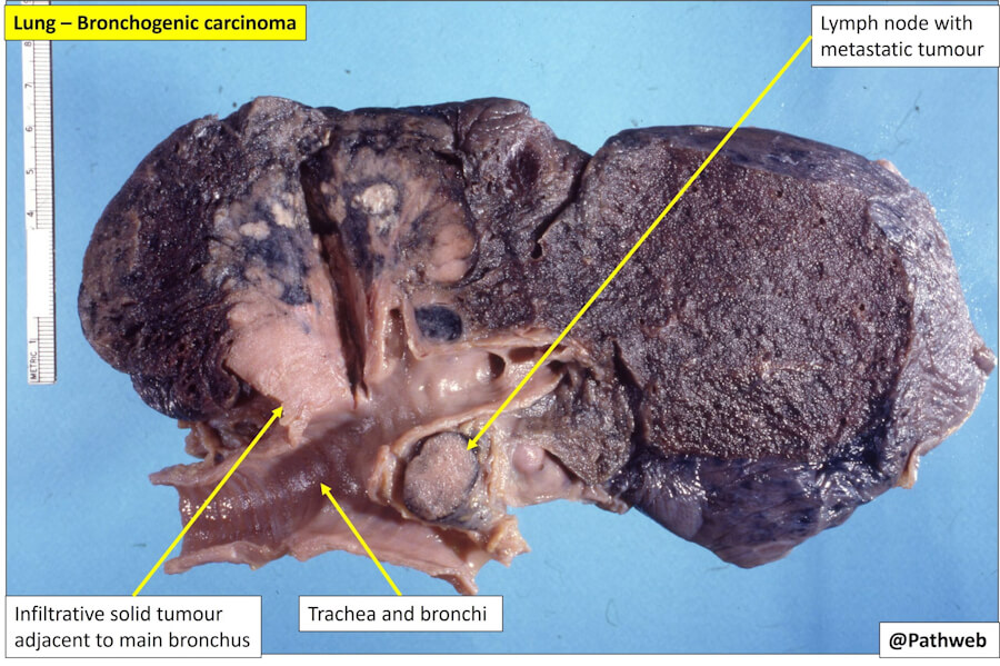
Bronchogenic Carcinoma (Bronchus Cancer) — A Comprehensive Clinical and Public Health Review. 500+ questions and answers.
Bronchogenic carcinoma, commonly referred to as lung cancer or bronchus cancer, is a malignant neoplasm arising from the bronchial epithelium or the lung parenchyma. It remains the leading cause of cancer‑related death worldwide, accounting for an estimated 2 million new diagnoses and 1.8 million deaths annually. Despite substantial advances in early detection, molecular profiling, and targeted therapeutics over the past decade, the overall prognosis remains guarded because most patients are diagnosed at an advanced stage. This long‑read article provides an evidence‑based, Wikipedia‑style synthesis of the epidemiology, etiology, prevention, diagnosis, staging, treatment, and survivorship of bronchogenic carcinoma, with reference to current international guidelines.
🏷️ ICD‑10 Classification
Bronchogenic carcinoma is classified under the International Classification of Diseases, Tenth Revision (ICD‑10), within the block C34 — Malignant neoplasm of bronchus and lung. The relevant subcodes are:
-
C34.0 — Malignant neoplasm of main bronchus (including carina tracheae and hilus of the lung)
-
C34.1 — Malignant neoplasm of upper lobe, bronchus, or lung
-
C34.2 — Malignant neoplasm of middle lobe, bronchus, or lung
-
C34.3 — Malignant neoplasm of lower lobe, bronchus, or lung
-
C34.8 — Malignant neoplasm with overlapping lesion of bronchus and lung
-
C34.9 — Malignant neoplasm of bronchus or lung, unspecified
Secondary malignant neoplasms of the lung (metastases from other primary sites) are coded separately under C78.0.
⚠️ Causes and Risk Factors
Tobacco smoking is the dominant etiological factor, responsible for approximately 9 out of 10 cases in men and 8 out of 10 cases in women. The carcinogenic effect of tobacco smoke is dose‑dependent and is measured in pack‑years; the risk remains elevated for years after cessation but declines progressively thereafter.
Additional established risk factors include:
-
Radon exposure — the second leading cause of lung cancer in many high‑income countries, particularly in uranium‑mining regions and poorly ventilated basements.
-
Occupational carcinogens — asbestos, arsenic, beryllium, cadmium, chromium, diesel exhaust, and silica.
-
Air pollution — fine particulate matter (PM₂.₅) is increasingly recognised as a significant contributor, especially in rapidly industrialising regions.
-
Prior thoracic radiotherapy — particularly in survivors of Hodgkin lymphoma or breast cancer.
-
Genetic susceptibility — inherited variants in genes involved in carcinogen metabolism (e.g., cytochrome P450 polymorphisms) and DNA repair.
-
Chronic lung disease — pre‑existing chronic obstructive pulmonary disease (COPD), pulmonary fibrosis, and prior tuberculosis are associated with modestly increased risk.
-
Infectious co‑factors — human papillomavirus (HPV) and Epstein‑Barr virus (EBV) have been investigated as possible co‑carcinogens in bronchial carcinogenesis, although their role remains less firmly established than that of smoking.
🛡️ Prevention and Avoidance
Primary prevention is centred on tobacco control. Comprehensive strategies include:
-
Smoking prevention and cessation — the single most effective intervention. Population‑level measures such as taxation, smoke‑free legislation, plain packaging, and mass media campaigns reduce initiation and promote quitting.
-
Radon mitigation — testing of homes and workplaces and installation of ventilation systems in high‑risk areas.
-
Occupational health surveillance — enforcement of exposure limits for asbestos, silica, and diesel exhaust; use of personal protective equipment.
-
Air quality improvement — reduction of ambient PM₂.₅ through clean‑energy policies and urban planning.
-
Lung cancer screening in high‑risk individuals — annual low‑dose computed tomography (LDCT) is recommended for adults aged 50–80 years with a ≥20 pack‑year smoking history who currently smoke or have quit within the past 15 years. The ESMO 2025 guideline recommends LDCT screening for individuals aged 55–74 with >30 pack‑years of exposure. Screening shifts diagnosis to earlier, more curable stages and has been shown to reduce lung cancer mortality in randomised trials.
🔬 Types (Histological Classification)
Bronchogenic carcinoma is broadly divided into two major clinicopathological groups, each with distinct biology, staging systems, and therapeutic paradigms.
Small‑Cell Lung Carcinoma (SCLC)
SCLC accounts for approximately 15 % of all lung cancers. It is characterised by rapid doubling time, early widespread dissemination, and initial high responsiveness to chemotherapy and radiotherapy. SCLC is strongly associated with smoking and is typically central in location. It is staged using a two‑tier system: limited stage (confined to one hemithorax and regional nodes that can be encompassed in a single radiotherapy portal) and extensive stage (disease beyond these boundaries).
Non‑Small‑Cell Lung Carcinoma (NSCLC)
NSCLC constitutes 80–85 % of cases. It comprises several histological subtypes:
-
Adenocarcinoma — the most common subtype overall, especially in never‑smokers and women; typically peripheral in location and often associated with actionable driver mutations (EGFR, ALK, ROS1, BRAF, KRAS, MET, RET, NTRK).
-
Squamous cell carcinoma — strongly smoking‑related, usually central, and historically associated with cavitation.
-
Large‑cell carcinoma — a diagnosis of exclusion after other subtypes have been ruled out.
-
Adenosquamous carcinoma and other rare variants.
Histological classification is mandatory for treatment selection, as SCLC and NSCLC follow fundamentally different algorithms. In NSCLC, molecular testing for EGFR mutations, ALK and ROS1 rearrangements, BRAF V600E, KRAS G12C, MET exon 14 skipping, RET fusions, NTRK fusions, and PD‑L1 expression is now standard of care.
🚨 Symptoms and Early Signs
Early‑stage bronchogenic carcinoma is frequently asymptomatic; symptoms often appear only when the tumour has grown sufficiently to obstruct airways, invade adjacent structures, or metastasise. When present, symptoms may include:
-
Persistent cough that does not resolve after 3 weeks or that changes in character
-
Haemoptysis (coughing up blood)
-
Dyspnoea (shortness of breath)
-
Chest pain, particularly on deep breathing, coughing, or laughing
-
Wheezing or stridor due to airway narrowing
-
Hoarseness from recurrent laryngeal nerve involvement
-
Unexplained weight loss and anorexia
-
Fatigue and general weakness
-
Paraneoplastic syndromes — including hypercalcaemia, Cushing syndrome, syndrome of inappropriate antidiuretic hormone (SIADH), Lambert–Eaton myasthenic syndrome, and hypertrophic pulmonary osteoarthropathy
-
Symptoms of metastatic spread — bone pain, headache or neurological deficits (brain metastases), jaundice (liver metastases), and subcutaneous nodules.
Because early symptoms are non‑specific and often attributed to smoking‑related chronic bronchitis or respiratory infections, a low threshold for chest imaging is essential in at‑risk individuals.
🩻 Diagnosis
The diagnostic pathway follows a structured sequence: clinical assessment → imaging → histological confirmation → molecular profiling → staging.
Imaging
-
Chest radiograph (X‑ray) — often the first test in symptomatic patients; may reveal a mass, nodule, hilar enlargement, or pleural effusion.
-
Contrast‑enhanced computed tomography (CT) of the chest, liver, adrenals, and lower neck — the cornerstone of diagnosis and initial staging. CT delineates tumour size, local invasion, and mediastinal lymphadenopathy and screens for distant metastases.
-
Positron emission tomography–CT (PET‑CT) — recommended for all patients considered for curative‑intent treatment to detect occult nodal and distant metastases and to guide biopsy.
-
Magnetic resonance imaging (MRI) — not used routinely for T‑staging of NSCLC, but essential for assessing superior sulcus tumours, spinal cord invasion, and brain metastases.
-
Ultrasound — used for guiding biopsy of peripheral lung lesions, pleural effusions, and supraclavicular lymph nodes; endobronchial ultrasound (EBUS) and endoscopic ultrasound (EUS) enable minimally invasive mediastinal nodal sampling.
Tissue Acquisition and Pathology
-
Bronchoscopy — central lesions; allows direct visualisation and biopsy.
-
EBUS‑TBNA — preferred for paratracheal and peri‑bronchial lesions and mediastinal nodal staging.
-
Transthoracic needle aspiration/biopsy — for peripheral lesions, often under CT or ultrasound guidance.
-
Surgical biopsy — mediastinoscopy, thoracoscopy, or open biopsy when less invasive methods are non‑diagnostic.
-
Liquid biopsy — circulating tumour DNA analysis is increasingly used when tissue is insufficient, particularly for genotyping EGFR, ALK, and other drivers.
Molecular and Biomarker Testing
All patients with advanced non‑squamous NSCLC should undergo broad molecular profiling. PD‑L1 immunohistochemistry is mandatory to guide immunotherapy decisions.
💊 Treatment Types
Treatment is determined by histological subtype, stage, molecular profile, PD‑L1 status, and patient performance status. A multidisciplinary team (MDT) approach is standard.
Surgery
Surgical resection (lobectomy with systematic nodal dissection, or sublobar resection in selected early‑stage patients) is the mainstay of curative treatment for stage I–II NSCLC and selected stage IIIA tumours. Preoperative mediastinal staging is mandatory. Surgery is rarely appropriate for SCLC, except in the uncommon situation of a solitary pulmonary nodule with no nodal involvement.
Chemotherapy
Platinum‑based doublet chemotherapy (e.g., cisplatin or carboplatin combined with pemetrexed, gemcitabine, taxanes, or vinorelbine) is used in the adjuvant, neoadjuvant, and advanced settings. In SCLC, platinum–etoposide is the backbone. Chemotherapy is also a key component of concurrent chemoradiation for locally advanced NSCLC and limited‑stage SCLC.
Radiotherapy
-
Stereotactic ablative radiotherapy (SABR) — an alternative to surgery for medically inoperable early‑stage NSCLC.
-
Concurrent chemoradiotherapy — standard of care for unresectable stage III NSCLC and limited‑stage SCLC.
-
Prophylactic cranial irradiation (PCI) — considered in selected SCLC patients who achieve a complete response.
-
Palliative radiotherapy — for bone metastases, brain metastases, and symptomatic local disease.
Targeted Therapy
In NSCLC harbouring actionable driver alterations, tyrosine kinase inhibitors (TKIs) have transformed outcomes. Examples include:
-
EGFR mutations — osimertinib (third‑generation TKI) is preferred first‑line therapy and is also used as adjuvant treatment after resection of EGFR‑mutant stage IB–IIIA NSCLC.
-
ALK rearrangements — alectinib, brigatinib, lorlatinib.
-
ROS1 rearrangements — crizotinib, entrectinib.
-
BRAF V600E — dabrafenib plus trametinib.
-
KRAS G12C — sotorasib, adagrasib.
-
MET exon 14 skipping — capmatinib, tepotinib.
-
RET fusions — selpercatinib, pralsetinib.
-
NTRK fusions — larotrectinib, entrectinib.
Immunotherapy
Immune checkpoint inhibitors (ICIs) targeting PD‑1 (pembrolizumab, nivolumab, cemiplimab), PD‑L1 (atezolizumab, durvalumab), and CTLA‑4 (ipilimumab) have become integral to NSCLC management. In advanced NSCLC with PD‑L1 tumour proportion score (TPS) ≥50 %, single‑agent pembrolizumab is a standard first‑line option. In PD‑L1‑low or negative tumours, combination chemoimmunotherapy is used. Perioperative immunotherapy (neoadjuvant or adjuvant) is now a category 1 recommendation for resectable stage II–III NSCLC.
Other Modalities
-
Ablative therapies — radiofrequency ablation, microwave ablation, and cryoablation for small peripheral lesions in non‑surgical candidates.
-
Endobronchial therapies — laser, electrocautery, cryotherapy, and stenting for central airway obstruction.
-
Photodynamic therapy — for early endobronchial lesions.
📊 Staging
Staging follows the TNM classification (8th edition, UICC/AJCC), which incorporates tumour size (T), nodal involvement (N), and distant metastasis (M) into prognostic stage groups I–IV.
-
Stage 0 — carcinoma in situ (Tis N0 M0).
-
Stage I — T1–T2a N0 M0; subdivided into IA1, IA2, IA3, IB.
-
Stage II — T1–T2b N1 M0 (IIB), T2b N0 M0 (IIA), T3 N0 M0 (IIB).
-
Stage III — locally advanced disease with ipsilateral mediastinal or subcarinal nodal involvement (N2) or invasion of adjacent structures (T3–T4); subdivided into IIIA, IIIB, IIIC.
-
Stage IV — any T, any N, M1a (contralateral lung nodule, pleural/pericardial effusion) or M1b (single extrathoracic metastasis) or M1c (multiple extrathoracic metastases).
SCLC is staged separately using the limited vs. extensive system, although TNM is increasingly applied for consistency.
🌐 Metastasis
Bronchogenic carcinoma has a propensity for early haematogenous and lymphatic dissemination. Common metastatic sites include:
-
Brain — particularly in adenocarcinoma and SCLC; may present with headache, seizures, or focal deficits. Brain MRI is standard for staging in stage II–III NSCLC and for symptomatic patients.
-
Bones — axial skeleton most frequently; bone pain and pathological fractures.
-
Liver — often asymptomatic but associated with poor prognosis.
-
Adrenal glands — frequently involved; usually asymptomatic.
-
Contralateral lung — intrapulmonary metastases.
-
Pleura and pericardium — malignant effusions.
Metastatic spread profoundly influences prognosis and treatment intent: stage IV disease is generally considered incurable, although selected patients with oligometastatic disease (e.g., solitary brain or adrenal metastasis) may benefit from aggressive local therapy with curative intent.
📈 Cure Rate and Survival Rate
Survival in bronchogenic carcinoma is strongly stage‑dependent. Contemporary population‑based data from the Surveillance, Epidemiology, and End Results (SEER) programme show:
-
Localised disease (confined to the lung): 5‑year relative survival ≈ 65–71 %.
-
Regional disease (spread to regional nodes): 5‑year relative survival ≈ 37–43 %.
-
Distant disease (metastatic): 5‑year relative survival ≈ 9–12 %.
-
All stages combined: 5‑year relative survival ≈ 28 % in recent US data, reflecting gradual improvements from historically ~15 %.
For SCLC, outcomes are poorer: limited‑stage disease has a 3‑year survival of approximately 20 %, while extensive‑stage disease has a 3‑year survival of <5 %.
“Cure” is defined as long‑term survival free of recurrence. In stage I NSCLC treated with complete surgical resection, 5‑year survival can exceed 70 % in contemporary series, and many patients are effectively cured. In stage IV disease, cure is not currently achievable with standard therapies, although durable remissions occur in a subset of patients with oncogene‑addicted tumours treated with TKIs or with high PD‑L1 expression treated with immunotherapy.
⏳ Life Expectancy With and Without Treatment
Life expectancy varies enormously by stage, histology, molecular profile, performance status, and treatment received.
-
Early‑stage NSCLC (stage I–II) with curative‑intent surgery — median survival often exceeds 5–10 years in completely resected cases; 5‑year survival approximates 60–80 % for stage I and 40–50 % for stage II.
-
Locally advanced NSCLC (stage III) treated with concurrent chemoradiation followed by durvalumab — median overall survival in the PACIFIC trial exceeded 47 months, with a substantial plateau in the survival curve.
-
Advanced NSCLC (stage IV) with targeted therapy — median overall survival can range from 2 to 5 years or more depending on the driver alteration (e.g., ALK‑positive disease treated with next‑generation TKIs).
-
Advanced NSCLC without actionable drivers treated with chemoimmunotherapy — median overall survival typically 12–24 months.
-
SCLC — limited‑stage median survival approximately 15–20 months with chemoradiation; extensive‑stage median survival 8–12 months with platinum–etoposide and immunotherapy.
-
Untreated bronchogenic carcinoma — median survival from diagnosis is generally 4–8 months for advanced disease, with 1‑year survival <20 %. Early‑stage disease may remain indolent for months to years before progression, but curative outcomes are lost if treatment is delayed until symptoms compel diagnosis.
These figures are population‑level averages and should not be used to predict individual outcomes; personalised prognostic discussions should incorporate molecular, immunological, and clinical variables.
🕊️ Palliative Care
Palliative care is an essential component of comprehensive lung cancer management, appropriate at any stage and delivered alongside disease‑directed therapy. The 2025 German S3 guideline and other international frameworks emphasise early integration of palliative care to address physical, psychological, social, and spiritual needs.
Key domains of palliative care include:
-
Symptom control — opioids for pain and dyspnoea; bronchodilators, corticosteroids, and oxygen for airway obstruction; antiemetics; laxatives; and management of malignant effusions (thoracentesis, pleurodesis, indwelling pleural catheters).
-
Psychological support — treatment of anxiety and depression; cognitive‑behavioural therapy; mindfulness‑based interventions.
-
Social and financial support — assistance with activities of daily living, caregiver support, and advance care planning.
-
Spiritual care — respecting patient values and preferences.
-
End‑of‑life care — timely discussion of goals of care, hospice referral when appropriate, and bereavement support for families.
Early palliative care has been shown in randomised trials to improve quality of life, reduce aggressive end‑of‑life care, and in some studies prolong survival compared with standard oncological care alone.
❓ Five Questions and Answers
1. Is bronchogenic carcinoma always caused by smoking?
No. Smoking is the dominant risk factor, but approximately 10–20 % of lung cancers occur in never‑smokers, particularly adenocarcinoma in women. Radon, air pollution, occupational exposures, and genetic predisposition contribute to non‑smoking‑related cases.
2. Can bronchogenic carcinoma be cured?
Yes, in a substantial proportion of patients diagnosed at an early stage and treated with complete surgical resection or stereotactic radiotherapy. Cure rates for stage I NSCLC exceed 70 % in contemporary series. Advanced‑stage disease is generally not curable, but long‑term remission is increasingly achievable with targeted therapy and immunotherapy.
3. What is the difference between SCLC and NSCLC?
SCLC is a fast‑growing, smoking‑related neuroendocrine tumour that is usually disseminated at diagnosis and treated primarily with chemotherapy and radiotherapy. NSCLC is a heterogeneous group of epithelial tumours (adenocarcinoma, squamous, large cell) for which surgery, targeted therapy, and immunotherapy play major roles depending on stage and molecular profile.
4. Does lung cancer screening work?
Yes. Annual low‑dose CT screening in high‑risk individuals (age 50–80, ≥20 pack‑years, current or former smoker within 15 years) reduces lung cancer mortality by detecting disease at earlier, more treatable stages. Screening is recommended by multiple international bodies, including the NCCN and ESMO.
5. What are the side effects of modern lung cancer treatment?
Surgery may cause pain, fatigue, and reduced pulmonary function. Chemotherapy causes myelosuppression, nausea, neuropathy, and alopecia. Radiotherapy can produce oesophagitis, pneumonitis, and fatigue. Targeted therapies have class‑specific toxicities (e.g., EGFR TKIs — rash and diarrhoea; ALK TKIs — bradycardia and hepatotoxicity). Immunotherapy can cause immune‑related adverse events affecting the skin, gut, liver, lungs, and endocrine organs. Most side effects are manageable with supportive care.
📌 Key Facts
-
Lung cancer is the most commonly diagnosed cancer globally (≈2.5 million new cases annually) and the leading cause of cancer death (≈1.8 million deaths annually).
-
Tobacco smoking accounts for approximately 85–90 % of cases.
-
SCLC accounts for ≈15 % of cases; NSCLC ≈85 %.
-
Adenocarcinoma is the most common histological subtype in never‑smokers and women.
-
Only about 28 % of lung cancers in the United States are diagnosed at a localised stage, where 5‑year survival is approximately 65 %.
-
The 5‑year relative survival for all stages combined has improved from ≈15 % in the 1990s to ≈28 % in recent years, largely due to targeted therapy, immunotherapy, and earlier detection.
-
Actionable driver mutations are found in approximately 30–40 % of NSCLC adenocarcinomas in Western populations, with higher proportions in Asian never‑smokers.
-
Immunotherapy has become a cornerstone of treatment for advanced NSCLC and is now moving into earlier stages.
✅ Key Takeaways and Conclusions
Bronchogenic carcinoma remains a formidable global health challenge, but the therapeutic landscape has changed dramatically over the past decade. The key messages are:
-
Prevention works: tobacco control and radon mitigation remain the most effective long‑term strategies.
-
Screening saves lives: low‑dose CT in high‑risk populations shifts diagnosis to earlier, curable stages.
-
Histology and molecular profiling are mandatory: treatment is no longer “one size fits all”; SCLC and NSCLC require distinct algorithms, and NSCLC must be interrogated for targetable drivers and PD‑L1 expression.
-
Multidisciplinary care is essential: surgeons, radiation oncologists, medical oncologists, pulmonologists, pathologists, radiologists, and palliative care specialists must collaborate.
-
Survival is improving: stage‑for‑stage outcomes have improved, and durable long‑term survival is now achievable in subsets of patients with advanced disease treated with targeted therapy or immunotherapy.
-
Palliative care is not a last resort: early integration improves quality of life and aligns treatment with patient values.
📚 References
-
World Health Organization. Cancer fact sheet. Updated February 2025. https://www.who.int/news-room/fact-sheets/detail/cancer
-
Siegel RL, Kratzer TB, Giaquinto AN, et al. Cancer statistics, 2025. CA Cancer J Clin. 2025;75:10–45. https://acsjournals.onlinelibrary.wiley.com/doi/10.3322/caac.21871
-
Sung H, Ferlay J, Siegel RL, et al. Global Cancer Statistics 2020: GLOBOCAN estimates of incidence and mortality worldwide for 36 cancers in 185 countries. CA Cancer J Clin. 2021;71:209–249. https://acsjournals.onlinelibrary.wiley.com/doi/10.3322/caac.21660
-
National Cancer Institute. SEER Cancer Stat Facts: Lung and Bronchus Cancer. https://seer.cancer.gov/statfacts/html/lungb.html
-
American Cancer Society. Lung cancer survival rates. https://www.cancer.org/cancer/types/lung-cancer/detection-diagnosis-staging/survival-rates.html
-
National Comprehensive Cancer Network. NCCN Clinical Practice Guidelines in Oncology: Non‑Small Cell Lung Cancer. Version 3.2025. https://www.nccn.org/professionals/physician_gls/pdf/nscl.pdf
-
European Society for Medical Oncology. Early and locally advanced non‑small‑cell lung cancer: ESMO Clinical Practice Guideline. Ann Oncol. 2025;36(11):1245–1262. https://www.sciencedirect.com/science/article/pii/S0923753425003489
-
National Institute for Health and Care Excellence. Lung cancer: diagnosis and management. NICE guideline [NG122]. https://www.nice.org.uk/guidance/ng122
-
Cancer Research UK. Tests for lung cancer. https://about-cancer.cancerresearchuk.org/about-cancer/lung-cancer/getting-diagnosed/tests-for-lung-cancer
-
Detterbeck FC, Boffa DJ, Kim AW, et al. The eighth edition lung cancer stage classification. Chest. 2017;151(1):193–203. https://journal.chestnet.org/article/S0012-3692(16)61243-0/fulltext
-
Goldstraw P, Chansky K, Crowley J, et al. The IASLC Lung Cancer Staging Project: proposals for revision of the TNM stage groupings in the forthcoming (eighth) edition of the TNM classification for lung cancer. J Thorac Oncol. 2016;11(1):39–51. https://www.jto.org/article/S1556-0864(15)30598-8/fulltext
-
Rami‑Porta R, Bolejack V, Giroux DJ, et al. The IASLC lung cancer staging project: the new database to inform the eighth edition of the TNM classification of lung cancer. J Thorac Oncol. 2014;9(11):1618–1624. https://www.jto.org/article/S1556-0864(15)30378-3/fulltext
-
Howlader N, Forjaz G, Mooradian MJ, et al. The effect of advances in lung‑cancer treatment on population mortality. N Engl J Med. 2020;383(7):640–649. https://www.nejm.org/doi/10.1056/NEJMoa1916623
-
Gandhi L, Rodríguez‑Abreu D, Gadgeel S, et al. Pembrolizumab plus chemotherapy in metastatic non‑small‑cell lung cancer. N Engl J Med. 2018;378(22):2078–2092. https://www.nejm.org/doi/10.1056/NEJMoa1801005
-
Antonia SJ, Villegas A, Daniel D, et al. Durvalumab after chemoradiotherapy in stage III non‑small‑cell lung cancer. N Engl J Med. 2017;377(20):1919–1929. https://www.nejm.org/doi/10.1056/NEJMoa1709937
-
Soria JC, Ohe Y, Vansteenkiste J, et al. Osimertinib in untreated EGFR‑mutated advanced non‑small‑cell lung cancer. N Engl J Med. 2018;378(2):113–125. https://www.nejm.org/doi/10.1056/NEJMoa1713137
-
Solomon BJ, Mok T, Kim DW, et al. First‑line crizotinib versus chemotherapy in ALK‑positive lung cancer. N Engl J Med. 2014;371(23):2167–2177. https://www.nejm.org/doi/10.1056/NEJMoa1408440
-
Shaw AT, Bauer TM, de Marinis F, et al. First‑line lorlatinib or crizotinib in advanced ALK‑positive lung cancer. N Engl J Med. 2020;383(21):2018–2029. https://www.nejm.org/doi/10.1056/NEJMoa2027187
-
Drilon A, Oxnard GR, Tan DSW, et al. Efficacy of selpercatinib in RET fusion–positive non–small‑cell lung cancer. N Engl J Med. 2020;383(9):813–824. https://www.nejm.org/doi/10.1056/NEJMoa2005653
-
Hong DS, DuBois SG, Kummar S, et al. Larotrectinib in patients with TRK fusion–positive solid tumours: a pooled analysis of three phase 1/2 clinical trials. Lancet Oncol. 2020;21(4):531–540. https://www.thelancet.com/journals/lanonc/article/PIIS1470-2045(19)30856-3/fulltext
-
Wolf AMD, Oeffinger KC, Shih TY, et al. Screening for lung cancer: 2023 guideline update from the American Cancer Society. CA Cancer J Clin. 2024;74(1):50–81. https://acsjournals.onlinelibrary.wiley.com/doi/10.3322/caac.21811
-
de Koning HJ, van der Aalst CM, de Jong PA, et al. Reduced lung‑cancer mortality with volume CT screening in a randomized trial. N Engl J Med. 2020;382(6):503–513. https://www.nejm.org/doi/10.1056/NEJMoa1911793
-
National Lung Screening Trial Research Team. Reduced lung‑cancer mortality with low‑dose computed tomographic screening. N Engl J Med. 2011;365(5):395–409. https://www.nejm.org/doi/10.1056/NEJMoa1102873
-
Travis WD, Brambilla E, Nicholson AG, et al. The 2015 World Health Organization classification of lung tumors: impact of genetic, clinical and radiologic advances since the 2004 classification. J Thorac Oncol. 2015;10(9):1243–1260. https://www.jto.org/article/S1556-0864(15)30557-5/fulltext
-
Nicholson AG, Tsao MS, Beasley MB, et al. The 2021 WHO classification of lung tumors: impact of advances since 2015. J Thorac Oncol. 2022;17(3):362–387. https://www.jto.org/article/S1556-0864(21)03254-2/fulltext
-
Planchard D, Popat S, Kerr K, et al. Metastatic non‑small cell lung cancer: ESMO Clinical Practice Guidelines for diagnosis, treatment and follow‑up. Ann Oncol. 2018;29(Suppl 4):iv192–iv237. https://www.annalsofoncology.org/article/S0923-7534(19)36256-8/fulltext
-
Jett JR, Schild SE, Kesler KA, et al. Treatment of small cell lung cancer: Diagnosis and management of lung cancer, 3rd ed: American College of Chest Physicians evidence‑based clinical practice guidelines. Chest. 2013;143(5 Suppl):e400S–e419S. https://journal.chestnet.org/article/S0012-3692(13)60137-0/fulltext
-
Früh M, De Ruysscher D, Popat S, et al. Small‑cell lung cancer (SCLC): ESMO Clinical Practice Guidelines for diagnosis, treatment and follow‑up. Ann Oncol. 2013;24(Suppl 6):vi99–vi105. https://www.annalsofoncology.org/article/S0923-7534(19)37568-7/fulltext
-
Reck M, Rabe KF. Precision diagnosis and treatment for advanced non‑small‑cell lung cancer. N Engl J Med. 2017;377(9):849–861. https://www.nejm.org/doi/10.1056/NEJMra1703413
-
Hirsch FR, Scagliotti GV, Mulshine JL, et al. Lung cancer: current therapies and new targeted treatments. Lancet. 2017;389(10066):299–311. https://www.thelancet.com/journals/lancet/article/PIIS0140-6736(16)30958-8/fulltext
-
Herbst RS, Morgensztern D, Boshoff C. The biology and management of non‑small cell lung cancer. Nature. 2018;553(7689):446–454. https://www.nature.com/articles/nature25183
-
Gridelli C, Rossi A, Carbone DP, et al. Non‑small‑cell lung cancer. Nat Rev Dis Primers. 2015;1:15009. https://www.nature.com/articles/nrdp20159
-
Gazdar AF, Bunn PA, Minna JD. Small‑cell lung cancer: what we know, what we need to know and the path forward. Nat Rev Cancer. 2017;17(12):725–737. https://www.nature.com/articles/nrc.2017.87
-
Sabari JK, Lok BH, Laird JH, et al. Unravelling the biology of SCLC: implications for therapy. Nat Rev Clin Oncol. 2017;14(9):549–561. https://www.nature.com/articles/nrclinonc.2017.71
-
Byers LA, Rudin CM. Small cell lung cancer: where do we go from here? Cancer. 2015;121(5):664–672. https://acsjournals.onlinelibrary.wiley.com/doi/10.1002/cncr.29098
-
Wang L, Wang H, Li Y, et al. The role of palliative care in lung cancer: a systematic review. Ann Palliat Med. 2021;10(4):4789–4800. https://apm.amegroups.org/article/view/65123
-
Temel JS, Greer JA, Muzikansky A, et al. Early palliative care for patients with metastatic non–small‑cell lung cancer. N Engl J Med. 2010;363(8):733–742. https://www.nejm.org/doi/10.1056/NEJMoa1000678
-
Zimmermann C, Swami N, Krzyzanowska M, et al. Early palliative care for patients with advanced cancer: a cluster‑randomised controlled trial. Lancet. 2014;383(9930):1721–1730. https://www.thelancet.com/journals/lancet/article/PIIS0140-6736(13)62416-2/fulltext
-
Ferrell BR, Temel JS, Temin S, et al. Integration of palliative care into standard oncology care: American Society of Clinical Oncology clinical practice guideline update. J Clin Oncol. 2017;35(1):96–112. https://ascopubs.org/doi/10.1200/JCO.2016.70.1474
-
Institute of Medicine. Dying in America: Improving Quality and Honoring Individual Preferences Near the End of Life. Washington, DC: National Academies Press; 2015. https://nap.nationalacademies.org/catalog/19476/dying-in-america-improving-quality-and-honoring-individual-preferences-near-the-end-of-life
-
National Cancer Institute. PDQ Adult Treatment Editorial Board. Non‑Small Cell Lung Cancer Treatment (PDQ®). https://www.cancer.gov/types/lung/hp/non-small-cell-lung-treatment-pdq
-
National Cancer Institute. PDQ Adult Treatment Editorial Board. Small Cell Lung Cancer Treatment (PDQ®). https://www.cancer.gov/types/lung/hp/small-cell-lung-treatment-pdq
-
International Agency for Research on Cancer. GLOBOCAN 2022: Lung cancer fact sheet. https://gco.iarc.who.int/media/globocan/factsheets/cancers/11-lung-fact-sheet.pdf
-
Bray F, Laversanne M, Sung H, et al. Global cancer statistics 2022: GLOBOCAN estimates of incidence and mortality worldwide for 36 cancers in 185 countries. CA Cancer J Clin. 2024;74(3):229–263. https://acsjournals.onlinelibrary.wiley.com/doi/10.3322/caac.21834
-
Thai AA, Solomon BJ, Sequist LV, et al. Lung cancer. Lancet. 2021;398(10299):535–554. https://www.thelancet.com/journals/lancet/article/PIIS0140-6736(21)00312-3/fulltext
-
Leiter A, Veluswamy RR, Wisnivesky JP. The global burden of lung cancer: current status and future trends. Nat Rev Clin Oncol. 2023;20(9):624–639. https://www.nature.com/articles/s41571-023-00798-3
-
Bade BC, Dela Cruz CS. Lung cancer 2020: epidemiology, etiology, and prevention. Clin Chest Med. 2020;41(1):1–24. https://www.chestmed.theclinics.com/article/S0272-5231(19)30101-2/fulltext
-
Schabath MB, Cote ML. Cancer progress and priorities: lung cancer. Cancer Epidemiol Biomarkers Prev. 2019;28(10):1563–1579. https://aacrjournals.org/cebp/article/28/10/1563/71364/Cancer-Progress-and-Priorities-Lung-Cancer
-
Malhotra J, Malvezzi M, Negri E, et al. Risk factors for lung cancer worldwide. Eur Respir J. 2016;48(3):889–902. https://erj.ersjournals.com/content/48/3/889
-
de Groot PM, Wu CC, Carter BW, et al. The epidemiology of lung cancer. Transl Lung Cancer Res. 2018;7(3):220–233. https://tlcr.amegroups.org/article/view/20896
Was it helpfull? Rate this article:

 (9 votes, average: 4.44 out of 5)
 Loading...