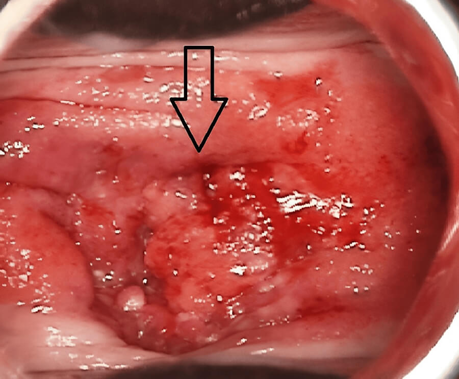
Cervical Squamous Cell Carcinoma is the most common histological type of cervical cancer, a malignancy arising from the cells of the cervix uteri. It is a preventable disease with a well-established viral etiology, primarily linked to persistent infection with high-risk human papillomavirus (HPV). Squamous cell carcinoma (SCC) accounts for approximately 80–90% of all invasive cervical cancers, while adenocarcinoma constitutes most of the remainder. Global epidemiological data indicate that SCC represents about 82.72% of cervical cancer cases, with the highest burden observed in low- and middle-income countries where access to vaccination and screening programs remains limited.
📊 ICD-10 Classification
In the International Classification of Diseases, Tenth Revision (ICD-10), malignant neoplasms of the cervix uteri are coded under the C53 category. Cervical squamous cell carcinoma is specifically classified under C53.1 (malignant neoplasm of the exocervix) and C53.8 (overlapping or other specified sites of the cervix), depending on the precise anatomical origin and extent. The ICD-11 code for cervical squamous cell carcinoma is 2C77.0. In the SNOMED CT terminology, the disorder is designated by the code 254886006 and is mapped to ICD-10-CM code C53.9 (malignant neoplasm of cervix uteri, unspecified) in the 2025 edition.
🦠 What Causes Cervical Squamous Cell Carcinoma?
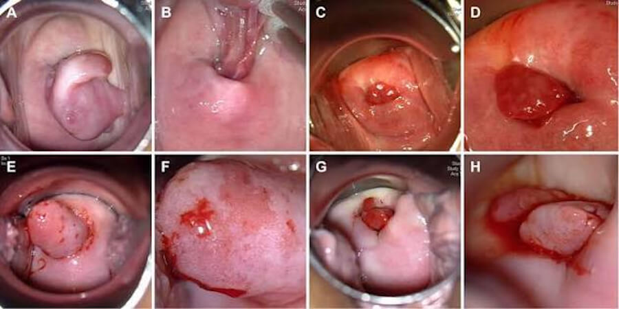
The development of cervical SCC is fundamentally driven by persistent infection with oncogenic, or high-risk, types of human papillomavirus. Nearly all cases of cervical cancer—approximately 99.7%—are linked to HPV infection, and two viral types, HPV 16 and HPV 18, are responsible for about 70% of cervical cancers globally. Most genital HPV infections are transient and cleared by the immune system within two years; however, in approximately 10% of cases, the infection persists and can lead to precancerous lesions and invasive carcinoma over a 10- to 20-year period.
Several cofactors accelerate viral persistence and progression to malignancy. These include immunosuppression (particularly HIV infection, which increases cervical cancer risk by a factor of six), tobacco smoking, increased parity and early age at first full-term pregnancy, and long-term use of oral hormonal contraceptives. Co-infection with other sexually transmitted infections and a history of vulvar, vaginal, or anal squamous intraepithelial neoplasia are also recognized risk factors.
🛡️ How to Avoid and Prevent Cervical Squamous Cell Carcinoma
Cervical SCC is highly preventable through a combination of primary and secondary prevention strategies. The World Health Organization (WHO) has established a global elimination strategy with three key targets for 2030: 90% of girls fully vaccinated with the HPV vaccine by age 15, 70% of women screened with a high-performance test by age 35 and again by age 45, and 90% of women identified with cervical disease receiving appropriate treatment.
Primary prevention centers on HPV vaccination. The availability of highly efficacious vaccines against oncogenic HPV types has made cervical cancer a vaccine-preventable disease. Emerging strategies include gender-neutral vaccination and single-dose schedules, which offer cost-effective and scalable approaches, particularly in low-resource settings. Secondary prevention relies on organized screening programs. While the Pap smear (cervical cytology) was historically the standard, HPV-based testing has now replaced cytology as the preferred screening method due to its higher sensitivity for detecting high-grade precancerous lesions. Screening allows for the detection and treatment of precancerous changes before they progress to invasive cancer. Additional risk-reduction measures include tobacco cessation and effective management of HIV infection with antiretroviral therapy, which reduces the risk of HPV persistence.
🔬 Types of Cervical Carcinoma
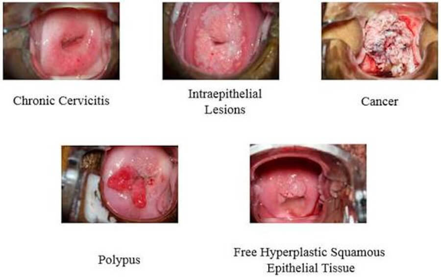
Cervical carcinomas are classified histologically into several types, with two dominant categories accounting for the vast majority of cases.
-
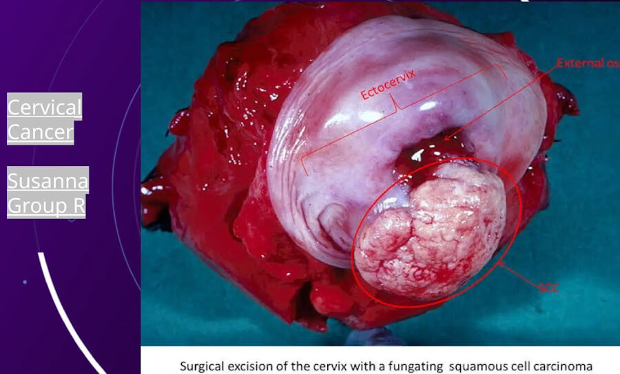
Squamous Cell Carcinoma (SCC): The most common type, arising from the squamous epithelium of the ectocervix. It typically develops from a high-grade squamous intraepithelial lesion (HSIL) and is strongly associated with HPV infection. SCC constitutes approximately 80–90% of invasive cervical cancers.
-
Adenocarcinoma: Arises from the glandular epithelium of the endocervical canal. It accounts for 10–20% of cases and, on average, presents in slightly younger patients (mean age 40–42 years) compared to SCC (median age 51 years).
-
Adenosquamous Carcinoma: A mixed histology containing both squamous and glandular malignant components.
-
Other Rare Types: These include small cell carcinoma (2–5% of cases), neuroendocrine tumors, sarcoma, and lymphoma of the cervix.
⚠️ Symptoms and Early Signs
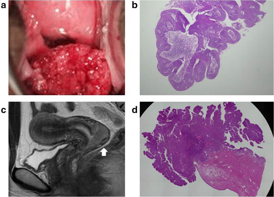
Early-stage cervical SCC is frequently asymptomatic, which underscores the critical importance of regular screening. When symptoms do occur, the most common presentation is abnormal vaginal bleeding. This may manifest as bleeding between menstrual periods, bleeding during or after sexual intercourse (post-coital bleeding), or bleeding after menopause. The tumor tissue is often friable and bleeds easily upon contact.
Other early signs and symptoms include unusual vaginal discharge (which may be watery, bloody, or malodorous), pain during sexual intercourse, and pelvic pain. Menstrual periods may become longer or heavier than usual. As the disease progresses, symptoms related to local invasion or metastasis may appear, such as pain in the lower back, leg swelling, difficulty urinating, or rectal bleeding.
🩸 Note on Colorectal Cancer Bleeding as a Differential Symptom
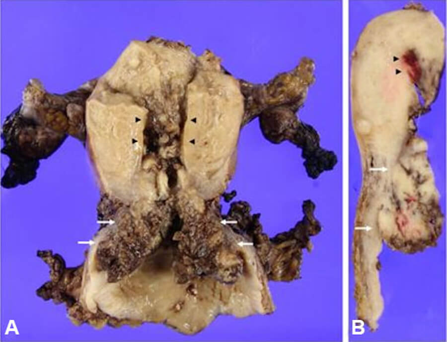
It is clinically important to distinguish cervical cancer-related bleeding from bleeding originating in the lower gastrointestinal tract. Colorectal cancer commonly presents with blood in the stool (hematochezia). The blood may be bright red, typically indicating a distal source in the rectum or sigmoid colon, or dark brown or black (melena), suggesting a more proximal, right-sided colonic lesion where blood has been digested. While rectal bleeding is often attributed to benign anorectal conditions such as hemorrhoids, it is a hallmark symptom of colorectal malignancy and warrants evaluation. The most serious cause of lower gastrointestinal bleeding is colorectal cancer, and colonoscopy is the primary diagnostic tool for investigating unexplained rectal bleeding, allowing for direct visualization and biopsy of suspicious lesions. In patients with cervical cancer, the presence of rectal bleeding may also indicate direct tumor invasion into the rectum, particularly in locally advanced stages (FIGO stage IVA).
🔍 Diagnosis
The diagnostic workup for cervical SCC involves a systematic evaluation to confirm the diagnosis, determine the histological type, and accurately stage the disease.
Initial Evaluation: Diagnosis typically begins with a pelvic examination and a cervical biopsy guided by colposcopy. A biopsy provides the definitive histological diagnosis.
Imaging Modalities: Imaging is essential for staging and treatment planning, and the 2018 FIGO staging system formally incorporated imaging and pathological findings into the staging classification.
-
Magnetic Resonance Imaging (MRI): Pelvic MRI is the imaging modality of choice for evaluating the locoregional extent of the tumor. It provides superior soft-tissue contrast, allowing accurate assessment of tumor size, parametrial invasion, and involvement of adjacent organs such as the bladder or rectum.
-
Computed Tomography (CT): CT of the chest, abdomen, and pelvis is used to detect lymph node enlargement and distant metastases, particularly in the lungs and liver.
-
Positron Emission Tomography–Computed Tomography (PET-CT): Whole-body FDG-PET/CT is recommended for initial staging, especially in patients with suspected stage IB1 disease or higher. It has high sensitivity for detecting nodal and distant metastases and has been shown to change staging in a significant proportion of cases, thereby modifying treatment plans.
-
Ultrasound: Transvaginal or transrectal ultrasound can be a useful adjunct for assessing tumor size and local extent, particularly in resource-limited settings where MRI may not be readily available.
💊 Treatment Types
Treatment for cervical SCC is stage-dependent and is best delivered by a multidisciplinary team. The primary modalities are surgery, radiation therapy, chemotherapy, and—more recently—immunotherapy.
Surgery: Surgical excision is the standard of care for early-stage disease (FIGO stages IA to IIA). For microinvasive disease (stage IA1), a simple hysterectomy or cone biopsy may be sufficient. For stages IA2 to IIA, a radical hysterectomy with bilateral pelvic lymph node dissection is typically performed. Fertility-sparing procedures, such as radical trachelectomy, may be an option for carefully selected young patients with early-stage disease.
Radiation Therapy: Radiation is a cornerstone of treatment for both early and locally advanced disease. It is typically delivered as external beam radiation therapy (EBRT) to the pelvis, followed by brachytherapy (internal radiation), which allows for a high dose of radiation to be delivered directly to the tumor while sparing surrounding normal tissues.
Chemotherapy: Chemotherapy is used in several settings. Concurrent chemoradiation—the administration of a platinum-based chemotherapy agent (typically cisplatin) alongside radiation—is the standard of care for locally advanced cervical cancer (stages IB3, IIA2, IIB, III, and IVA). This combined approach has been shown to improve both progression-free and overall survival compared to radiation alone. Chemotherapy is also used as adjuvant treatment after surgery in patients with high-risk pathological features, and as systemic therapy for recurrent or metastatic disease.
Immunotherapy and Targeted Therapy: The treatment landscape for recurrent and metastatic cervical cancer has been transformed by immunotherapy. Pembrolizumab, an immune checkpoint inhibitor targeting PD-1, has demonstrated significant benefit in patients with PD-L1-positive tumors. It is now recommended in combination with chemotherapy and as maintenance therapy for patients with advanced, persistent, or recurrent disease. This represents a major advance for a disease that historically had limited systemic treatment options.
📈 Staging (FIGO 2018)
The International Federation of Gynecology and Obstetrics (FIGO) staging system is the global standard for classifying the extent of cervical cancer. The 2018 revision was a landmark update that, for the first time, allowed imaging and pathological findings to be used for staging, rather than relying solely on clinical examination.
-
Stage I: Carcinoma is strictly confined to the cervix.
-
IA: Invasive carcinoma diagnosed only by microscopy, with a maximum depth of invasion ≤5 mm.
-
IB: Invasive carcinoma with measured deepest invasion >5 mm, limited to the cervix. Substages (IB1, IB2, IB3) are defined by tumor size.
-
-
Stage II: Carcinoma extends beyond the uterus but has not reached the pelvic wall or the lower third of the vagina.
-
IIA: Involvement of the upper two-thirds of the vagina without parametrial involvement.
-
IIB: With parametrial involvement but not up to the pelvic wall.
-
-
Stage III: Carcinoma involves the lower third of the vagina and/or extends to the pelvic wall and/or causes hydronephrosis or non-functioning kidney, and/or involves pelvic or para-aortic lymph nodes.
-
IIIA: Involvement of the lower third of the vagina, with no extension to the pelvic wall.
-
IIIB: Extension to the pelvic wall and/or hydronephrosis or non-functioning kidney.
-
IIIC: Involvement of pelvic or para-aortic lymph nodes, regardless of tumor size or extent (a major addition in the 2018 revision).
-
-
Stage IV: Carcinoma has extended beyond the true pelvis or has involved the mucosa of the bladder or rectum.
-
IVA: Spread to adjacent pelvic organs.
-
IVB: Distant metastasis.
-
For Stage 1 Cervical Squamous Cell Carcinoma (CSCC) patients who wish to preserve fertility, surgical de-escalation depends primarily on tumor size, depth of invasion, and lymphovascular space invasion (LVSI). All three procedures remove the cancerous portion of the cervix while leaving the uterine body and ovaries intact, though they differ substantially in how much surrounding tissue is resected and their impact on future pregnancy.
| Feature | Cold-Knife Cone Biopsy (Conization) | Simple Trachelectomy | Radical Trachelectomy |
| What Is Removed | A cone-shaped wedge of the cervix containing the lesion; upper cervical canal and parametria remain intact. | The entire cervix (vaginal and supravaginal portions) without removing parametrial tissue or the upper vagina. | The entire cervix, surrounding parametrial tissue (ligaments), and the upper 1–2 cm of the vagina. |
| Lymph Node Assessment | Not required for Stage 1A1 without LVSI; required (sentinel node mapping/dissection) if LVSI is present or Stage 1A2+. | Required (sentinel lymph node biopsy and/or pelvic lymphadenectomy). | Required (sentinel lymph node biopsy and/or pelvic lymphadenectomy). |
| Primary Eligibility (FIGO Stage) |
• Stage 1A1 (invasion ≤3 mm) without LVSI (standard).
• Select Stage 1A2 or low-risk Stage 1B1 (≤2 cm, shallow invasion, no LVSI) per ConCerv/SHAPE trial criteria. |
• Stage 1A1 with LVSI.
• Stage 1A2 (invasion >3 to ≤5 mm).
• Select low-risk Stage 1B1 (≤2 cm, <10 mm stromal invasion, negative nodes). |
• Stage 1B1 (≤2 cm) with deeper invasion or LVSI.
• Select Stage 1B2 (2–4 cm) at specialized centers (sometimes after neoadjuvant chemo). |
| 5-Year Recurrence & Survival |
• Recurrence: <1%–2% in properly selected Stage 1A1.
• 5-year survival: >98%. |
• Recurrence: ~2%–4% in tumors ≤2 cm with negative nodes.
• 5-year survival: >95%–98%. |
• Recurrence: ~3%–5% (for tumors ≤2 cm; rises to 10%–17% if >2 cm).
• 5-year survival: >95%. |
| Pregnancy & Live Birth Rates |
• Highest natural conception rate (~70%–85% among those trying).
• Live birth rate: ~70%–80%. |
• Conception rate: ~60%–75%.
• Live birth rate: ~65%–75% (lower cervical stenosis risk than radical). |
• Conception rate: ~50%–65% (may require IUI/IVF due to cervical stenosis or reduced mucus).
• Live birth rate: ~60%–70%. |
| Obstetric Risks & Delivery |
• Lowest risk of preterm birth (~10%–15%, increases if deep or repeat cones are done).
• Vaginal delivery is usually possible. |
• Moderate risk of second-trimester miscarriage or preterm birth (~15%–25%).
• Often requires a permanent cerclage; C-section is typically recommended. |
• Highest risk of cervical insufficiency, preterm premature rupture of membranes (PPROM), and preterm birth (~25%–35%).
• Requires permanent cerclage and mandatory C-section. |
| Surgical Complications | Minimal: mild bleeding, infection, or rare cervical stenosis. | Low: mild bleeding, cervical stenosis (~5%–10%), or pelvic infection. | Moderate: bladder/ureteral injury, temporary urinary retention, dyspareunia, and cervical stenosis (~10%–15%). |
🌐 Metastasis
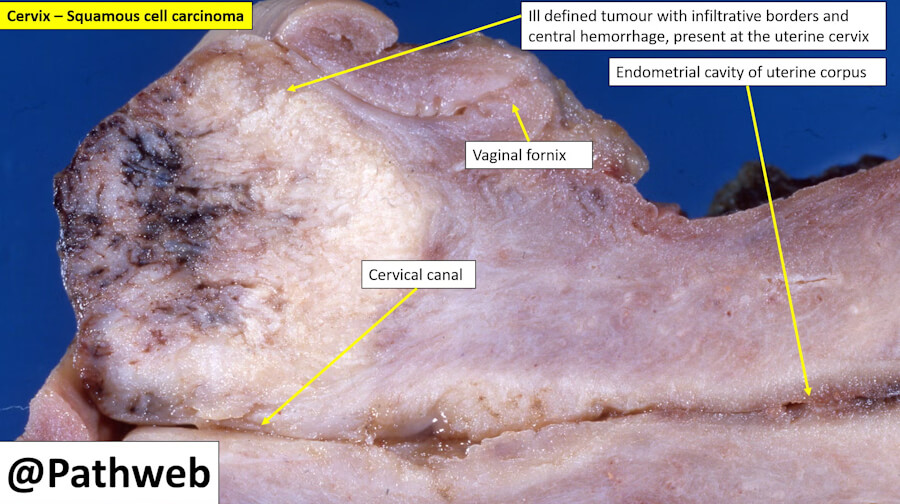
Cervical SCC metastasizes primarily through direct local extension and lymphatic spread, with hematogenous (blood-borne) dissemination occurring in more advanced stages. Distant metastasis is the major pattern of failure in both SCC and adenocarcinoma.
The most common sites of distant metastasis are the lungs, para-aortic lymph nodes, liver, and bone. Metastasis to multiple sites is not uncommon; nearly one in four cases of distant organ metastasis involves two or more sites. The presence of hematogenous metastases is associated with a significantly poorer prognosis compared to lymphatic spread alone, with a 5.3-fold higher risk of death. Median overall survival for patients with liver metastases is particularly poor, reported in the range of 6.8 to 10.9 months.
📉 Cure Rate and Survival Rate
Survival outcomes for cervical SCC are highly stage-dependent. The Surveillance, Epidemiology, and End Results (SEER) database provides population-based 5-year relative survival rates based on the extent of disease at diagnosis.
Based on patients diagnosed between 2014 and 2020, the 5-year relative survival rates are:
-
Localized disease (confined to the cervix or uterus): 91%
-
Regional disease (spread to nearby lymph nodes): 61%
-
Distant disease (metastasis to distant organs): 19%
-
All stages combined: 67%
These figures underscore the critical importance of early detection through screening. When cervical cancer is caught at a localized stage, the cure rate is very high. It is important to note that these are population-level estimates and cannot predict the outcome for an individual patient, as many other factors—including age, overall health, tumor biology, and response to treatment—influence prognosis.
⏳ Life Expectancy With and Without Treatment
Life expectancy for cervical SCC varies widely based on stage and treatment. With treatment, patients with early-stage disease have an excellent prognosis, with the vast majority achieving long-term survival and cure. For locally advanced disease, concurrent chemoradiation offers a realistic chance of cure, though the prognosis is more guarded. In the metastatic setting, treatment is generally not curative, but systemic therapy (chemotherapy and immunotherapy) can extend survival and improve quality of life.
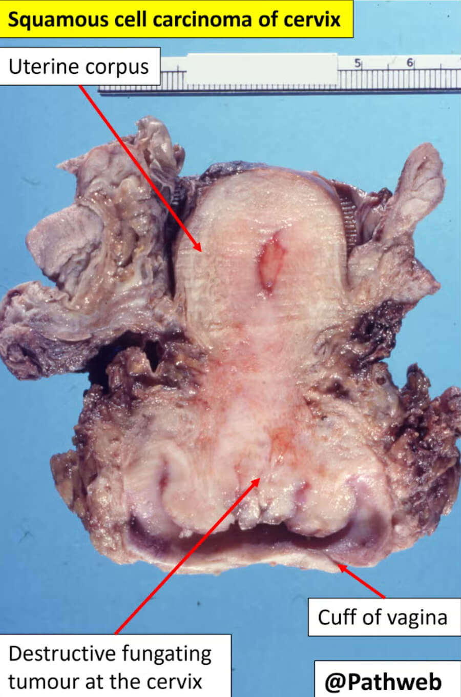
Without treatment, invasive cervical cancer is progressive and ultimately fatal. The natural history of the disease involves local tumor growth causing pain, bleeding, and organ dysfunction (e.g., ureteral obstruction leading to kidney failure), followed by metastatic spread. The time course can vary, but the disease is life-limiting, and palliative care becomes the primary focus of management to control symptoms and maintain dignity.
🕊️ Palliative Care
Palliative care is an essential component of comprehensive cervical cancer management, particularly for patients with advanced or recurrent disease. The WHO defines palliative care as an approach that improves the quality of life of patients and their families facing life-threatening illness, through the prevention and relief of suffering by means of early identification, impeccable assessment, and treatment of pain and other problems—physical, psychosocial, and spiritual.
Women with advanced cervical cancer often experience a complex symptom burden. Pain is highly prevalent and may be neuropathic due to sacral nerve infiltration, requiring a multimodal analgesic approach that may include opioids. Other distressing symptoms include vaginal bleeding, malodorous discharge, fistulae (abnormal connections between the vagina and bladder or rectum), fatigue, anxiety, and depression. Palliative care services should address nutritional support, wound care, and the management of these complex symptoms. Early integration of palliative care into oncology services has been shown to support symptom control, reduce avoidable hospital admissions, and help patients maintain daily functioning.
❓ 5 Questions and Answers
Definition & Basics (Informational Intent)
What is cervical squamous cell carcinoma?
It is a cancer that begins in the flat, scale-like squamous cells covering the outer surface of the cervix. It is the most common type of cervical cancer, accounting for roughly 70% of cases, and is almost always linked to persistent infection with high-risk types of HPV.
Is cervical squamous cell carcinoma curable?
Yes, especially when caught early. For stage IA disease, 5-year survival is 97–99% with conservative surgery. Even for later stages, cure is possible with chemoradiation, though survival rates decline as stage increases.
How common is it?
It is the most common histological subtype of cervical cancer, representing about 70% of all cervical cancers in the United States and similar proportions globally.
What is the difference between squamous cell carcinoma and adenocarcinoma of the cervix?
Squamous cell carcinoma arises from the flat cells on the outer cervix (ectocervix). Adenocarcinoma arises from the glandular cells lining the cervical canal (endocervix). They differ in risk factors (adenocarcinoma is less consistently linked to HPV-16 and more often HPV-18) and in how readily they are detected by Pap smears, with adenocarcinoma being harder to catch early.
What does “invasive” squamous cell carcinoma mean?
“Invasive” means the cancer cells have broken through the surface layer (epithelium) of the cervix and grown into the deeper supporting tissue (stroma). This is distinct from in situ disease (CIS), where abnormal cells remain confined to the surface layer.
What is squamous cell carcinoma in situ (CIS) of the cervix?
CIS is a pre-invasive, non-cancerous condition (also called high-grade squamous intraepithelial lesion, HSIL) where abnormal squamous cells are present but have not invaded deeper tissues. It is highly treatable and is considered the last step before invasive cancer.
How fast does cervical squamous cell carcinoma grow?
It typically develops slowly, over 10 to 20 years from initial HPV infection through precancerous changes to invasive cancer. This long window is why regular screening is so effective at prevention.
Is cervical squamous cell carcinoma always caused by HPV?
Essentially yes. More than 99% of cervical cancers are caused by persistent infection with high-risk HPV types, most commonly HPV-16 and HPV-18. Cases without HPV are exceedingly rare.
What is non-keratinizing squamous cell carcinoma?
This is the most common variant. Under the microscope, the cancer cells form nests or sheets and do not produce large amounts of the tough protein keratin. It is generally associated with moderate to poor differentiation.
What is keratinizing squamous cell carcinoma?
In this less common variant, the cancer cells produce keratin, a protein that makes them appear more like mature squamous cells. It is often classified as “well-differentiated.” Some studies suggest the keratinizing variant may carry a slightly poorer prognosis than non-keratinizing types, though findings are not uniform.
What does “poorly differentiated” mean?
“Differentiation” describes how much the cancer cells resemble normal squamous cells. “Poorly differentiated” (or high-grade) means the cells look very abnormal and are growing aggressively, which can be associated with a more aggressive clinical course.
What does “moderately differentiated” mean?
It means the cancer cells have some features of normal squamous cells but are more abnormal than “well-differentiated” cells. It represents an intermediate grade.
Is cervical squamous cell carcinoma hereditary?
No, it is not considered a hereditary cancer. The primary driver is acquired HPV infection, not inherited genetic mutations. Risk is related to exposure to HPV and immune status, not family lineage.
Can you get cervical squamous cell carcinoma without HPV?
It is extremely rare. Virtually all cases are HPV-associated. When HPV-negative cases occur, they are often unusual histological subtypes and require expert pathological review.
What are the risk factors?
The dominant risk factor is persistent high-risk HPV infection. Other factors that increase the risk of progression from HPV infection to cancer include cigarette smoking, a weakened immune system (e.g., from HIV or immunosuppressive drugs), and lack of regular cervical screening.
Symptoms & Early Warning Signs
What are the first signs?
Very early-stage disease often causes no symptoms. When symptoms appear, the most common early sign is abnormal vaginal bleeding, particularly after sexual intercourse, between periods, or after menopause. Unusual vaginal discharge (watery, bloody, or foul-smelling) is another early warning sign.
Does it cause pain?
Early cancer usually does not cause pain. Pain in the pelvic area or lower back can occur as the tumor grows and begins to affect surrounding tissues or nerves.
What does bleeding from cervical cancer look like?
It can range from light spotting or a bloody discharge to heavier bleeding. It is most characteristically contact bleeding—bleeding after intercourse, douching, or a pelvic exam.
Is bleeding after intercourse a sign?
Yes. Post-coital bleeding is one of the classic early symptoms of cervical cancer and should always be evaluated by a clinician.
What kind of vaginal discharge is associated?
The discharge may be watery, blood-tinged, or have an unpleasant, foul odor. It can occur between periods or after menopause.
Can it have no symptoms?
Yes. Early-stage cervical cancer is frequently asymptomatic and is detected only through routine screening (Pap/HPV testing).
Does it cause lower back pain?
Lower back pain can occur, particularly as the disease advances and involves the pelvic sidewalls or nerves. It is not typically an early symptom.
What are the symptoms of advanced disease?
Advanced disease can cause more severe pelvic or back pain, weight loss, fatigue, loss of appetite, and problems with urination or bowel movements if the tumor presses on the bladder or rectum.
Can it cause pelvic pressure or bloating?
Yes, as the tumor enlarges, it can create a sensation of pelvic fullness, pressure, or bloating.
Does it cause fatigue and weight loss?
Yes, these are constitutional symptoms that typically appear with more advanced or metastatic disease.
Diagnosis, Screening & Pathology
Can a Pap smear detect squamous cell carcinoma?
A Pap smear is a screening test designed to detect pre-cancerous changes and early cancers, including squamous cell carcinoma. It is very effective but is not a diagnostic test; an abnormal result leads to further evaluation with colposcopy and biopsy.
What is the difference between HSIL and squamous cell carcinoma?
HSIL (high-grade squamous intraepithelial lesion) is a pre-cancer. The abnormal cells are confined to the surface layer of the cervix (in situ). Squamous cell carcinoma is invasive cancer, meaning the cells have broken through the surface and grown into the deeper cervical tissue.
How is it diagnosed?
Diagnosis is made by biopsy. This is typically done during a colposcopy, where a doctor visualizes the cervix and takes small tissue samples from abnormal areas. Sometimes a larger excision, such as a LEEP or cone biopsy, is needed to make the diagnosis and assess the depth of invasion.
What happens during a colposcopy?
A colposcope (a magnifying instrument) is used to examine the cervix closely. The doctor applies a dilute vinegar solution, which makes abnormal areas turn white (acetowhite). Biopsies are then taken from these suspicious areas.
How long for biopsy results?
Results are usually available within one to two weeks, depending on the pathology laboratory’s processing time.
What is a LEEP procedure and can it cure early cancer?
LEEP (loop electrosurgical excision procedure) uses a thin wire loop with electrical current to remove a portion of the cervix. For microinvasive squamous cell carcinoma (stage IA1), a LEEP with clear margins can be curative, especially if fertility preservation is desired.
What is a cone biopsy (conization)?
A cone biopsy removes a larger, cone-shaped piece of cervical tissue, including the transformation zone and part of the cervical canal. It is used for diagnosis, to assess the depth of invasion, and sometimes as definitive treatment for very early-stage disease.
What blood tests check for it?
There is no routine blood test that diagnoses cervical cancer. However, squamous cell carcinoma antigen (SCC-Ag) can be measured in the blood. It is not used for screening but can be useful for monitoring response to treatment and detecting recurrence in some patients.
What is the SCC antigen blood test?
SCC-Ag is a tumor marker. Elevated levels can be seen in squamous cell carcinomas of the cervix, but it is not specific to cervical cancer and is not a stand-alone diagnostic tool. Its main role is in surveillance after treatment.
Do I need a PET scan?
A PET scan (often combined with CT) is not needed for early-stage disease. It is primarily used for staging locally advanced disease (to check for lymph node or distant spread) or for evaluating suspected recurrence.
What does an MRI show?
MRI is excellent for showing the local extent of the tumor—how large it is and whether it has invaded the parametrium (tissue next to the cervix) or pelvic sidewall. It is a key part of staging to determine if surgery is feasible.
What do “positive margins” mean after a cone biopsy?
It means that cancer cells were found at the cut edge of the removed tissue specimen. This suggests that some tumor may have been left behind, and further treatment (such as a second procedure or radiation) may be needed.
What is lymphovascular space invasion (LVSI)?
LVSI means cancer cells are seen inside small blood vessels or lymphatic channels within the cervical tissue. Its presence is a risk factor for lymph node metastasis and may influence treatment decisions, such as whether to recommend adjuvant therapy after surgery.
Staging & Metastasis
What are the stages?
Cervical cancer is staged using the FIGO system (I–IV). Stage I is confined to the cervix; Stage II extends beyond the cervix but not to the pelvic wall or lower vagina; Stage III involves the lower vagina or pelvic wall, or causes kidney problems; Stage IV involves the bladder/rectum (IVA) or distant organs (IVB).
Stage 1A1 vs. 1A2?
Both are microinvasive (only visible under a microscope). 1A1 means invasion is ≤3 mm deep and ≤7 mm wide. 1A2 means invasion is >3 mm but ≤5 mm deep and ≤7 mm wide.
Stage 1B1, 1B2, and 1B3?
These are clinically visible lesions confined to the cervix. 1B1 is ≤2 cm; 1B2 is >2 cm but ≤4 cm; 1B3 is >4 cm.
What does Stage 2B mean?
Stage IIB means the tumor has invaded the parametrium (the connective tissue next to the cervix) but has not reached the pelvic wall.
What is Stage 3C1 and 3C2?
These are determined by lymph node involvement (detected by imaging or biopsy). 3C1 means pelvic lymph nodes are positive; 3C2 means para-aortic lymph nodes are positive. The “r” and “p” modifiers indicate whether the node status was determined by imaging (r) or pathology (p).
Stage 4A vs. 4B?
IVA means the tumor has spread to adjacent organs—the bladder or rectum. IVB means it has spread to distant organs (e.g., lungs, liver, bones).
Where does it spread first?
It spreads first through direct extension into nearby tissues (parametrium, vagina) and through the lymphatic system to pelvic lymph nodes. Hematogenous (blood-borne) spread to distant organs like the lungs, liver, or bones typically occurs later.
Can it spread to the lungs?
Yes, the lungs are one of the common sites for distant metastasis in Stage IVB disease.
What are the symptoms of lymph node spread?
Pelvic lymph node metastasis may be asymptomatic or cause pelvic pain. Para-aortic node involvement can sometimes cause back or abdominal pain, but often it is detected on imaging rather than by symptoms.
Can it metastasize to bones or liver?
Yes, Stage IVB disease can spread to the bones (causing pain) and the liver.
How is FIGO staging different from TNM?
FIGO is the standard staging system used for gynecologic cancers. TNM (Tumor, Node, Metastasis) is a more general system used across cancers. For cervical cancer, FIGO staging incorporates clinical findings, imaging, and sometimes pathology, and it is the system that guides treatment decisions in guidelines.
Treatment Options & Protocols
What is the standard treatment?
It depends on stage. Early-stage (IA–IIA1) is typically treated with surgery (radical hysterectomy + lymph node dissection or fertility-sparing trachelectomy). Locally advanced (IIB–IVA) is treated with concurrent chemoradiation (cisplatin + pelvic radiation + brachytherapy). Metastatic/recurrent disease is treated with systemic therapy (chemotherapy ± bevacizumab ± pembrolizumab, or tisotumab vedotin).
Can Stage 1 be treated without a hysterectomy?
Yes. A radical trachelectomy removes the cervix and surrounding tissue but preserves the uterus, allowing for future pregnancy. It is an option for carefully selected patients with small, early-stage tumors (usually ≤2 cm).
Radical vs. simple hysterectomy?
A simple hysterectomy removes the uterus and cervix. A radical hysterectomy removes the uterus, cervix, and surrounding tissue (parametrium) and a portion of the upper vagina, and is the standard surgery for early-stage invasive cervical cancer.
What is a trachelectomy?
It is a fertility-sparing surgery that removes the cervix and surrounding tissue but leaves the uterine body intact. It is offered to young women with early-stage disease who wish to preserve fertility.
When is chemotherapy needed?
Chemotherapy is used in two main settings: (1) concurrently with radiation (chemoradiation) as a radiosensitizer for locally advanced disease; (2) as systemic therapy for metastatic or recurrent disease.
What chemo drugs are used?
The backbone is cisplatin. For metastatic/recurrent disease, common regimens include cisplatin or carboplatin + paclitaxel, often with bevacizumab. Pembrolizumab is added for PD-L1-positive tumors.
How does chemoradiation work?
Chemotherapy (usually weekly cisplatin) is given during the course of radiation. It makes the cancer cells more sensitive to radiation, improving the chance of killing tumor cells in the pelvis.
What is brachytherapy?
Brachytherapy is a form of internal radiation where a radioactive source is placed directly into or near the tumor (in the cervix/uterus). It is a required component of curative-intent radiation for cervical cancer and has been shown to significantly improve local control and survival.
How many sessions of radiation are needed?
External beam radiation is typically given daily (Monday–Friday) for about 5 weeks (25–28 fractions). This is followed by brachytherapy, usually given in a few sessions (e.g., 4–5 fractions). The entire course should ideally be completed within 7–8 weeks.
What immunotherapy drugs are approved?
Pembrolizumab (Keytruda) is approved for PD-L1-positive (CPS ≥1) persistent, recurrent, or metastatic cervical cancer, in combination with chemotherapy ± bevacizumab.
What is targeted therapy for recurrent cervical cancer?
Bevacizumab (Avastin) is an anti-angiogenic antibody that blocks blood vessel growth to the tumor. Tisotumab vedotin (Tivdak) is an antibody-drug conjugate targeting Tissue Factor, approved for recurrent/metastatic disease.
What is PD-L1 testing?
PD-L1 testing measures the Combined Positive Score (CPS) on a tumor biopsy. A CPS ≥1 indicates that pembrolizumab may be beneficial. It is standard practice to test for this biomarker before starting immunotherapy in advanced disease.
Are there clinical trials?
Yes, numerous trials are ongoing for metastatic and recurrent cervical cancer, including trials of new immunotherapies, TIL therapy, therapeutic vaccines, and novel ADCs.
Prognosis, Survival Rates & Recurrence
What is the survival rate?
Survival varies widely by stage. Using FIGO 2018 staging, 5-year overall survival is approximately 88% for Stage I, 71% for Stage II, 74% for Stage III (this can vary; some older series report lower rates for stage III), and 40% for Stage IV in recent series. Other data using older staging reported stage IB survival around 80% and stage III around 33–39%.
5-year survival for Stage 1?
It is generally excellent, around 88–90% for all Stage I combined. Sub-stage 1B1 may be around 90%, 1B2 around 87%, and 1B3 around 81%.
Survival for Stage 2B?
Stage II overall survival is around 71%. Stage 2B specifically may be somewhat lower within that group.
Prognosis for Stage 3?
It is more guarded. Recent series report around 55–74% 5-year survival, depending on the cohort and whether nodes are involved.
Can you survive Stage 4?
Yes, but it is less common. Stage IVA (bladder/rectum involvement) may be potentially curable with exenteration in selected cases. Stage IVB (distant metastasis) is generally not curable, but patients can live for extended periods with modern systemic therapy. 5-year survival is roughly 16–40% overall for Stage IV.
What is the recurrence rate?
Recurrence rates vary by stage but are significant. In one large series, 14.2% of patients recurred at a median of 1.9 years. Most recurrences (over 90%) occur within the first 2–3 years after treatment.
Where does it usually recur?
Recurrences can be local (pelvis) or distant. In one series, distant relapses (62%) were more common than loco-regional relapses (38%).
What are the signs of recurrence?
Signs can include new pelvic pain, vaginal bleeding or discharge, unexplained weight loss, or symptoms related to the site of spread (e.g., cough if lung). Surveillance with exams and imaging is used to detect recurrence.
How is recurrent disease treated?
It depends on the site and prior treatment. Central pelvic recurrence after radiation may be treated with pelvic exenteration (radical surgery removing pelvic organs) with curative intent in selected patients. Distant recurrence is treated with systemic therapy (chemotherapy, immunotherapy, bevacizumab, or tisotumab vedotin).
What is pelvic exenteration?
It is a very radical operation that removes the bladder, rectum, and reproductive organs en bloc. It is only appropriate for a small subset of patients with central pelvic recurrence and no distant spread. 5-year survival in contemporary series is around 40–50% when performed with clear margins.
Does keratinizing vs. non-keratinizing affect prognosis?
There is some evidence that the keratinizing variant may have a poorer prognosis than non-keratinizing types, but this is not a consistent finding across all studies, and stage remains the dominant prognostic factor.
7. Recovery, Fertility & Life After Treatment
Can I get pregnant after treatment?
Yes, if you undergo a fertility-sparing trachelectomy. This procedure is designed to preserve the uterus. However, it carries risks for future pregnancy, including a higher rate of second-trimester miscarriage (around 10%) and preterm delivery. It is generally limited to tumors ≤2 cm.
How long is recovery after radical hysterectomy?
Recovery typically takes 4–6 weeks for return to normal activities, though it can vary. It is major abdominal or minimally invasive surgery.
Long-term side effects of pelvic radiation?
These can include vaginal stenosis (narrowing and shortening of the vagina), radiation proctitis (rectal bleeding/urgency), cystitis (bladder irritation), bowel changes, and lymphedema (leg swelling) if lymph nodes were also removed.
How to use a vaginal dilator?
Dilators are used regularly (often starting a few weeks after radiation) to gently stretch the vagina and prevent stenosis. Your radiation oncology team will give you a specific schedule and size to use.
Does treatment cause early menopause?
Pelvic radiation can damage the ovaries and cause immediate or early menopause if the ovaries are in the radiation field. Surgery (removal of ovaries) also causes surgical menopause. Ovarian transposition (surgically moving the ovaries out of the radiation field) can sometimes preserve ovarian function.
Is HRT safe after treatment?
For most patients with squamous cell carcinoma, HRT is generally considered safe and is often recommended to manage menopausal symptoms, as this cancer is not typically hormone-driven (unlike some endometrial or breast cancers). This should be discussed with your oncologist.
How often do I need follow-up?
Follow-up is typically every 3–4 months for the first 2 years, then every 6 months for the next 3 years, then annually. It includes a pelvic exam and sometimes imaging or HPV testing.
What is the best diet?
There is no single “cancer diet.” A balanced diet rich in fruits, vegetables, lean protein, and whole grains is recommended. During treatment, managing side effects (like diarrhea or nausea) may require specific dietary adjustments guided by a dietitian.
How to manage lymphedema?
Lymphedema is managed with compression garments, manual lymphatic drainage (a specialized massage technique), exercise, and skin care. It is best managed with a lymphedema therapist.
Can I still get the HPV vaccine after diagnosis?
Yes, the HPV vaccine is preventive, not therapeutic—it will not treat existing cancer. However, it may still be recommended to protect against other HPV types you have not been exposed to, and it is generally safe.
Specialized Topics (Chemoradiation, Immunotherapy, ADCs, Trials, TIL, Vaccines)
Chemoradiation
-
How is it different from separate chemo and radiation? Concurrent chemoradiation uses chemotherapy as a radiosensitizer—it makes cancer cells more vulnerable to radiation, improving local control and survival compared to radiation alone.
-
Why weekly cisplatin? Weekly cisplatin at 40 mg/m² is the standard radiosensitizing schedule. It is given on days 1, 8, 15, 22, and 29 of external beam radiation.
-
If kidneys cannot handle cisplatin? Carboplatin is an acceptable alternative, though some data suggest it may be slightly less effective as a radiosensitizer. It is used when cisplatin is contraindicated (e.g., renal impairment).
-
How long is EBRT? Typically 5 weeks (25–28 daily fractions). The overall treatment course including brachytherapy should be completed within 7–8 weeks .
-
IMRT vs. standard radiation? IMRT (Intensity-Modulated Radiation Therapy) conforms the radiation dose more precisely to the tumor, sparing nearby normal tissues (bowel, bladder, bone marrow) and reducing toxicity.
-
Why brachytherapy after EBRT? Brachytherapy delivers a high, concentrated dose directly to the tumor, which is essential for eradicating central disease. Omitting it is associated with significantly worse survival.
-
Does HDR brachytherapy hurt? It is usually done with anesthesia (general or spinal) for patient comfort. You will not feel the radiation during treatment.
-
Am I radioactive? After external beam radiation, you are not radioactive. After brachytherapy, the radioactive source is removed, and you are not radioactive. You can safely be around children and pets.
-
Why finish within 7–8 weeks? Prolonging treatment allows tumor cells to repopulate, reducing the chance of cure. Every effort is made to complete treatment on time.
-
What if white count drops? Chemotherapy may cause myelosuppression. Your counts are monitored weekly. If they are too low, chemotherapy may be held for a week, but radiation usually continues.
-
Hair loss from cisplatin? Cisplatin is not typically associated with significant hair loss (alopecia). Some patients may experience mild thinning.
-
Fatigue? Fatigue is very common during pelvic chemoradiation. It often peaks toward the end of treatment and improves in the weeks after.
-
Managing diarrhea/cystitis? Dietary changes, medications (e.g., loperamide), and hydration are used. Bladder irritation (cystitis) may require pain relievers and hydration.
-
“Full bladder, empty bowel” protocol? For daily radiation, a full bladder helps push the bowel out of the radiation field, and an empty bowel reduces gas and movement, improving targeting accuracy.
-
Cisplatin hearing loss/neuropathy? Cisplatin can cause tinnitus, high-frequency hearing loss, and peripheral neuropathy (numbness/tingling in hands/feet). These can be permanent. Kidney function is also monitored.
-
Immediate menopause from radiation? If ovaries are in the field, yes, radiation can cause immediate and permanent menopause. Ovarian transposition can prevent this.
-
Vaginal stenosis and dilators? Vaginal stenosis (narrowing) is common after radiation. Regular use of a vaginal dilator, starting a few weeks after treatment, is essential to maintain vaginal caliber.
-
Will sex feel normal again? Many women experience changes (dryness, shortening), but with dilator use, lubricants, and sometimes topical estrogen, sexual function can be improved.
-
Late radiation toxicity? Can include bowel obstruction, radiation proctitis (rectal bleeding), and fistulas (abnormal connections between organs). These are uncommon but serious late effects.
Immunotherapy
-
How does it work? Pembrolizumab is a checkpoint inhibitor. It blocks the PD-1 receptor on immune T-cells, “releasing the brakes” and allowing the immune system to attack HPV-driven cancer cells.
-
When is it added? It is added to first-line chemotherapy for persistent, recurrent, or metastatic disease with PD-L1 CPS ≥1. It is also being studied in the locally advanced setting with chemoradiation.
-
PD-L1 CPS <1—can I still get it? The approved indication is for CPS ≥1. Benefit in CPS <1 is less clear, and eligibility should be discussed with your oncologist.
-
MSI-H/TMB testing? While pembrolizumab is approved for MSI-H/dMMR solid tumors regardless of site, this is rare in cervical cancer. TMB testing is not routinely required for cervical cancer immunotherapy.
-
Other checkpoint inhibitors? Cemiplimab and Nivolumab have also shown activity in cervical cancer trials, though pembrolizumab is the most established.
-
How long on immunotherapy? Treatment is typically continued until disease progression or unacceptable toxicity, often for up to 2 years in responders.
-
When to know if it’s working? Response may take a few months to become apparent on scans. Sometimes tumors can appear to grow initially (pseudoprogression) before shrinking.
-
Pseudoprogression? This is a temporary increase in tumor size on imaging due to immune cell infiltration and inflammation, followed by shrinkage. It is uncommon but can occur.
-
Side effects different from chemo? Immunotherapy side effects are immune-related and can affect any organ (skin, colon, lungs, thyroid, liver, pituitary). They are managed with steroids and other immunosuppressants.
-
Warning signs of pneumonitis/colitis? Pneumonitis: new cough, shortness of breath. Colitis: diarrhea, abdominal pain, blood in stool. These require urgent medical attention.
-
Permanent damage to thyroid/pituitary/adrenal? Yes, immune-related endocrinopathies (thyroiditis, hypophysitis, adrenal insufficiency) can be permanent and require lifelong hormone replacement.
-
Steroids stop immunotherapy from working? Steroids are used to manage side effects. They may slightly dampen the anti-tumor effect, but this is necessary to manage toxicity. The risk-benefit is individualized.
-
Pre-existing autoimmune disease? Immunotherapy can exacerbate autoimmune conditions. It may be used with caution and close monitoring in collaboration with a rheumatologist.
-
Can it cure metastatic disease? Immunotherapy can produce durable responses and long-term survival in a subset of patients, but cure is not guaranteed. It is a significant advance in controlling the disease.
Targeted Therapy & ADCs
-
Targeted vs. chemo/immunotherapy? Targeted therapies (like bevacizumab) attack specific features of cancer cells or their environment (e.g., blood vessel growth). ADCs are “smart bombs” that deliver chemotherapy directly to cancer cells expressing a specific surface protein.
-
Bevacizumab (Avastin)? It is an anti-angiogenic antibody that blocks VEGF, cutting off the tumor’s blood supply. It is used in combination with chemotherapy for recurrent/metastatic disease.
-
Bevacizumab risks? Serious risks include high blood pressure, blood clots, bleeding, bowel perforation, and fistulas. It is not given soon after surgery or in patients with certain conditions.
-
Tisotumab vedotin (Tivdak)? It is an ADC targeting Tissue Factor (TF), a protein highly expressed on cervical cancer cells. It delivers a chemotherapy payload (monomethyl auristatin E) directly into the cancer cell.
-
Eye care with tisotumab? It can cause ocular toxicity (dry eyes, corneal damage, blurred vision). Patients require preservative-free eye drops, cold compresses, and regular eye exams during treatment.
-
HER2-targeted therapy? Trastuzumab deruxtecan is approved for HER2-positive solid tumors, including cervical cancer, but HER2 positivity is more common in adenocarcinoma than in squamous cell carcinoma. Testing may be considered in recurrent disease.
Clinical Trials
-
When to look? It is appropriate to consider clinical trials at any stage, including first-line metastatic disease or after standard treatments fail. Discussing this early with your oncologist is ideal.
-
Performance status (ECOG 0/1)? ECOG 0 means fully active; ECOG 1 means restricted in strenuous activity but ambulatory and able to carry out light work. Trials require good performance status because patients must be well enough to tolerate experimental treatments.
-
Prior pembrolizumab/bevacizumab disqualify me? Not necessarily, but it depends on the trial. Some trials exclude prior checkpoint inhibitors; others specifically study novel agents after progression on these drugs.
-
Washout period? This is the time required between your last treatment and starting the trial drug to allow effects to wear off. It varies by trial (often 3–4 weeks).
-
Phase 1, 2, 3? Phase 1: tests safety and dosing. Phase 2: tests efficacy in a specific cancer. Phase 3: compares the new treatment to the standard of care in a large population.
-
Placebo in randomized trials? In cancer trials, a placebo is almost never used alone. It is usually added to the standard of care so all patients receive at least the best available treatment.
-
Insurance coverage? Routine care costs (scans, labs) are often covered by insurance. The trial sponsor usually covers the investigational drug and trial-specific tests. Financial counselors can help clarify.
-
Grants/travel assistance? Many organizations (e.g., Lazarex Cancer Foundation, NIH) offer travel grants. Major cancer centers may have patient housing resources.
-
Can I drop out? Yes, participation is voluntary. You can withdraw at any time if side effects are too severe or the treatment is not working.
TIL Therapy
-
What is TIL therapy? Tumor-Infiltrating Lymphocyte therapy is a personalized cell therapy. Immune cells (T-cells) are harvested from your tumor, grown in large numbers in the lab, and then re-infused into you to attack the cancer.
-
Why a surgical biopsy? A piece of tumor tissue is needed to extract the T-cells. The biopsy should be from a site that is safe to access (e.g., a lymph node or pelvic mass).
-
How long to grow T-cells? It takes approximately 4 weeks to expand the T-cells in the lab. During this time, you may receive bridging therapy or no treatment.
-
Lymphodepleting chemotherapy? Before the TIL infusion, you receive chemotherapy (cyclophosphamide + fludarabine) to deplete your existing lymphocytes, creating “space” for the new T-cells to expand and persist.
-
Hospitalization? You will be hospitalized for the TIL infusion and the high-dose Interleukin-2 (IL-2) that follows. This typically requires 1–2 weeks in an intensive care or specialized unit.
-
IL-2 side effects? IL-2 can cause capillary leak syndrome (fluid leakage from blood vessels leading to low blood pressure), high fevers, and organ dysfunction. It requires close monitoring.
-
Is it a one-time treatment? Yes, TIL therapy is designed as a one-time infusion. It has produced long-term complete responses in some patients with metastatic cervical cancer.
Therapeutic HPV Vaccines
-
Does Gardasil treat existing cancer? No. Gardasil is a preventive vaccine. It prevents new HPV infections but does not treat existing HPV-related cancer.
-
How do therapeutic vaccines work? They are designed to train your immune system to recognize and attack HPV E6 and E7 proteins, which are expressed in HPV-positive cancer cells and drive their growth.
-
Do I need to know my HPV subtype? For some trials, yes. Many therapeutic vaccines target HPV-16, so knowing your subtype may be an eligibility requirement.
-
FDA-approved therapeutic vaccines? No. There are currently no FDA-approved therapeutic HPV vaccines for cervical cancer. They are available only in clinical trials.
-
How are they given? Various delivery methods are being tested, including DNA vaccines, mRNA vaccines, synthetic long peptide vaccines, and viral vector vaccines. Some may use special devices like electroporation to enhance uptake.
-
Combined with checkpoint inhibitors? Yes, therapeutic vaccines are often combined with pembrolizumab or cemiplimab in trials to boost the immune response.
-
Can they prevent recurrence? This is an active area of research. Adjuvant therapeutic vaccination after surgery or chemoradiation is being studied as a strategy to prevent recurrence.
Antibody-Drug Conjugates (ADCs)
-
How does an ADC work? An ADC is a monoclonal antibody linked to a potent chemotherapy payload. The antibody seeks out a specific protein on cancer cells, and the payload is delivered directly into the cell, minimizing damage to normal tissues.
-
Tisotumab vedotin target? It targets Tissue Factor (TF), a protein involved in blood clotting that is highly expressed on cervical cancer cells.
-
Biomarker test for tisotumab? No, routine biomarker testing is not required because Tissue Factor is expressed in the vast majority of cervical squamous cell carcinomas.
-
New ADCs in trials? ADCs targeting Trop-2 (sacituzumab govitecan, datopotamab deruxtecan), Nectin-4, and HER2 are being studied in cervical cancer.
-
HER2-low eligibility? Trastuzumab deruxtecan is approved for HER2-low solid tumors, including cervical cancer, making HER2 testing relevant for some patients with recurrent disease.
-
Tisotumab eye problems? Ocular toxicity is a known side effect, requiring prophylactic eye drops, cold compresses, and ophthalmology exams. If changes occur, treatment may be held or dose-reduced.
-
Common ADC side effects? Depending on the ADC, side effects can include peripheral neuropathy, low blood counts, nausea, fatigue, and interstitial lung disease (with some agents).
-
Switch ADCs? If one ADC stops working, switching to an ADC with a different target or different payload may be an option, though this is an area of active research.
📝 Facts
-
Cervical cancer is the fourth most common cancer in women worldwide, with an estimated 604,000 new cases and 342,000 deaths annually.
-
Over 90% of cervical cancer deaths occur in low- and middle-income countries, reflecting global inequities in access to prevention and treatment.
-
Persistent infection with HPV 16 and HPV 18 is responsible for approximately 70% of all cervical cancers.
-
The 2018 FIGO staging system was a paradigm shift, allowing imaging and pathological findings to be incorporated into staging for the first time.
-
Immunotherapy with pembrolizumab has emerged as a practice-changing treatment for PD-L1-positive recurrent and metastatic cervical cancer.
🔑 Key Takeaways and Conclusions
Cervical squamous cell carcinoma is a largely preventable disease with a well-understood viral cause. The burden of this cancer is disproportionately borne by women in low-resource settings, where access to HPV vaccination and screening is limited. Primary prevention through HPV vaccination and secondary prevention through organized screening are the pillars of the WHO’s global elimination strategy. For patients who develop invasive disease, outcomes are highly stage-dependent, with excellent cure rates for early-stage disease and more guarded prognoses for advanced stages. Recent advances in immunotherapy have provided new hope for patients with recurrent or metastatic disease. Early integration of palliative care remains essential for optimizing quality of life in advanced stages. Continued global efforts to improve vaccine and screening coverage, alongside equitable access to modern treatment, are critical to reducing the morbidity and mortality of this disease.
📚 References
-
World Health Organization. Strategic framework for the comprehensive prevention and control of cervical cancer in the Western Pacific Region 2023–2030. https://iris.who.int/bitstream/handle/10665/367303/9789290620068-eng.pdf
-
Davies JC, Cao J, Baker-Rand H, et al. Cervical cancer elimination: what, how and WHO? The Obstetrician & Gynaecologist. 2025;27:241-251. https://obgyn.onlinelibrary.wiley.com/doi/pdfdirect/10.1111/tog.12984
-
Fischerova D, Sousa N, Scovazzi U. Cervical cancer. ISUOG VISUOG. https://www.isuog.org/education/visuog/gynecology/uterine-pathology/cervix/cervical-cancer.html
-
National Cancer Institute. Squamous cell carcinoma of cervix (Code 254886006). NCI Enterprise Vocabulary Services. https://evsexplore.semantics.cancer.gov/evsexplore/concept/snomedct_us/254886006
-
Orphanet. Squamous cell carcinoma of the cervix uteri. https://www.orpha.net/en/disease/detail/213767
-
Pathology Outlines. HPV associated cervical squamous cell carcinoma. https://www.pathologyoutlines.com/topic/cervixsquamouscellcarcinoma.html
-
Journal of Epidemiology and Global Health. Global incidence of cervical cancer by histology. PMC. 2024. https://pmc.ncbi.nlm.nih.gov/articles/PMC10907423/
-
Family Health Service, Hong Kong. What are the early symptoms of cervical cancer? https://www.fhs.gov.hk/english/health_info/faq/cervical_screening/32120.html
-
Royal Marsden NHS Foundation Trust. Cervical cancer awareness: signs, symptoms and screening. https://www.royalmarsden.nhs.uk/cervical-cancer-awareness-signs-symptoms-and-screening
-
Teixeira LMB, Abud C, Ferreira M, et al. Pre- and post-treatment imaging assessment of locally advanced cervical cancer. ECR 2025. https://epos.myesr.org/poster/esr/ecr2025/C-18670
-
B.C. Cancer. FIGO staging of cancer of the cervix uteri (2018). http://www.bccancer.bc.ca/health-professionals/clinical-resources/cancer-management-guidelines/gynecology/cervix
-
Advances in Cervical Cancer. Hematology/Oncology Clinics of North America. 2025. https://www.sciencedirect.com/science/article/abs/pii/S0889858825000541
-
American Cancer Society. Cervical Cancer Survival Rates. https://qa.cancer.org/cancer/types/cervical-cancer/detection-diagnosis-staging/survival.html
-
Comparison of Outcomes and Prognostic Factors Between Early-Stage Cervical Adenocarcinoma and Adenosquamous Carcinoma. Taylor & Francis. https://www.tandfonline.com/doi/full/10.2147/IJGM.S374514
-
Dynamic Nomogram for Metastatic Cervical Cancer. Taylor & Francis. 2022. https://www.tandfonline.com/doi/full/10.2147/IJGM.S383131
-
Principles of palliative care for advanced gynaecological cancers. ScienceDirect. 2025. https://www.sciencedirect.com/science/article/pii/S0950355225000064
-
World Health Organization. Palliative care. https://platform.who.int/docs/default-source/mca-documents/policy-documents/operational-guidance/WHO-Palliative-Care.pdf
-
American Cancer Society. Colorectal Cancer Signs and Symptoms. https://www.cancer.org/cancer/types/colon-rectal-cancer/detection-diagnosis-staging/signs-and-symptoms.html
-
Johns Hopkins Medicine. Colon Cancer Symptoms. https://www.hopkinsmedicine.org/health/conditions-and-diseases/colon-cancer/colon-cancer-symptoms
-
StatPearls. Rectal Bleeding. NCBI Bookshelf. https://www.statpearls.com/point-of-care/28567
-
Colonoscopic Evaluation of Lower Gastrointestinal Bleeding. PMC. https://pmc.ncbi.nlm.nih.gov/articles/PMC7175835/
-
World Health Organization. Cervical cancer screening. https://www.who.int/news-room/fact-sheets/detail/cervical-cancer
-
National Cancer Institute. Cervical Cancer Treatment (PDQ®). https://www.cancer.gov/types/cervical/hp/cervical-treatment-pdq
-
National Comprehensive Cancer Network. NCCN Guidelines for Cervical Cancer. https://www.nccn.org/guidelines/category_1
-
International Federation of Gynecology and Obstetrics. FIGO Cancer Report 2021. https://www.figo.org/figo-cancer-report-2021
-
World Health Organization. WHO guideline for screening and treatment of cervical pre-cancer lesions. https://www.who.int/publications/i/item/9789240030824
-
Arbyn M, Weiderpass E, Bruni L, et al. Estimates of incidence and mortality of cervical cancer in 2018. Lancet Global Health. 2020. https://www.thelancet.com/journals/langlo/article/PIIS2214-109X(19)30482-6/fulltext
-
Schiffman M, Doorbar J, Wentzensen N, et al. Carcinogenic human papillomavirus infection. Nature Reviews Disease Primers. 2016. https://www.nature.com/articles/nrdp201686
-
Cohen PA, Jhingran A, Oaknin A, Denny L. Cervical cancer. The Lancet. 2019. https://www.thelancet.com/journals/lancet/article/PIIS0140-6736(18)32470-X/fulltext
-
Small W Jr, Bacon MA, Bajaj A, et al. Cervical cancer: A global health crisis. Cancer. 2017. https://acsjournals.onlinelibrary.wiley.com/doi/10.1002/cncr.30667
-
Bray F, Ferlay J, Soerjomataram I, et al. Global cancer statistics 2018: GLOBOCAN estimates. CA: A Cancer Journal for Clinicians. 2018. https://acsjournals.onlinelibrary.wiley.com/doi/10.3322/caac.21492
-
Siegel RL, Miller KD, Jemal A. Cancer statistics, 2020. CA: A Cancer Journal for Clinicians. 2020. https://acsjournals.onlinelibrary.wiley.com/doi/10.3322/caac.21590
-
Colombo N, Carinelli S, Colombo A, et al. Cervical cancer: ESMO Clinical Practice Guidelines. Annals of Oncology. 2012. https://www.annalsofoncology.org/article/S0923-7534(19)39250-7/fulltext
-
Marth C, Landoni F, Mahner S, et al. Cervical cancer: ESMO Clinical Practice Guidelines. Annals of Oncology. 2017. https://www.annalsofoncology.org/article/S0923-7534(19)36964-0/fulltext
-
Koh WJ, Abu-Rustum NR, Bean S, et al. Cervical Cancer, Version 3.2019, NCCN Clinical Practice Guidelines. Journal of the National Comprehensive Cancer Network. 2019. https://jnccn.org/view/journals/jnccn/17/1/article-p64.xml
-
Bhatla N, Aoki D, Sharma DN, Sankaranarayanan R. Cancer of the cervix uteri: 2021 update. International Journal of Gynecology & Obstetrics. 2021. https://obgyn.onlinelibrary.wiley.com/doi/10.1002/ijgo.13865
-
World Health Organization. Comprehensive cervical cancer control: a guide to essential practice. 2nd edition. https://www.who.int/publications/i/item/9789241548953
-
de Martel C, Plummer M, Vignat J, Franceschi S. Worldwide burden of cancer attributable to HPV. International Journal of Cancer. 2017. https://onlinelibrary.wiley.com/doi/10.1002/ijc.30587
-
Forman D, de Martel C, Lacey CJ, et al. Global burden of human papillomavirus and related diseases. Vaccine. 2012. https://www.sciencedirect.com/science/article/pii/S0264410X12010067
-
Serrano B, Brotons M, Bosch FX, Bruni L. Epidemiology and burden of HPV-related disease. Best Practice & Research Clinical Obstetrics & Gynaecology. 2018. https://www.sciencedirect.com/science/article/pii/S1521693417301457
-
Bosch FX, Broker TR, Forman D, et al. Comprehensive control of human papillomavirus infections and related diseases. Vaccine. 2013. https://www.sciencedirect.com/science/article/pii/S0264410X13012924
-
Doorbar J, Egawa N, Griffin H, et al. Human papillomavirus molecular biology and disease association. Reviews in Medical Virology. 2015. https://onlinelibrary.wiley.com/doi/10.1002/rmv.1822
-
zur Hausen H. Papillomaviruses and cancer: from basic studies to clinical application. Nature Reviews Cancer. 2002. https://www.nature.com/articles/nrc798
-
Walboomers JM, Jacobs MV, Manos MM, et al. Human papillomavirus is a necessary cause of invasive cervical cancer worldwide. Journal of Pathology. 1999. https://pathsocjournals.onlinelibrary.wiley.com/doi/10.1002/(SICI)1096-9896(199909)189:1<12::AID-PATH431>3.0.CO;2-F
-
Muñoz N, Bosch FX, de Sanjosé S, et al. Epidemiologic classification of human papillomavirus types associated with cervical cancer. New England Journal of Medicine. 2003. https://www.nejm.org/doi/full/10.1056/NEJMoa021641
-
Smith JS, Lindsay L, Hoots B, et al. Human papillomavirus type distribution in invasive cervical cancer and high-grade cervical lesions: a meta-analysis update. International Journal of Cancer. 2007. https://onlinelibrary.wiley.com/doi/10.1002/ijc.22527
-
Guan P, Howell-Jones R, Li N, et al. Human papillomavirus types in 115,789 HPV-positive women: a meta-analysis from cervical infection to cancer. International Journal of Cancer. 2012. https://onlinelibrary.wiley.com/doi/10.1002/ijc.27485
-
Clifford GM, Smith JS, Plummer M, et al. Human papillomavirus types in invasive cervical cancer worldwide: a meta-analysis. British Journal of Cancer. 2003. https://www.nature.com/articles/6600688
-
Li N, Franceschi S, Howell-Jones R, et al. Human papillomavirus type distribution in 30,848 invasive cervical cancers worldwide: Variation by geographical region, histological type and year of publication. International Journal of Cancer. 2011. https://onlinelibrary.wiley.com/doi/10.1002/ijc.25396
-
de Sanjose S, Quint WG, Alemany L, et al. Human papillomavirus genotype attribution in invasive cervical cancer: a retrospective cross-sectional worldwide study. Lancet Oncology. 2010. https://www.thelancet.com/journals/lanonc/article/PIIS1470-2045(10)70230-8/fulltext
Was it helpfull? Rate this article:

 (4 votes, average: 4.75 out of 5)
 Loading...