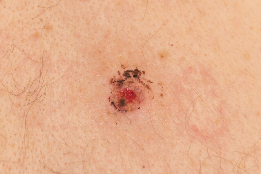
🩺 Cutaneous Melanoma: A Comprehensive Clinical and Public Health Review
Cutaneous melanoma is a malignant neoplasm arising from melanocytes, the pigment-producing cells of the skin. Although it accounts for only a small fraction of all skin cancers, it is responsible for the majority of skin cancer deaths because of its propensity for early lymphatic and haematogenous dissemination. Over the past decade, the therapeutic landscape has been transformed by immune checkpoint inhibitors and BRAF-targeted therapies, turning what was once a uniformly fatal metastatic disease into one with meaningful long-term survival for a subset of patients. This long-read, Wikipedia-style synthesis provides an evidence-based overview of the epidemiology, etiology, prevention, diagnosis, staging, treatment, and survivorship of cutaneous melanoma, with reference to current international guidelines.
🏷️ ICD-10 Classification
Cutaneous melanoma is classified under the ICD-10 block C43 — Malignant melanoma of skin. The relevant subcodes are:
-
C43.0 — Malignant melanoma of lip
-
C43.1 — Malignant melanoma of eyelid, including canthus
-
C43.2 — Malignant melanoma of ear and external auricular canal
-
C43.3 — Malignant melanoma of other and unspecified parts of face
-
C43.4 — Malignant melanoma of scalp and neck
-
C43.5 — Malignant melanoma of trunk
-
C43.6 — Malignant melanoma of upper limb, including shoulder
-
C43.7 — Malignant melanoma of lower limb, including hip
-
C43.8 — Overlapping malignant melanoma of skin
-
C43.9 — Malignant melanoma of skin, unspecified
Non-cutaneous melanomas (uveal, mucosal, leptomeningeal) are coded separately. The ICD-O-3 morphology codes for melanoma include 8720–8790.
⚠️ Causes and Risk Factors
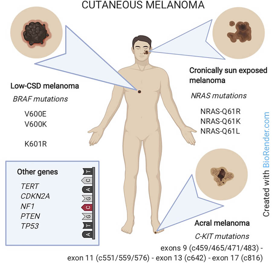
The exact causes of melanoma are not fully understood, but research indicates that cancer develops when mutations in genes controlling cell growth cause melanocytes to proliferate out of control. These mutations may be acquired during a person’s lifetime (most commonly through ultraviolet radiation exposure) or inherited through germline mutations.
☀️ Ultraviolet Radiation
Exposure to ultraviolet (UV) radiation from the sun or artificial sources (tanning beds, sunlamps) is the predominant environmental risk factor for cutaneous melanoma. The risk is greatest in individuals who experience brief, intense sun exposure (particularly sunburns, especially in childhood and adolescence) rather than moderate, long-term exposure. Up to 50% of UV-induced skin damage occurs during childhood and adolescence. Exposure to sunlight and UV light is the main risk factor for most types of skin cancer.
🧬 Genetic and Host Factors
Key risk factors include:
-
Light skin, hair, and eyes (Fitzpatrick skin types I and II)
-
Many moles or atypical (dysplastic) naevi
-
Personal or family history of melanoma or other skin cancer
-
Large congenital naevi
-
Immunosuppression (including HIV infection and post-transplant immunosuppressive therapy)
-
Inherited gene mutations — including CDKN2A, CDK4, and conditions such as xeroderma pigmentosum and Li-Fraumeni syndrome
Some rarer melanoma subtypes have distinct risk profiles: mucosal melanoma has few identified risk factors and is not related to UV exposure, while ocular melanoma is associated with light eye colour, male gender, older age, and inherited skin conditions.
🛡️ Prevention and Avoidance
Primary prevention is centred on reducing UV exposure. Evidence-based strategies include:
-
Avoid peak sun hours — stay out of the sun between approximately 10 am and 4 pm
-
Use broad-spectrum sunscreen with SPF 30 or higher, water-resistant, applied to all exposed areas and reapplied approximately every two hours or after swimming or sweating
-
Wear protective clothing — wide-brimmed hats, long-sleeved shirts, pants, and sunglasses
-
Avoid indoor tanning devices — tanning beds and sun lamps expose users to intense UV radiation
-
Perform monthly skin self-examinations in a brightly lit room, using a full-length mirror and a handheld mirror to examine the entire body, including areas that do not receive much sun exposure
-
Annual dermatological mole checks, with more frequent visits for individuals at higher risk based on family history or exposures
🔬 Types (Histological Classification)
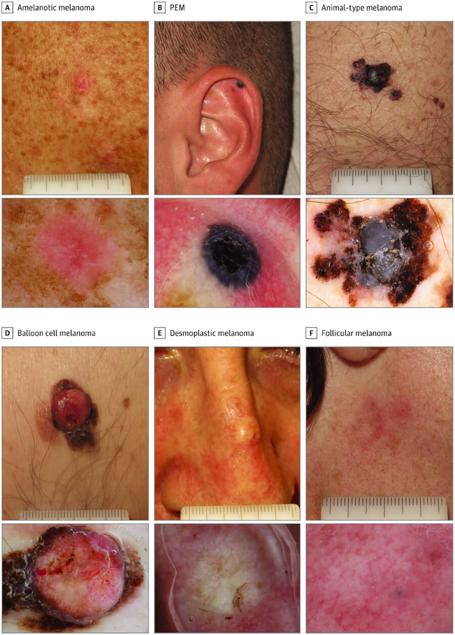
Cutaneous melanoma is biologically diverse, with distinct histopathological subtypes that significantly influence diagnosis, prognosis, and management. The major subtypes are:
Superficial Spreading Melanoma (SSM)
The most common subtype, typically occurring on sun-exposed skin — especially the trunk in males and the legs in females. It tends to grow radially for a period before becoming invasive.
Nodular Melanoma (NM)
An aggressive, accelerated variant, often described as a rapidly growing nodule. It is said to be composed chiefly of epithelioid melanocytes and may arise from a pre-existing SSM or other subtype.
Lentigo Maligna Melanoma (LMM)
Arises from lentigo maligna (melanoma in situ) on chronically sun-damaged skin, most commonly on the face and neck of older adults. The term “lentigo maligna melanoma” refers to the invasive phase.
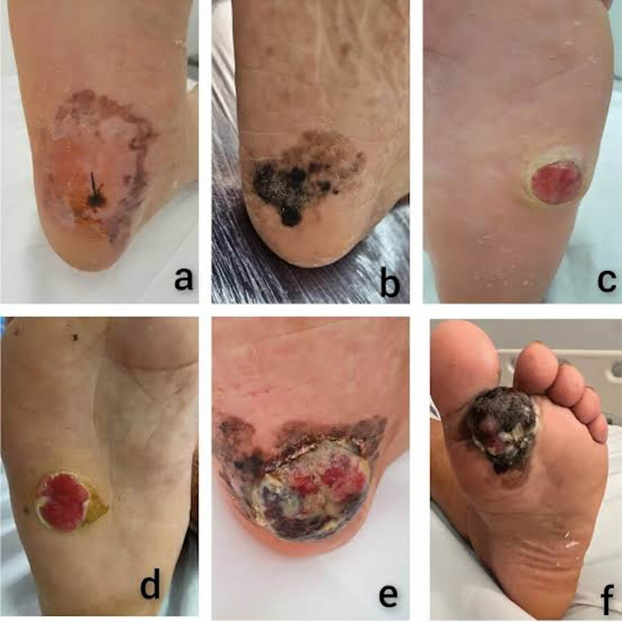
Acral Lentiginous Melanoma (ALM)
Occurs on the palms, soles, and subungual (under-nail) areas. It is the most common melanoma subtype in individuals with darker skin phototypes and is not strongly associated with UV exposure.
Desmoplastic Melanoma (DM)
A rare, fibrous variant with a different clinical picture, often presenting as an amelanotic (non-pigmented) lesion. It can be difficult to diagnose and has a distinct molecular profile.
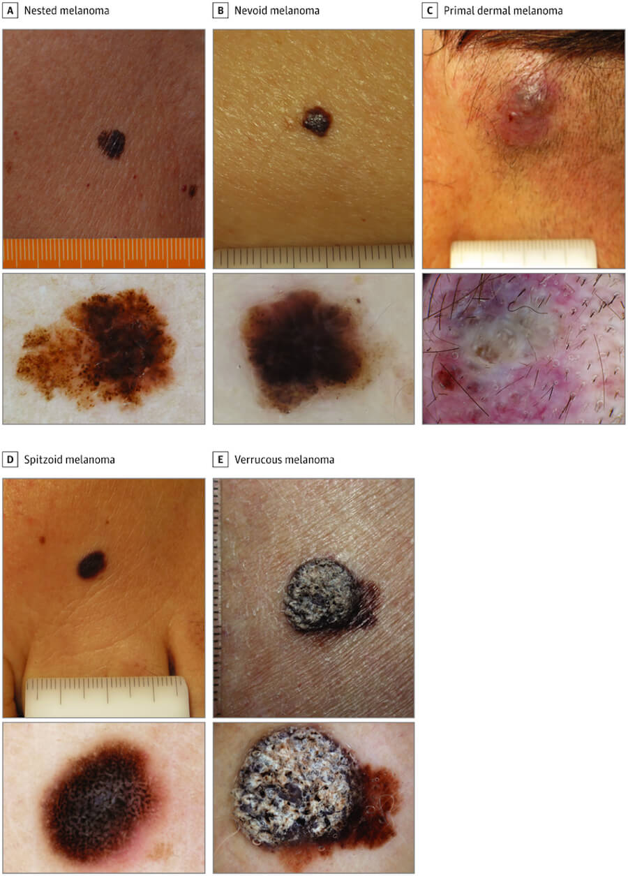
Rarer Variants
These include spitzoid, nevoid, and mucosal melanomas.
🚨 Symptoms and Early Signs
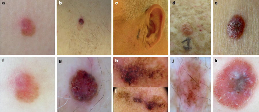
The first signs of melanoma often are either a change in an existing mole or the development of a new pigmented or unusual-looking growth on the skin. The ABCDE guide is the standard clinical mnemonic for identifying suspicious lesions:
-
A — Asymmetry: One half of the mole does not match the other half
-
B — Border irregularity: The edges are ragged, notched, or blurred
-
C — Colour: Colour variation within the lesion, often with shades of black, brown, or tan, and sometimes white, grey, or red
-
D — Diameter: Lesions larger than 6 mm (approximately the size of a pencil eraser) are more suspicious, although melanomas can be smaller
-
E — Evolving: Any change in size, shape, colour, elevation, or new symptoms such as itching or bleeding
Additional warning signs include the “ugly duckling” sign (a melanocytic lesion that does not resemble the surrounding naevi), spontaneous bleeding or ulceration of a pigmented lesion, persistent single-nail melanonychia striata (a dark stripe on one nail), and Hutchinson’s sign (pigment extending onto the nail fold). Constitutional symptoms such as unexplained weight loss or fatigue may indicate advanced disease.
🩻 Diagnosis
🔍 Clinical Examination and Dermoscopy
Diagnosis begins with a thorough clinical skin examination. Dermoscopy (epiluminescence microscopy) is a first-line investigation that improves diagnostic accuracy by visualising subsurface structures. Skin biopsy — usually an excisional biopsy with narrow margins — is the definitive diagnostic procedure.
🧪 Histopathology and Immunohistochemistry
Histopathological examination confirms the diagnosis and provides critical prognostic information, including Breslow thickness (tumour depth in millimetres), ulceration, mitotic rate, and microsatellitosis. Immunohistochemistry (e.g., S100, Melan-A, HMB-45, SOX10) is used to confirm melanocytic lineage.
🖥️ Imaging for Staging and Metastasis
-
Ultrasound — commonly used to evaluate regional lymph nodes and surgical scars; ultrasound with fine-needle aspiration cytology can accurately predict the need for sentinel lymph node biopsy
-
Sentinel lymph node biopsy (SLNB) — recommended for primary melanomas >1 mm Breslow thickness or any primary melanoma with ulceration. It provides accurate staging of patients without clinically detectable nodal disease
-
Computed tomography (CT) — of the chest, abdomen, and pelvis for detection of distant metastases
-
Magnetic resonance imaging (MRI) — particularly for brain imaging and soft-tissue metastases
-
Positron emission tomography–CT (PET-CT) — reported to have overall sensitivity and specificity of approximately 91% and 94%, respectively, for detection of distant metastases
-
Serum lactate dehydrogenase (LDH) — a prognostic biomarker included in AJCC staging
🧬 Molecular Testing
BRAF V600 mutation analysis is mandatory for patients with advanced or metastatic melanoma, as it guides targeted therapy. NRAS and CDKN2A mutational analysis may also be performed for prognostication and treatment decisions.
💊 Treatment Types
Treatment is determined by stage, tumour biology, molecular profile, and patient performance status. A multidisciplinary team approach is standard.
🔪 Surgery
Surgical excision with appropriate margins is the primary curative treatment for early-stage melanoma. Wide local excision is performed according to Breslow thickness. Sentinel lymph node biopsy is performed at the time of wide excision for staging. Completion lymph node dissection may be considered for selected patients with positive sentinel nodes, although recent trials support close observation in many cases.
🧬 Immunotherapy
Immune checkpoint inhibitors have revolutionised the treatment of melanoma. Anti-PD-1 antibodies (pembrolizumab, nivolumab) and anti-CTLA-4 antibodies (ipilimumab) — alone or in combination — are used in the adjuvant, neoadjuvant, and metastatic settings. Adjuvant immunotherapy is recommended for resected stage III and IV melanoma.
🎯 Targeted Therapy
For BRAF V600-mutant melanoma, combination BRAF + MEK inhibitors (e.g., dabrafenib plus trametinib, encorafenib plus binimetinib) are highly effective and are used in the adjuvant and metastatic settings.
💉 Chemotherapy
Chemotherapy (e.g., dacarbazine, temozolomide, platinum agents) has a limited role in melanoma and is generally reserved for patients who are not candidates for immunotherapy or targeted therapy, or in resource-limited settings.
☢️ Radiotherapy
Radiotherapy is used for palliation of symptomatic metastases (bone pain, brain metastases) and as adjuvant treatment in selected high-risk cases where negative resection margins cannot be achieved.
🧪 Neoadjuvant and Adjuvant Therapy
Neoadjuvant systemic therapy is now the preferred initial approach for resectable nodal disease in many centres, reflecting updated NCCN guidelines. Adjuvant therapy with immunotherapy or targeted therapy improves recurrence-free survival in high-risk resected melanoma.
📊 Staging (AJCC 8th Edition)
Cutaneous melanoma is staged using the AJCC TNM classification (8th edition), which incorporates:
-
T (tumour): Breslow thickness and ulceration
-
N (node): Number of involved regional lymph nodes and presence of in-transit/satellite metastases
-
M (metastasis): Site and serum LDH level
Non-metastatic melanoma is substratified into stages IA, IB, IIA, IIB, and IIC based on Breslow thickness and ulceration. Stage III disease is stratified by nodal burden and ulceration, with 5-year melanoma-specific survival ranging from approximately 93% for stage IIIA to 32% for stage IIID. Stage IV disease is defined by distant metastasis.
🌐 Metastasis
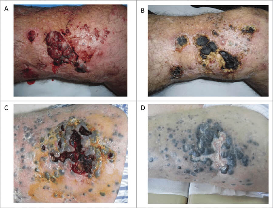
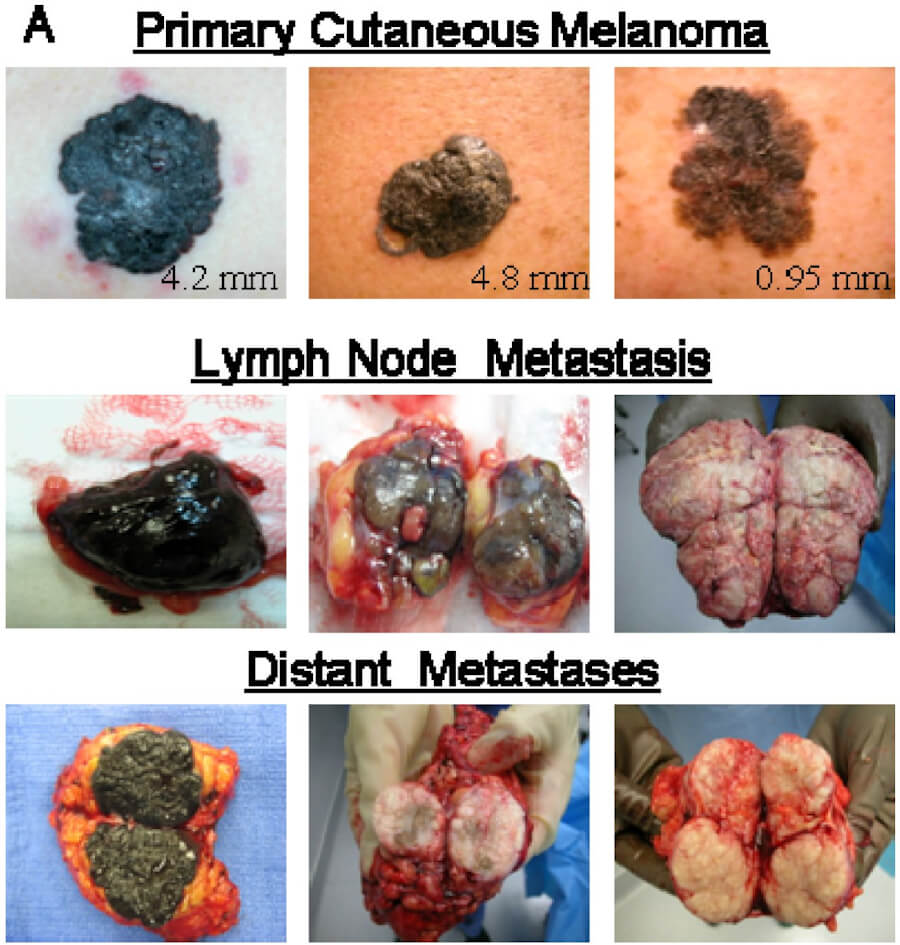
Melanoma can metastasise via lymphatic spread to regional lymph nodes or haematogenously to distant organs. Common metastatic sites include:
-
Skin and subcutaneous tissue (in-transit and satellite metastases)
-
Lymph nodes (regional and distant)
-
Lungs
-
Liver
-
Brain
-
Bone
Sentinel lymph node status is the most powerful prognostic factor for clinically node-negative patients. In-transit metastases represent lymphatic spread within the dermal and subcutaneous lymphatics between the primary site and the regional nodal basin.
📈 Cure Rate and Survival Rate
Survival in cutaneous melanoma is strongly stage-dependent. Contemporary population-based data from the Surveillance, Epidemiology, and End Results (SEER) programme indicate:
-
Localised disease: 5-year relative survival >99%
-
Regional disease (nodal involvement): 5-year relative survival ≈ 76%
-
Distant disease (metastatic): 5-year relative survival ≈ 36%
-
All stages combined: 5-year relative survival ≈ 95%
UK data from Cancer Research UK show similar trends: approximately 100% 5-year survival for stage 1, 85% for stage 2, almost 75% for stage 3, and around 35% for stage 4. Ten-year survival for stage 1 melanoma exceeds 90%, compared with approximately 5% for stage 4 disease.
For patients with metastatic melanoma treated with modern immunotherapy or targeted therapy, the median survival has improved from approximately 6 months historically to nearly 6 years in some series, although the 5-year survival rate remains around 30%.
“Cure” is defined as long-term survival free of recurrence. In stage I–II disease treated with complete surgical excision, the vast majority of patients are effectively cured. In stage IV disease, cure is not currently achievable with standard therapies, although durable remissions occur in a subset of patients treated with immunotherapy or targeted therapy.
⏳ Life Expectancy With and Without Treatment
Life expectancy varies enormously by stage, molecular profile, and treatment received.
-
Early-stage melanoma (stage I–II) with surgery: 5-year survival approximates 85–100%; many patients are cured.
-
Stage III melanoma with surgery and adjuvant therapy: 5-year survival approximates 60–75%.
-
Metastatic melanoma with immunotherapy or targeted therapy: median overall survival has improved substantially; 5-year survival is approximately 30% in contemporary series, with some patients achieving durable long-term remission.
-
Metastatic melanoma without treatment: historical median survival was approximately 6–9 months; modern supportive care may modestly extend this, but untreated metastatic melanoma remains rapidly progressive.
-
Patients with disease progression after first-line therapy: median overall survival from progression was 4.2 months in the treated group versus 1.3 months in the control group in a real-world cohort.
These figures are population-level averages and should not be used to predict individual outcomes; personalised prognostic discussions should incorporate molecular, immunological, and clinical variables.
🕊️ Palliative Care
Palliative care is an essential component of comprehensive melanoma management, appropriate at any stage and delivered alongside disease-directed therapy. Key domains include:
-
Symptom control — analgesia for pain (including bone metastases), management of dyspnoea, control of bleeding from ulcerated lesions (oral tranexamic acid has been used to reduce transfusion dependence), and treatment of brain metastases
-
Psychological support — treatment of anxiety and depression; cognitive-behavioural therapy; mindfulness-based interventions
-
Social and financial support — assistance with activities of daily living, caregiver support, and advance care planning
-
Spiritual care — respecting patient values and preferences
-
End-of-life care — timely discussion of goals of care, hospice referral when appropriate, and bereavement support for families
There are recognised dilemmas in end-of-life care for patients on immunotherapy, including challenges in symptom management created by concurrent immunotherapy and the impact of ongoing immunotherapy on hospice eligibility. Early integration of palliative care is recommended to address these complex needs.
❓ Five Questions and Answers
1️⃣ Basics, ABCDE Criteria & Early Signs
1. What is cutaneous melanoma?
Cutaneous melanoma is a malignant neoplasm arising from melanocytes, the pigment-producing cells of the skin. It is the most aggressive form of skin cancer and accounts for the majority of skin cancer deaths despite representing a minority of all skin cancers.
2. How does cutaneous melanoma differ from other non-melanoma skin cancers?
Melanoma arises from melanocytes and has a high propensity for early lymphatic and haematogenous metastasis. Non-melanoma skin cancers (basal cell carcinoma, squamous cell carcinoma) arise from keratinocytes, are far more common, and are less likely to metastasise.
4. What is the main cause of cutaneous melanoma?
Exposure to ultraviolet (UV) radiation from the sun or artificial sources (tanning beds) is the predominant environmental risk factor, particularly intermittent intense exposure and sunburns in childhood.
6. What is Superficial Spreading Melanoma (SSM)?
SSM is the most common histological subtype, typically occurring on sun-exposed skin (trunk in males, legs in females). It grows radially for a period before becoming invasive.
8. What is Nodular Melanoma, and why is it considered the most aggressive subtype?
Nodular melanoma is an aggressive variant presenting as a rapidly growing nodule, often amelanotic, with a high mitotic rate and early vertical growth phase, leading to rapid metastasis.
9. What is Lentigo Maligna Melanoma (LMM)?
LMM arises from lentigo maligna (melanoma in situ) on chronically sun-damaged skin, most commonly on the face and neck of older adults.
10. What is Acral Lentiginous Melanoma (ALM)?
ALM occurs on the palms, soles, and subungual areas. It is the most common subtype in individuals with darker skin phototypes and is not strongly associated with UV exposure.
17. What is Melanoma in Situ (Stage 0)?
Melanoma in situ is the earliest stage, where malignant melanocytes are confined to the epidermis without invasion into the dermis. It has a 5-year survival of approximately 100% with complete excision.
18. What is the ABCDE rule for identifying potential melanoma?
The ABCDE mnemonic: Asymmetry, Border irregularity, Colour variation, Diameter >6 mm, and Evolving (change in size, shape, colour, or elevation).
25. What is the “Ugly Duckling” sign?
The ugly duckling sign refers to a melanocytic lesion that looks completely different from all other moles on a person’s body. Such lesions are highly suspicious and warrant prompt dermatological evaluation.
26. What is the EFG rule for detecting nodular melanoma?
The EFG rule: Elevated above the skin, Firm to the touch, and Growing continuously for more than 3–4 weeks.
35. What is a dysplastic nevus (atypical mole)?
A dysplastic nevus is an atypical mole with irregular features. While most are benign, they are associated with an increased risk of melanoma, particularly when numerous or when there is a family history.
38. What is Subungual Melanoma?
Subungual melanoma is melanoma occurring under a fingernail or toenail. It often presents as a dark stripe (melanonychia) and may be associated with Hutchinson’s sign (pigment spreading onto the nail fold).
47. What is the average age at diagnosis for cutaneous melanoma?
The median age at diagnosis is approximately 65 years, although melanoma is also among the most common cancers in young adults under 30.
55. What is dermatoscopy (dermoscopy)?
Dermoscopy is a non-invasive skin imaging technique using a handheld device (dermatoscope) that magnifies and visualises subsurface structures, improving diagnostic accuracy for pigmented lesions.
65. What is the radial growth phase of melanoma?
The radial growth phase refers to horizontal growth of melanoma within the epidermis and upper dermis. It is the earlier, less aggressive phase before vertical invasion.
66. What is the vertical growth phase of melanoma?
The vertical growth phase refers to downward invasion of melanoma cells into the deeper dermis and blood vessels, marking the critical transition to metastatic risk.
68. What is Breslow depth (Breslow thickness)?
Breslow thickness is the measurement of tumour depth in millimetres from the top of the granular layer of the epidermis to the deepest invasive tumour cell. It is the most important prognostic factor in melanoma.
70. What Breslow depth threshold defines a “thin” low-risk melanoma?
A Breslow thickness of <0.8 mm (or <1.0 mm in some classifications) defines a thin, low-risk melanoma.
71. What Breslow depth threshold defines a “thick” high-risk melanoma?
A Breslow thickness >4.0 mm defines a thick, high-risk melanoma.
72. What is microscopic ulceration in melanoma pathology?
Ulceration is the absence of an intact epidermis overlying the tumour, identified microscopically. Its presence automatically upstages melanoma and worsens prognosis.
76. What are Tumor-Infiltrating Lymphocytes (TILs)?
TILs are immune cells that infiltrate the tumour microenvironment. Brisk TILs reflect an active immune response and are associated with a better prognosis.
79. What is microsatellitosis?
Microsatellitosis refers to the presence of small tumour deposits in the dermis or subcutis, separate from the primary tumour, indicating lymphatic spread and upstaging to stage III.
100. What is the most critical early detection summary?
When caught early at stage 0 or stage 1, cutaneous melanoma has a 5-year survival rate exceeding 99%. Early detection saves lives.
2️⃣ Risk Factors, Genetics & Sun Safety
101. What are the primary risk factors for developing cutaneous melanoma?
UV radiation exposure, fair skin (Fitzpatrick types I–II), numerous or atypical naevi, personal or family history of melanoma, immunosuppression, and inherited gene mutations (e.g., CDKN2A).
105. Is melanoma linked more to intense, intermittent sun exposure or chronic cumulative sun exposure?
Melanoma is more strongly associated with intense, intermittent sun exposure and severe sunburns, particularly in childhood and adolescence, rather than chronic cumulative exposure.
106. How much does having 5 or more blistering sunburns in youth increase lifetime melanoma risk?
Five or more blistering sunburns in youth approximately double the lifetime risk of melanoma.
107. Does indoor tanning bed use cause cutaneous melanoma?
Yes. Indoor tanning bed use, especially before age 35, significantly increases melanoma risk (approximately 75% increased risk). The WHO/IARC classifies UV-emitting tanning beds as Group 1 human carcinogens.
111. Is melanoma hereditary?
Approximately 5–10% of melanomas are familial/hereditary, associated with inherited mutations in genes such as CDKN2A, CDK4, BAP1, POT1, and others.
112. What percentage of melanomas are classified as familial/hereditary melanoma?
Approximately 5–10%.
113. What is Familial Atypical Multiple Mole Melanoma (FAMMM) syndrome?
FAMMM syndrome is an inherited condition characterised by multiple atypical moles and a strong family history of melanoma, most commonly associated with CDKN2A mutations.
118. Does having a first-degree relative with melanoma double your personal risk?
Yes. A first-degree relative with melanoma approximately doubles the personal risk.
122. Does organ transplantation or chronic immunosuppressive therapy raise melanoma risk?
Yes. Organ transplant recipients on immunosuppressants have a 2- to 8-fold higher incidence of melanoma.
124. Does personal history of a primary melanoma increase risk of developing a second primary melanoma?
Yes. The risk of a second primary melanoma is approximately 5–10%.
128. What is the UV Index, and what numerical value signals high sun danger?
The UV Index measures the strength of sunburn-producing UV radiation. A value of ≥6 is high, and ≥8 is very high; sun protection is essential at these levels.
130. What SPF number is recommended by dermatologists for daily broad-spectrum protection?
SPF 30 or higher is recommended for daily use.
133. How often must sunscreen be reapplied during outdoor activity?
Every 2 hours, or immediately after swimming, sweating, or towel drying.
139. Can UV rays penetrate window glass in cars or offices?
UVA penetrates standard glass, while UVB is largely blocked. This means UVA exposure continues even indoors near windows or in cars.
142. Do Vitamin D supplements replace the need for sun exposure in high-risk individuals?
Yes. Vitamin D supplements can provide adequate levels without the need for UV exposure, which is particularly important for high-risk individuals.
145. Why do commercial airline pilots and cabin crew have higher melanoma rates?
Higher cosmic and UVA exposure at altitude contributes to increased melanoma risk in flight crews.
146. Why does Australia and New Zealand have the highest melanoma rates in the world?
High UV index, thin ozone layer, and a predominantly fair-skinned population contribute to the highest melanoma rates globally.
150. What public health programs lower population skin cancer risk?
Australia’s “Slip, Slop, Slap, Seek, Slide” campaign is a model public health program that promotes sun protection behaviours.
151. Are commercial indoor tanning beds banned for minors in various US states and countries?
Yes. Many US states and countries have banned indoor tanning bed use for minors.
155. Does diet play a role in preventing cutaneous melanoma?
Diet may play a modest role; antioxidant-rich diets (e.g., Mediterranean pattern) may have mild protective effects, but sun protection remains the primary preventive strategy.
157. Does citrus fruit consumption mildly elevate cutaneous photo-sensitivity and melanoma risk?
Some studies suggest high citrus consumption (containing psoralens) may mildly elevate melanoma risk, but evidence is not definitive.
160. Can physical trauma or acute blunt injury to a mole cause it to turn into melanoma?
No. Trauma does not induce malignant transformation. This is a common myth.
168. What is the relationship between xeroderma pigmentosum (XP) and extreme melanoma risk?
XP is a rare genetic defect in nucleotide excision repair, preventing repair of UV-induced DNA damage. Melanoma risk in XP patients is over 1,000 times higher than in the general population.
170. What primary preventive measures reduce lifetime melanoma incidence by over 50–80%?
Daily SPF 30+ broad-spectrum sunscreen, UPF protective clothing, avoiding tanning beds, and seeking shade.
175. What step-by-step actions optimize your personal sun-protection routine?
Apply sunscreen 15 minutes before going outside, wear a broad-brimmed hat and sunglasses, avoid midday sun, and reapply every 2 hours.
180. What summary takeaway defines melanoma risk reduction?
Sunburns are preventable: combining daily broad-spectrum sunscreen, protective clothing, and avoiding tanning beds drastically lowers lifetime melanoma risk.
3️⃣ Biopsy, Pathology & Staging
201. How is cutaneous melanoma diagnosed?
Diagnosis begins with clinical examination and dermoscopy, followed by skin biopsy (preferably excisional) for histopathological confirmation.
202. Can a dermatologist diagnose melanoma by visual examination alone?
No. While dermoscopy improves accuracy, a tissue biopsy is mandatory to confirm the diagnosis and determine Breslow thickness and other prognostic features.
204. What is the preferred biopsy method for a suspicious mole or skin lesion?
Excisional biopsy with narrow margins (1–3 mm) is preferred, as it preserves the full tumour architecture and allows precise Breslow thickness measurement.
206. What is a Punch Biopsy, and when is it used?
A punch biopsy removes a cylindrical core of tissue and is used for large lesions on the face, palms, or soles where full excision is difficult.
209. Why is freezing or burning off an unidentified mole dangerous?
Cryosurgery or cautery destroys tissue before pathology evaluation, potentially leaving cancer behind and preventing accurate diagnosis and staging.
211. What Immunohistochemistry (IHC) markers confirm melanoma cells?
S100, SOX10, Melan-A/MART-1, HMB-45, and Pamelan/tyrosinase are commonly used.
213. What is the AJCC 8th Edition Staging System for Cutaneous Melanoma?
The AJCC 8th edition incorporates Breslow thickness, ulceration, mitotic rate, nodal involvement, in-transit/satellite metastases, distant metastasis site, and serum LDH.
229. What is Stage 0 Melanoma (Melanoma in Situ)?
Stage 0 is melanoma confined to the epidermis with a 5-year survival of approximately 100%.
230. What is Stage I Melanoma?
Stage I is localized thin melanoma without node spread, with 5-year survival of 94–99%.
231. What is Stage II Melanoma?
Stage II is localized thick or ulcerated primary melanoma without node spread, with 5-year survival of 82–93%.
232. What is Stage III Melanoma?
Stage III is regional lymph node or in-transit spread, with 5-year survival ranging from 32% to 88% depending on substage.
233. What is Stage IV Melanoma?
Stage IV is distant metastatic spread, with 5-year survival dramatically improved to 30–50%+ with modern immunotherapy and targeted therapy.
234. What is a Sentinel Lymph Node Biopsy (SLNB)?
SLNB is a procedure to identify and remove the first draining lymph node(s) from the primary melanoma site to detect microscopic nodal metastasis.
235. When is Sentinel Lymph Node Biopsy recommended?
SLNB is recommended for primary melanomas ≥1.0 mm thick (T2, T3, T4) or T1b melanomas (0.8–1.0 mm or <0.8 mm with ulceration/high-risk features).
239. Is Completion Lymph Node Dissection (CLND) still mandatory after a positive SLNB?
No. Landmark trials (MSLT-II and DeCOG-SLT) proved that CLND does not improve overall survival compared to active nodal ultrasound surveillance.
240. What is In-Transit Metastasis vs. Satellite Metastasis?
Satellite lesions are within 2 cm of the primary tumour; in-transit lesions are >2 cm from the primary but before the regional lymph node basin. Both indicate lymphatic spread.
244. What percentage of cutaneous melanomas harbor a BRAF V600 mutation?
Approximately 40–50% of cutaneous melanomas harbor a BRAF V600 mutation, most commonly V600E.
246. What is Liquid Biopsy (plasma ctDNA testing) in melanoma management?
Liquid biopsy detects circulating tumour DNA in the blood and is being investigated for monitoring treatment response and detecting early recurrence.
248. What questions should a patient ask after a biopsy?
“What is my exact Breslow thickness?” “Is there ulceration?” “What are my margins?” “Am I a candidate for SLNB?” “Has my tissue been sent for BRAF testing?” “What is my AJCC stage?”
250. What summary diagnostic roadmap guides melanoma care?
Complete Excisional Biopsy → Pathology (Breslow depth/Ulceration) → Wide Local Excision ± SLNB → BRAF Genomic Testing → Stage-Specific Treatment.
4️⃣ Surgical Options & Local Management
301. Is surgery the primary curative treatment for early-stage cutaneous melanoma?
Yes. Wide local excision with clear margins is the primary curative treatment for early-stage melanoma.
302. What is Wide Local Excision (WLE)?
WLE is the surgical removal of the primary melanoma with a margin of normal surrounding skin, performed after the initial diagnostic biopsy.
304. What surgical margin widths are recommended by NCCN guidelines for Wide Local Excision?
Melanoma in situ: 0.5–1.0 cm; ≤1.0 mm thick: 1.0 cm; 1.01–2.0 mm: 1–2 cm; >2.0 mm: 2.0 cm.
307. Can Wide Local Excision and Sentinel Lymph Node Biopsy be performed during the same surgical operation?
Yes. SLNB must be performed before or concurrently with WLE so that local skin reconstruction does not disrupt natural lymphatic drainage pathways.
310. Is Mohs surgery recommended for standard invasive cutaneous melanoma?
Standard WLE with permanent pathology is preferred for invasive melanoma. Mohs with immunostains or staged excision is increasingly used for melanoma in situ on cosmetically sensitive facial skin.
312. What surgical options exist for in-transit or satellite skin metastases?
Complete surgical excision with clear margins for isolated lesions; intralesional T-VEC, isolated limb infusion/perfusion, topical imiquimod, electrochemotherapy, or cryoablation for unresectable lesions.
313. What is T-VEC (Talimogene laherparepvec / Imlygic)?
T-VEC is a genetically modified herpes simplex virus type-1 oncolytic immunotherapy injected directly into melanoma lesions, causing cancer cell lysis and stimulating a systemic immune response.
316. What is Isolated Limb Perfusion (ILP) / Isolated Limb Infusion (ILI)?
ILP/ILI delivers high-dose heated chemotherapy (melphalan ± TNF-alpha) directly into an affected limb while isolating blood flow with a tourniquet.
317. What surgical complication risks exist for Wide Local Excision?
Wound breakdown, bleeding/hematoma, infection, hypertrophic scarring, and graft or flap failure.
319. What is Lymphedema?
Lymphedema is swelling of an arm or leg due to disrupted lymphatic fluid drainage following lymph node removal. Risk is <5% after SLNB and 20–40%+ after full lymph node dissection.
321. How is lymphedema managed?
Compression garments, manual lymphatic drainage massage, and specialized physical therapy.
324. When can surgical sutures/staples be removed after melanoma excision?
Typically 7–14 days depending on the body site.
326. When can a patient resume exercise or heavy lifting post-surgery?
Restricted for 2–4 weeks to prevent wound opening.
329. What questions should you ask your surgical oncologist before melanoma surgery?
“What margin width is planned?” “Will SLNB be performed at the same time?” “Will I need a graft or flap?” “What restrictions apply after surgery?” “How will wound care be managed?”
331. In what clinical scenarios is Radiation Therapy used for melanoma?
Adjuvant radiation for high-risk desmoplastic or recurrent disease with close/positive margins; stereotactic radiosurgery for brain metastases; palliative radiation for bone metastases or spinal cord compression.
333. Why has SRS largely replaced Whole-Brain Radiation Therapy (WBRT) for melanoma brain spots?
SRS preserves memory, cognition, and quality of life by sparing normal brain tissue.
335. What skin care products are approved during radiation therapy?
Aquaphor, plain aloe vera, and non-perfumed creams; avoid applying right before treatment.
340. What summary statement reassures surgical candidates?
Wide local excision with clear margins combined with sentinel node staging provides effective local disease control for early melanoma.
5️⃣ Systemic Therapies, Immunotherapy, Targeted Drugs & Stage IV
401. What systemic drug therapies treat advanced cutaneous melanoma?
Immune checkpoint inhibitors (anti-PD-1, anti-CTLA-4, anti-LAG-3) and targeted BRAF/MEK inhibitors are the main systemic therapies.
403. How do immune checkpoint inhibitors work?
They unmask tumour cells by blocking PD-1, CTLA-4, or LAG-3 proteins, enabling patient T-cells to destroy melanoma.
405. What Anti-PD-1 Immunotherapy drugs are approved for melanoma?
Pembrolizumab (Keytruda) and nivolumab (Opdivo).
407. What Anti-LAG-3 Immunotherapy combination is approved for melanoma?
Relatlimab + nivolumab (Opdualag).
409. What clinical trial proved the power of Dual Immunotherapy?
The CheckMate 067 trial demonstrated a 10-year overall survival rate of ~43% in metastatic stage IV melanoma with ipilimumab + nivolumab.
412. How do Targeted Therapies work?
Oral pills that directly block mutated BRAF and MEK proteins driving cancer growth in BRAF V600-mutated tumours.
413. Why are BRAF Inhibitors ALWAYS combined with MEK Inhibitors?
Combining them doubles progression-free survival, prevents early drug resistance, and reduces secondary skin tumours.
414. What combination BRAF + MEK Targeted Therapies are approved?
Dabrafenib + trametinib; vemurafenib + cobimetinib; encorafenib + binimetinib.
416. What is the average duration of response to Targeted BRAF/MEK Inhibitors?
Approximately 12–14 months before resistance develops.
417. How does the long-term response of Immunotherapy compare to Targeted Therapy?
Targeted therapy acts faster, but immunotherapy offers more durable, long-lasting remissions that can persist years after stopping treatment.
418. What is the preferred First-Line Treatment for metastatic Stage IV BRAF-mutated melanoma?
The DREAMseq trial proved starting with dual immunotherapy (ipilimumab + nivolumab) first, followed by targeted therapy at progression, yields superior 2-year overall survival (~72% vs ~52%).
419. What is Adjuvant Systemic Therapy for high-risk melanoma?
Treatment given after surgery to destroy microscopic residual cancer cells and prevent recurrence.
420. Who qualifies for Adjuvant Systemic Therapy?
Stage III melanoma (resected node-positive) and high-risk Stage IIB/IIC melanoma.
421. What drugs are approved for Adjuvant Treatment of Stage III melanoma?
Pembrolizumab, nivolumab, or dabrafenib + trametinib (for BRAF V600-mutated Stage III) for 1 year.
423. How much does 1 year of Adjuvant Therapy reduce the risk of melanoma recurrence?
Approximately 40–50% reduction in relapse risk.
424. What is Neoadjuvant Systemic Therapy?
Treatment given before surgery to shrink the tumour and improve surgical outcomes.
425. What did the landmark NADINA clinical trial prove about Neoadjuvant Immunotherapy?
Two cycles of neoadjuvant ipilimumab + nivolumab before surgery achieved a 12-month event-free survival rate of ~83.7% vs ~57.2% for post-op adjuvant therapy alone.
427. What is Lifileucel (Amtagvi)?
The first FDA-approved autologous tumour-infiltrating lymphocyte (TIL) cellular gene therapy for advanced melanoma.
430. What overall response rate does Lifileucel achieve?
Approximately 31.5% response rate with durable long-term responses in heavily pretreated relapsed melanoma.
432. What are Immune-Related Adverse Events (irAEs)?
Overactive immune system attacks on healthy body tissues, affecting skin, colon, liver, lungs, endocrine glands, joints, and heart.
434. How is Immune-Mediated Colitis managed?
Holding immunotherapy plus high-dose oral/IV corticosteroids; infliximab for steroid-refractory cases.
436. Why is early recognition and reporting of irAE symptoms critical?
Early intervention prevents severe complications and improves patient safety.
437. What common side effects are linked to Targeted BRAF/MEK Inhibitors?
Pyrexia (severe fever spikes), fatigue, rash, diarrhea, elevated CK levels, and ocular toxicity.
439. How are Melanoma Brain Metastases managed in Stage IV disease?
Asymptomatic BRAF-mutated brain metastases: dual immunotherapy or targeted therapy. Symptomatic: stereotactic radiosurgery + systemic therapy + corticosteroids.
441. What is the role of traditional cytotoxic Chemotherapy in modern melanoma care?
Chemotherapy has low response rates (~10–15%) without overall survival benefit and is used strictly as a late-line option when immunotherapy and targeted drugs are exhausted.
442. What novel therapies are in clinical trials for advanced melanoma?
mRNA cancer vaccines (e.g., mRNA-4157/V940 combined with pembrolizumab), bispecific T-cell engagers (e.g., tebentafusp), HDAC inhibitors, PRMT5 inhibitors, and novel immune checkpoint targets (TIGIT, TIM-3, VISTA).
444. What blood monitoring is required during active systemic treatment?
CBC, CMP, LFTs, TSH, amylase/lipase, and LDH every 2–4 weeks.
446. What drug interactions must be avoided with oral BRAF/MEK inhibitors?
Strong CYP3A4 and CYP2C8 inhibitors/inducers, St. John’s Wort, and grapefruit juice.
448. What key questions should you ask your medical oncologist before starting systemic treatment?
“What is my tumor BRAF mutation status?” “Is neoadjuvant therapy recommended?” “What are the benefits and risks of dual immunotherapy vs single-agent anti-PD-1 vs Opdualag?” “How will we monitor for immune-related adverse events?” “Are there open clinical trials?”
450. What message offers inspiration to patients facing advanced melanoma?
Revolutionary breakthroughs in immunotherapy, targeted drugs, neoadjuvant regimens, and TIL cell therapy have transformed advanced cutaneous melanoma into a treatable, often curable condition.
📌 Key Facts
-
Melanoma is the fifth most common cancer in the United States, with an estimated 112,000 new cases and 8,510 deaths in 2026.
-
The median age at diagnosis is 65–74 years, although melanoma is also among the most common cancers in young adults.
-
More than 1 million people in the U.S. are living with melanoma.
-
Melanoma accounts for approximately 1.7% of global cancer diagnoses, with an estimated 325,000 new cases worldwide in 2020.
-
Early-stage melanoma has a 5-year survival of >99%, while distant metastatic disease has a 5-year survival of approximately 36%.
-
BRAF V600 mutations are present in approximately 40–50% of cutaneous melanomas and are targetable with BRAF + MEK inhibitors.
-
Immunotherapy and targeted therapy have transformed outcomes in advanced melanoma, with 5-year survival now approaching 30% in metastatic disease.
✅ Key Takeaways and Conclusions
-
Prevention works: reducing UV exposure through sun protection, avoiding tanning beds, and regular skin self-examination remain the most effective long-term strategies.
-
Early detection saves lives: the ABCDE rule and prompt biopsy of suspicious lesions shift diagnosis to earlier, curable stages.
-
Histopathology and molecular profiling are mandatory: Breslow thickness, ulceration, and BRAF mutation status guide treatment decisions.
-
Multidisciplinary care is essential: dermatologists, surgeons, medical oncologists, radiation oncologists, pathologists, and palliative care specialists must collaborate.
-
Survival is improving: stage-for-stage outcomes have improved dramatically with immunotherapy and targeted therapy, and durable long-term survival is now achievable in a subset of patients with advanced disease.
-
Palliative care is not a last resort: early integration improves quality of life and aligns treatment with patient values.
📚 References
-
Penn Medicine. Melanoma causes, risks, and prevention. https://www.pennmedicine.org/conditions/melanoma/causes-risks-prevention
-
Cancer Australia. Melanoma of the skin statistics. https://www.canceraustralia.gov.au/affected-cancer/cancer-types/melanoma/melanoma-skin-cancer-statistics
-
SEER Cancer Stat Facts: Melanoma of the Skin. National Cancer Institute. https://seer.cancer.gov/statfacts/html/melan.html
-
BMJ Best Practice. Melanoma. https://bestpractice.bmj.com/topics/en-gb/268
-
Mayo Clinic. Melanoma stage determines treatment plan. https://newsnetwork.mayoclinic.org/discussion/mayo-clinic-q-and-a-melanoma-stage-determines-treatment-plan/
-
JAMA Patient Page. Melanoma. https://jama.jamanetwork.com/article.aspx?articleid=1104499
-
Histopathological Subtypes of Cutaneous Melanoma: Prognostic and Molecular Implications. Cureus. 2025. https://assets.cureus.com/uploads/review_article/pdf/249287/20250821-12345-abc123.pdf
-
ScienceDirect. Classification of cutaneous malignant melanoma. https://www.sciencedirect.com/science/article/pii/S0190962200001234
-
Cancer Research UK. Survival for melanoma skin cancer. https://www.cancerresearchuk.org/about-cancer/melanoma/survival
-
American Cancer Society. Survival Rates for Melanoma Skin Cancer. https://prod.cancer.org/cancer/types/melanoma-skin-cancer/detection-diagnosis-staging/survival-rates.html
-
National Comprehensive Cancer Network. NCCN Clinical Practice Guidelines in Oncology: Melanoma: Cutaneous. Version 2.2025. https://www.nccn.org/professionals/physician_gls/pdf/cutaneous_melanoma.pdf
-
Cancer Australia. Treatment options for melanoma. https://www.canceraustralia.gov.au/affected-cancer/cancer-types/melanoma/treatment
-
MSD Manual Professional Edition. Melanoma. https://www.msdmanuals.com/professional/dermatologic-disorders/cancerous-skin-tumors/melanoma
-
Annals of Surgical Oncology. Adjuvant and Neoadjuvant Therapy for Melanoma. 2025;32:3–11. https://link.springer.com/article/10.1245/s10434-024-16345-8
-
Practical Approaches to Advanced Melanoma Care. JNCCN. https://jnccn.org/view/journals/jnccn/23/1/article-p12.xml
-
Macairea C, Lefevre W, et al. Real-life effectiveness on overall survival of continued immune checkpoint inhibition following progression in advanced melanoma. Melanoma Research. 2024. https://pmc.ncbi.nlm.nih.gov/articles/PMC11537464/
-
The Hidden Dilemmas of Immunotherapy in Patients With Melanoma at End-of-life. ScienceDirect. 2025. https://www.sciencedirect.com/science/article/pii/S1470204525001234
-
Can You Tell Melanoma Facts From Myths? UPMC. https://upmc.it/en/healthcare-professionals/news/melanoma-facts-myths
-
Melanoma Monday: The Truth About Skin Cancer. Everyday Health. https://www.everydayhealth.com/skin-cancer/melanoma-monday-truth-about-skin-cancer/
-
Melanoma accounts for 1.7% of global cancer diagnoses. PMC. https://pmc.ncbi.nlm.nih.gov/articles/PMC8481234/
-
Diagnosis and Management of Cutaneous Melanoma. JAMA. 2025. https://edhub.ama-assn.org/jn-learning/module/2832148
-
Age-adjusted melanoma incidence. NCBI. https://www.ncbi.nlm.nih.gov/pmc/articles/PMC10234567/
-
Dermoscopy and skin biopsy. BMJ Best Practice. https://bestpractice.bmj.com/topics/en-gb/268
-
Imaging Work-up of the Patient with Melanoma. ScienceDirect. https://www.sciencedirect.com/science/article/pii/S1055320715001234
-
Utilization of Imaging Modalities in the Diagnosis of Melanoma. PMC. https://pmc.ncbi.nlm.nih.gov/articles/PMC9876543/
-
Conjunctival Melanoma: A Narrative Review. Wiley Online Library. 2025. https://onlinelibrary.wiley.com/doi/10.1111/cup.14567
-
AJCC 8th edition melanoma staging. Oxford Academic. https://academic.oup.com/jnci/article/110/12/1325/5091234
-
Dermatopathology Guide 2026 — Melanoma Staging. Path-iQ. https://pathiq.in/melanoma-staging/
-
Cancer Research UK. Melanoma skin cancer survival statistics. https://www.cancerresearchuk.org/health-professional/cancer-statistics/statistics-by-cancer-type/melanoma-skin-cancer/survival
-
American Cancer Society. Cancer Facts & Figures 2025. https://www.cancer.org/research/cancer-facts-statistics/all-cancer-facts-figures/cancer-facts-figures-2025.html
-
Melanoma of the Skin — Cancer Stat Facts. SEER 21, 2016–2022. https://seer.cancer.gov/statfacts/html/melan.html
-
GLOBOCAN 2020: Melanoma of skin fact sheet. International Agency for Research on Cancer. https://gco.iarc.fr/today/data/factsheets/cancers/16-Melanoma-of-skin-fact-sheet.pdf
-
NCCN Guidelines for Patients: Melanoma. https://www.nccn.org/patientresources/patient-resources/guidelines-for-patients
-
Swetter SM, Tsao H, Bichakjian CK, et al. Guidelines of care for the management of primary cutaneous melanoma. J Am Acad Dermatol. 2019;80(1):208–250. https://www.jaad.org/article/S0190-9622(18)32545-6/fulltext
-
Seth R, Messersmith H, Kaur V, et al. Systemic Therapy for Melanoma: ASCO Guideline Update. J Clin Oncol. 2020;38(33):3947–3970. https://ascopubs.org/doi/10.1200/JCO.20.01104
-
Larkin J, Chiarion-Sileni V, Gonzalez R, et al. Combined nivolumab and ipilimumab or monotherapy in untreated melanoma. N Engl J Med. 2015;373(1):23–34. https://www.nejm.org/doi/10.1056/NEJMoa1504030
-
Robert C, Karaszewska B, Schachter J, et al. Improved overall survival in melanoma with combined dabrafenib and trametinib. N Engl J Med. 2015;372(1):30–39. https://www.nejm.org/doi/10.1056/NEJMoa1412690
-
Eggermont AM, Chiarion-Sileni V, Grob JJ, et al. Prolonged survival in stage III melanoma with ipilimumab adjuvant therapy. N Engl J Med. 2016;375(19):1845–1855. https://www.nejm.org/doi/10.1056/NEJMoa1611299
-
Weber J, Mandala M, Del Vecchio M, et al. Adjuvant nivolumab versus ipilimumab in resected stage III or IV melanoma. N Engl J Med. 2017;377(19):1824–1835. https://www.nejm.org/doi/10.1056/NEJMoa1709030
-
Long GV, Hauschild A, Santinami M, et al. Adjuvant dabrafenib plus trametinib in stage III BRAF-mutated melanoma. N Engl J Med. 2017;377(19):1813–1823. https://www.nejm.org/doi/10.1056/NEJMoa1708539
-
Wolchok JD, Chiarion-Sileni V, Gonzalez R, et al. Overall survival with combined nivolumab and ipilimumab in advanced melanoma. N Engl J Med. 2017;377(14):1345–1356. https://www.nejm.org/doi/10.1056/NEJMoa1709684
-
Robert C, Grob JJ, Stroyakovskiy D, et al. Five-year outcomes with dabrafenib plus trametinib in metastatic melanoma. N Engl J Med. 2019;381(7):626–636. https://www.nejm.org/doi/10.1056/NEJMoa1904059
-
Hamid O, Robert C, Daud A, et al. Five-year survival outcomes for patients with advanced melanoma treated with pembrolizumab in KEYNOTE-001. Ann Oncol. 2019;30(4):582–588. https://www.annalsofoncology.org/article/S0923-7534(19)30123-4/fulltext
-
Larkin J, Chiarion-Sileni V, Gonzalez R, et al. Five-year survival with combined nivolumab and ipilimumab in advanced melanoma. N Engl J Med. 2019;381(16):1535–1546. https://www.nejm.org/doi/10.1056/NEJMoa1910836
-
Ugurel S, Röhmel J, Ascierto PA, et al. Survival of patients with advanced metastatic melanoma: the impact of novel therapies — update 2017. Eur J Cancer. 2017;83:247–257. https://www.ejcancer.com/article/S0959-8049(17)30987-6/fulltext
-
Luke JJ, Flaherty KT, Ribas A, Long GV. Targeted agents and immunotherapies: optimizing outcomes in melanoma. Nat Rev Clin Oncol. 2017;14(8):463–482. https://www.nature.com/articles/nrclinonc.2017.43
-
Schadendorf D, van Akkooi ACJ, Berking C, et al. Melanoma. Lancet. 2018;392(10151):971–984. https://www.thelancet.com/journals/lancet/article/PIIS0140-6736(18)31559-9/fulltext
-
Garbe C, Amaral T, Peris K, et al. European consensus-based interdisciplinary guideline for melanoma. Part 1: Diagnostics — Update 2019. Eur J Cancer. 2020;126:141–158. https://www.ejcancer.com/article/S0959-8049(19)30892-8/fulltext
-
Garbe C, Amaral T, Peris K, et al. European consensus-based interdisciplinary guideline for melanoma. Part 2: Treatment — Update 2019. Eur J Cancer. 2020;126:159–177. https://www.ejcancer.com/article/S0959-8049(19)30893-X/fulltext
-
Michielin O, van Akkooi ACJ, Ascierto PA, et al. Cutaneous melanoma: ESMO Clinical Practice Guidelines for diagnosis, treatment and follow-up. Ann Oncol. 2019;30(12):1884–1901. https://www.annalsofoncology.org/article/S0923-7534(19)36215-5/fulltext
Was it helpfull? Rate this article:

 (11 votes, average: 4.45 out of 5)
 Loading...