Lung Cancer: A Comprehensive Clinical and Public Health Guide
Lung cancer is a malignant neoplasm arising from the epithelial cells of the lower respiratory tract, most commonly the bronchi and bronchioles. It is the leading cause of cancer incidence and mortality worldwide, with an estimated 2.5 million new cases and 1.8 million deaths in 2022. Despite representing a major global health burden, lung cancer is also among the most preventable of malignancies, bec
ause the overwhelming majority of cases are attributable to modifiable environmental and behavioural risk factors—principally tobacco smoking, which accounts for 60–70% of cases globally and approximately 85% of cases in high-income settings. Lung cancer is frequently diagnosed at an advanced stage, when curative treatment is no longer possible, which explains its persistently high case-fatality rate. Nevertheless, advances in low-dose computed tomography (LDCT) screening, molecular profiling, immunotherapy, and targeted therapy have begun to reshape outcomes, particularly for the non-small cell lung cancer (NSCLC) subtypes that constitute approximately 85% of all cases.
📋 ICD-10 Classification
In the International Classification of Diseases, Tenth Revision (ICD-10), malignant neoplasms of the lung and bronchus are coded under C34, which is subdivided by anatomical location:
-
C34.0 — Main bronchus (carina, hilus)
-
C34.1 — Upper lobe, bronchus or lung
-
C34.2 — Middle lobe, bronchus or lung
-
C34.3 — Lower lobe, bronchus or lung
-
C34.8 — Overlapping lesion of bronchus and lung
-
C34.9 — Bronchus or lung, unspecified
The closely related code C33 refers to malignant neoplasms of the trachea. In the AJCC TNM staging system (8th edition, with the forthcoming 9th edition), lung cancer is staged using the T (primary tumour), N (regional lymph nodes), and M (distant metastasis) categories, which are combined into prognostic stage groups ranging from occult carcinoma through Stage 0 to Stage IV. Small cell lung cancer (SCLC) is traditionally staged using the simpler limited-stage versus extensive-stage dichotomy, although TNM staging is increasingly applied.
🧬 What Causes Lung Cancer?
The aetiology of lung cancer is dominated by cigarette smoking, which is responsible for approximately 85 out of every 100 cases in many populations. Smoking-related carcinogenesis involves more than 70 known carcinogens, including polycyclic aromatic hydrocarbons, nitrosamines, and radioactive polonium-210, which induce characteristic patterns of somatic mutations—most notably C:G to A:T transversions in the TP53 and KRAS genes.
Other established risk factors include:
-
Secondhand tobacco smoke (involuntary smoking), which raises risk by approximately 20–30% in never-smokers.
-
Outdoor and household air pollution, including fine particulate matter (PM₂.₅) and indoor combustion products from solid fuels.
-
Occupational carcinogens: asbestos, silica, diesel exhaust, arsenic, chromium, nickel, and polycyclic aromatic hydrocarbons.
-
Radon exposure, a radioactive gas that accumulates in basements and is the second leading cause of lung cancer in many countries.
-
Prior thoracic radiation therapy, particularly for Hodgkin lymphoma or breast cancer.
-
Chronic lung diseases, such as chronic obstructive pulmonary disease (COPD) and pulmonary fibrosis, which independently elevate risk.
-
Genetic susceptibility, including family history and inherited variants in DNA repair and detoxification genes.
In never-smokers, lung cancer is more frequently associated with oncogenic driver mutations—such as EGFR mutations, ALK and ROS1 rearrangements, and BRAF V600E—and is more commonly of the adenocarcinoma subtype.
🛡️ How to Avoid and Prevent Lung Cancer
Primary prevention is the most effective strategy. The single most important intervention is tobacco control: avoiding smoking initiation, supporting cessation, and protecting non-smokers from secondhand smoke. Smoking cessation at any age reduces subsequent lung cancer risk relative to continued smoking, although the risk does not return to that of a never-smoker.
Public health measures include smoke-free legislation, tobacco taxation, graphic warning labels, and mass-media campaigns. Occupational hygiene—enforcement of asbestos bans, silica dust control, and diesel exhaust mitigation—reduces exposure to workplace carcinogens. Radon mitigation in high-risk dwellings and air-quality regulation further lower population risk.
Secondary prevention through low-dose computed tomography (LDCT) screening is recommended for high-risk individuals—typically adults aged 50–80 years with a ≥20 pack-year smoking history who currently smoke or have quit within the past 15 years. Landmark trials have demonstrated that LDCT screening reduces lung cancer mortality by approximately 20% in high-risk populations. Screening is not recommended for average-risk individuals or for those with limited life expectancy.
🔬 Types of Lung Cancer
Lung cancer is broadly divided into two major histological categories, which differ in biology, staging, and treatment.
🟢 Non-Small Cell Lung Cancer (NSCLC)
NSCLC accounts for approximately 85% of all lung cancers. It comprises several subtypes:
-
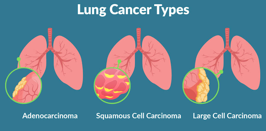
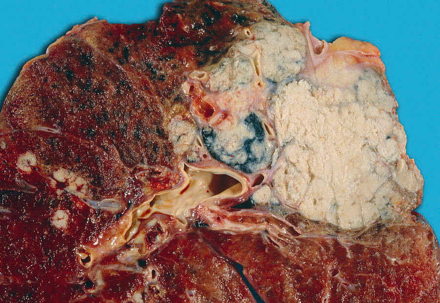
Adenocarcinoma: the most common subtype, accounting for approximately 40% of lung cancers and 60–70% of NSCLC. It typically arises in the peripheral lung, often in mucus-secreting glandular cells, and is the predominant subtype in never-smokers and younger patients.
-
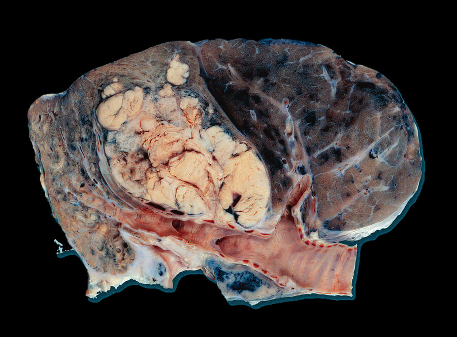
Squamous cell carcinoma: accounts for approximately 25–30% of NSCLC and typically arises centrally, near the major airways, from the flat squamous cells lining the bronchi.
-
Large cell carcinoma: accounts for approximately 10–15% of NSCLC and lacks the specific features of adenocarcinoma or squamous cell carcinoma; it tends to be peripheral and aggressive.
-
Rare subtypes: adenosquamous carcinoma, sarcomatoid carcinoma, salivary-gland-type tumours, and lung sarcoma.
🔴 Small Cell Lung Cancer (SCLC)
SCLC accounts for approximately 13–15% of lung cancers and is strongly associated with smoking. It is a neuroendocrine tumour that typically arises centrally and grows rapidly, with early dissemination to regional lymph nodes and distant sites. SCLC is traditionally staged as limited-stage (confined to one hemithorax and encompassable within a single radiation port) or extensive-stage (disease beyond these boundaries). Despite high initial chemosensitivity, SCLC relapse is common and prognosis remains poor.
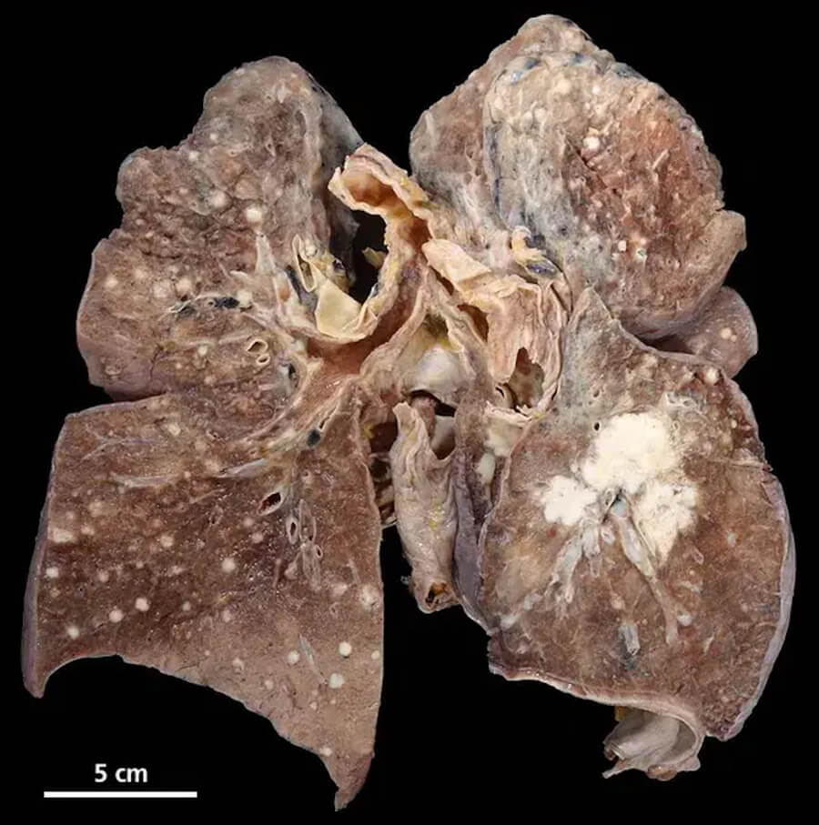
🩺 Symptoms and Early Signs
Lung cancer is often asymptomatic in its early stages; when symptoms do occur, they frequently reflect locally advanced or metastatic disease. This delayed symptomatology is a principal reason why lung cancer is the leading cause of cancer death.
Common respiratory symptoms include:
-
A new or changing cough that does not resolve within 3 weeks, or a chronic cough that worsens.
-
Haemoptysis (coughing up blood or rust-coloured sputum).
-
Shortness of breath (dyspnoea).
-
Chest pain, particularly pleuritic pain worsened by deep breathing, coughing, or laughing.
-
Recurrent or slowly resolving respiratory infections (bronchitis, pneumonia) due to airway obstruction.
-
Hoarseness from recurrent laryngeal nerve involvement.
-
Wheezing or stridor from central airway narrowing.
Systemic symptoms include unexplained weight loss, fatigue, loss of appetite, and fever.
Symptoms of metastatic spread depend on the site: bone pain (skeletal metastases), headache, nausea, seizures, or focal neurological deficits (brain metastases), jaundice or right upper quadrant pain (liver metastases), and swollen supraclavicular or cervical lymph nodes. Paraneoplastic syndromes—including hypercalcaemia (squamous cell carcinoma), SIADH (SCLC), Cushing syndrome (SCLC), and Lambert–Eaton myasthenic syndrome (SCLC)—may be the presenting feature.
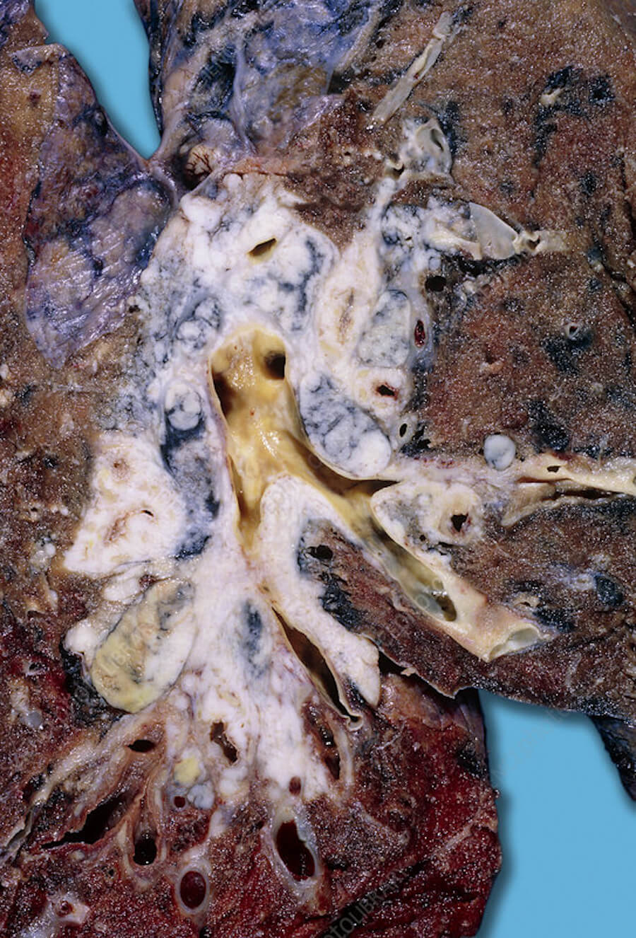
🖥️ Diagnosis: Imaging, Biopsy, and Biomarkers
The diagnostic pathway for suspected lung cancer integrates clinical assessment, imaging, tissue confirmation, and molecular profiling.
🩻 Imaging
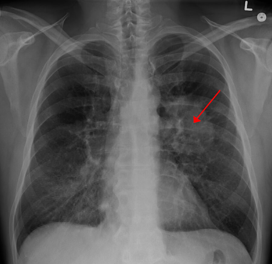
Chest radiography is often the first test but has limited sensitivity for small or centrally located tumours. Computed tomography (CT) of the chest and upper abdomen with intravenous contrast is the cornerstone of diagnosis and staging, providing detailed anatomical information about the primary tumour, mediastinal lymph nodes, and distant organs. Positron emission tomography combined with CT (FDG-PET/CT) is used for whole-body staging, detecting metabolically active metastases and guiding biopsy. Magnetic resonance imaging (MRI) of the brain with contrast is recommended for patients with neurological symptoms or those with stage III–IV disease at high risk of brain metastasis.
🔬 Tissue Confirmation
A tissue biopsy is mandatory to confirm the diagnosis and determine histology. Approaches include bronchoscopy (with endobronchial ultrasound-guided transbronchial needle aspiration, EBUS-TBNA, for mediastinal nodes), CT-guided percutaneous core needle biopsy, endoscopic ultrasound-guided biopsy (EUS), mediastinoscopy, and, less commonly, surgical biopsy via video-assisted thoracoscopic surgery (VATS). Sputum cytology and pleural fluid analysis may provide supportive evidence but are insufficient alone for a definitive diagnosis.
🧪 Biomarker Testing
Molecular profiling of the tumour tissue is now standard of care for all patients with advanced NSCLC, and increasingly for early-stage disease to guide adjuvant therapy. Key biomarkers include:
-
EGFR sensitising mutations (exons 19 deletion, L858R), which predict response to EGFR tyrosine kinase inhibitors (erlotinib, gefitinib, osimertinib).
-
ALK and ROS1 rearrangements, which predict response to ALK/ROS1 inhibitors (crizotinib, alectinib, lorlatinib).
-
BRAF V600E mutation, targetable with dabrafenib plus trametinib.
-
KRAS G12C mutation, targetable with sotorasib or adagrasib.
-
PD-L1 expression by immunohistochemistry, which predicts benefit from immune checkpoint inhibitors.
-
MET exon 14 skipping, RET fusions, NTRK fusions, and HER2 mutations, each with corresponding targeted agents.
Liquid biopsy—analysis of circulating tumour DNA (ctDNA) in plasma—is increasingly used when tissue is insufficient or for monitoring resistance mutations.
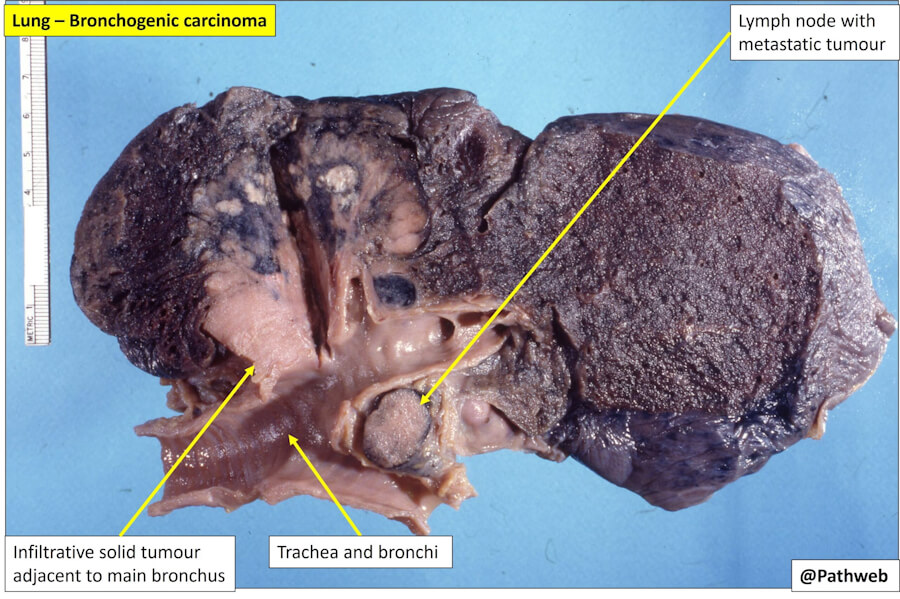
💊 Treatment Types
Treatment for lung cancer is multidisciplinary and depends on histology, stage, molecular profile, and patient fitness.
🔪 Surgery
Surgical resection is the primary curative modality for early-stage NSCLC (stages I–II, and selected IIIA). Procedures include lobectomy (the standard), segmentectomy or wedge resection for small peripheral tumours or patients with limited pulmonary reserve, and pneumonectomy for centrally located tumours. Video-assisted thoracoscopic surgery (VATS) and robot-assisted approaches reduce morbidity compared with open thoracotomy. Surgery is generally not indicated for SCLC, except in the rare case of a solitary pulmonary nodule without nodal or distant spread.
☢️ Radiation Therapy
Radiation therapy is used in multiple settings:
-
Definitive chemoradiotherapy for locally advanced, unresectable NSCLC (stage III).
-
Concurrent chemoradiotherapy for limited-stage SCLC, often followed by prophylactic cranial irradiation (PCI) to reduce brain metastases.
-
Stereotactic body radiotherapy (SBRT) for medically inoperable early-stage NSCLC or oligometastatic disease.
-
Palliative radiotherapy for symptom control in advanced disease (bone pain, haemoptysis, airway obstruction, brain metastases).
💉 Systemic Therapy
Cytotoxic chemotherapy remains a backbone for both NSCLC and SCLC. Platinum-based doublets (cisplatin or carboplatin combined with pemetrexed, gemcitabine, paclitaxel, docetaxel, or etoposide) are standard first-line regimens for advanced disease without actionable drivers. In SCLC, platinum plus etoposide is the standard backbone, combined with immunotherapy in extensive-stage disease.
Immunotherapy with immune checkpoint inhibitors (pembrolizumab, nivolumab, atezolizumab, durvalumab, ipilimumab) has transformed the treatment of NSCLC and SCLC. These agents are used as monotherapy in PD-L1-high NSCLC, in combination with chemotherapy in first-line metastatic NSCLC, as consolidation after chemoradiotherapy in stage III NSCLC, and in extensive-stage SCLC.
Targeted therapy is indicated for NSCLC harbouring actionable driver mutations. Oral tyrosine kinase inhibitors achieve high response rates with generally favourable toxicity profiles compared with chemotherapy. Examples include osimertinib (EGFR), alectinib and lorlatinib (ALK), crizotinib (ROS1), dabrafenib/trametinib (BRAF V600E), and sotorasib/adagrasib (KRAS G12C).
Antibody–drug conjugates (ADCs) and bispecific antibodies are emerging options for previously treated NSCLC.
📊 Stages and TNM Classification
The AJCC/UICC TNM staging system (8th edition) is the standard for lung cancer staging.
-
T categories describe the primary tumour size and local invasion: Tis (carcinoma in situ), T1 (≤3 cm), T2 (>3–5 cm or involving main bronchus, visceral pleura, or hilar nodes), T3 (>5–7 cm or invading chest wall, phrenic nerve, or parietal pericardium), T4 (>7 cm or invading mediastinum, heart, great vessels, trachea, oesophagus, vertebral body, or carina).
-
N categories describe regional lymph node involvement: N0 (none), N1 (ipsilateral peribronchial/hilar), N2 (ipsilateral mediastinal/subcarinal), N3 (contralateral mediastinal, hilar, or supraclavicular).
-
M categories describe distant metastasis: M0 (none), M1a (contralateral lung nodule, pleural/pericardial effusion, or pleural nodules), M1b (single extrathoracic metastasis), M1c (multiple extrathoracic metastases).
Stage groups range from occult carcinoma (TX N0 M0) through Stage 0 (Tis N0 M0), Stage I (IA1–IB), Stage II (IIA–IIB), Stage III (IIIA–IIIC), to Stage IV (IVA–IVB).
🌐 Metastasis
Lung cancer metastasises via lymphatic and haematogenous routes. Common sites of distant metastasis include the brain, bones, liver, adrenal glands, and contralateral lung. Brain metastases are particularly common in SCLC, ALK-rearranged NSCLC, and EGFR-mutated NSCLC, and routine brain MRI surveillance is recommended in these subgroups. Bone metastases may cause pain, pathological fracture, and hypercalcaemia. Liver metastases are associated with poor prognosis. Adrenal metastases are often asymptomatic and detected on staging imaging. Metastatic spread is the principal driver of lung cancer mortality and the reason why stage IV disease remains largely incurable, although systemic therapy can achieve durable disease control in a subset of patients.
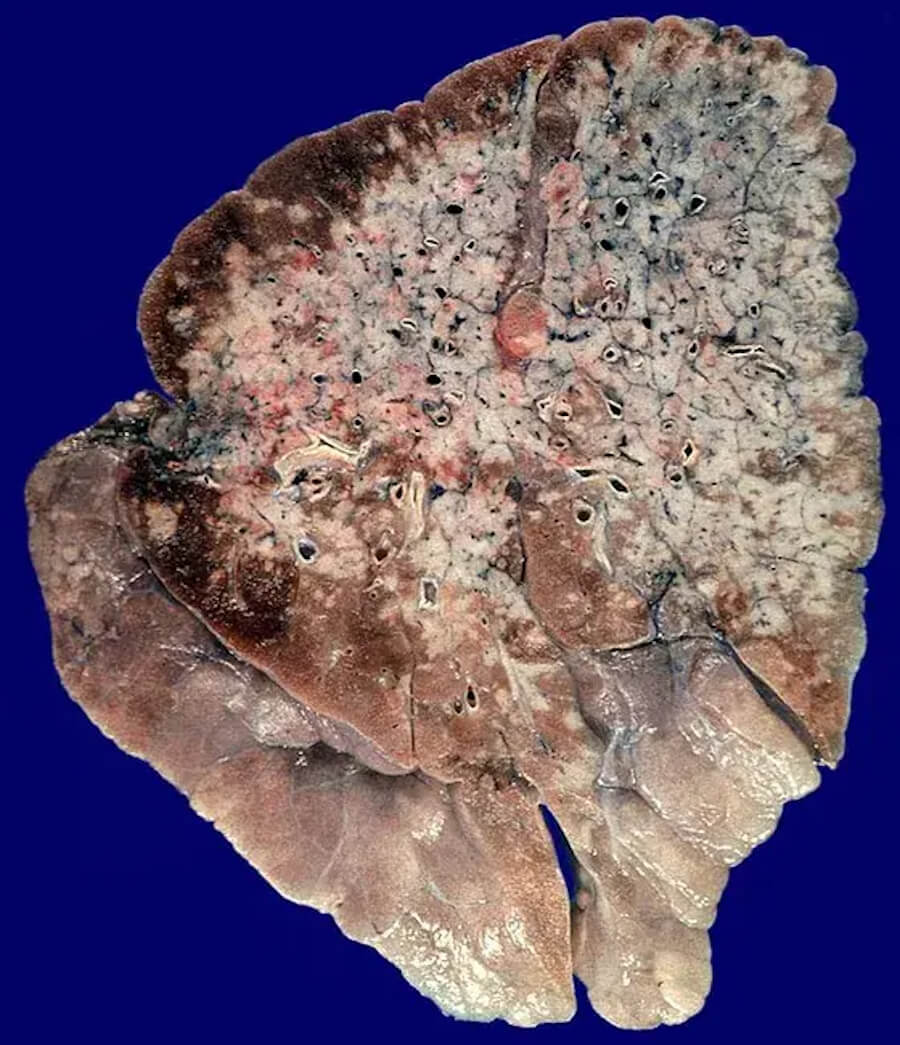
📈 Cure Rate, Survival Rate, and Life Expectancy
Lung cancer survival is strongly stage-dependent. According to SEER data for NSCLC:
-
Localised disease: 5-year relative survival ≈ 67%.
-
Regional disease: 5-year relative survival ≈ 40%.
-
Distant disease: 5-year relative survival ≈ 12%.
-
All stages combined: 5-year relative survival ≈ 32%.
For SCLC, outcomes are poorer: limited-stage 5-year survival ≈ 16–28%, and extensive-stage 5-year survival ≈ 12%.
Without treatment, the natural history of lung cancer is one of progressive local growth and metastatic dissemination, with median survival typically measured in months for advanced disease. With treatment, outcomes vary widely: early-stage NSCLC treated with surgical resection can achieve 5-year survival rates of 65–80% for stage I and 30–60% for stage II; locally advanced disease treated with chemoradiotherapy and immunotherapy achieves 5-year survival of approximately 13–36% for stage III; and metastatic disease, while generally incurable, has seen meaningful improvements with immunotherapy and targeted therapy, with 5-year survival for stage IV now reaching 5–6% in some contemporary series, and considerably higher for molecularly defined subgroups such as ALK-rearranged NSCLC treated with sequential TKIs.
Life expectancy for an individual patient depends on stage, histology, molecular profile, performance status, comorbidity, and response to therapy. Population-level median survival for metastatic NSCLC is approximately 8–12 months without targeted therapy, but can extend to several years in patients with actionable drivers treated with appropriate TKIs or in those achieving durable responses to immunotherapy.
🕊️ Palliative Care
Palliative care is an essential component of comprehensive lung cancer management, aimed at relieving symptoms, improving quality of life, and supporting patients and families, irrespective of curative or life-prolonging intent. Early integration of palliative care alongside standard oncology treatment has been shown to improve quality of life, mood, and even survival in advanced NSCLC. In a contemporary real-world analysis, patients receiving early palliative care had a median survival of 8.7 months compared with 5.9 months for those receiving late palliative care, and lower 2-year and 5-year mortality rates.
Palliative interventions include opioid and non-opioid analgesia for pain, palliative radiotherapy for bone pain, haemoptysis, airway obstruction, and brain metastases, pleurodesis for malignant pleural effusion, airway stenting for central airway obstruction, and psychosocial and spiritual support. Hospice care—a subset of palliative care for patients with limited life expectancy—focuses on comfort and dignity at the end of life. Advance care planning discussions should occur early, ideally while the patient retains decision-making capacity.
❓ Five Questions and Answers
1. What is lung cancer?
Lung cancer is a malignant tumor arising from the cells of the lung, most commonly the epithelial lining of the bronchi and bronchioles. It is the leading cause of cancer death worldwide.
2. What are the main types of lung cancer?
The two main types are non-small cell lung cancer (NSCLC), accounting for about 85% of cases, and small cell lung cancer (SCLC), accounting for about 15%.
3. What is Non-Small Cell Lung Cancer (NSCLC)?
NSCLC is a group of lung cancers that includes adenocarcinoma, squamous cell carcinoma, and large cell carcinoma. It generally grows more slowly than SCLC and is more often amenable to surgery and targeted therapy.
4. What is Small Cell Lung Cancer (SCLC)?
SCLC is a fast-growing, neuroendocrine lung cancer strongly associated with smoking. It tends to disseminate early and is usually treated with chemotherapy and radiation rather than surgery.
5. What percentage of lung cancers are NSCLC (~85%) vs SCLC (~15%)?
NSCLC accounts for approximately 85% of lung cancers, and SCLC for approximately 13–15%.
6. What is Adenocarcinoma of the lung?
Adenocarcinoma is the most common subtype of NSCLC, arising from glandular cells, often in the peripheral lung. It is the predominant subtype in never-smokers and younger patients.
7. What is Squamous Cell Carcinoma of the lung?
Squamous cell carcinoma is an NSCLC subtype that typically arises centrally in the major bronchi from squamous epithelial cells. It is strongly associated with smoking.
8. What is Large Cell Carcinoma?
Large cell carcinoma is an NSCLC subtype that lacks the specific features of adenocarcinoma or squamous cell carcinoma. It tends to be peripheral and aggressive.
9. What is Adenosquamous Carcinoma of the lung?
Adenosquamous carcinoma is a rare NSCLC subtype containing both adenocarcinoma and squamous cell carcinoma components.
10. What is Sarcomatoid Carcinoma of the lung?
Sarcomatoid carcinoma is a rare, aggressive NSCLC subtype with spindle-shaped or giant cells, often associated with poor prognosis.
11. What is Bronchial Carcinoid Tumor (typical vs atypical)?
Bronchial carcinoid tumors are neuroendocrine tumors of the lung. Typical carcinoids are low-grade and slow-growing; atypical carcinoids are more aggressive and have a higher risk of metastasis.
12. What is Mesothelioma, and how does it differ from true lung cancer?
Mesothelioma is a cancer of the pleura (the lining around the lungs), usually caused by asbestos exposure. It is not a primary lung cancer, although it can invade the lung.
13. Where in the lungs do adenocarcinomas typically arise (outer/peripheral edges)?
Adenocarcinomas typically arise in the peripheral lung tissue, often near the pleura.
14. Where in the lungs do squamous cell carcinomas typically arise (central airways/bronchi)?
Squamous cell carcinomas typically arise centrally in the major bronchi.
15. What is Adenocarcinoma in Situ (AIS), formerly known as bronchioloalveolar carcinoma (BAC)?
AIS is a pre-invasive lesion in which malignant glandular cells grow along the alveolar walls without invasion. It is classified as stage 0.
16. What is Minimally Invasive Adenocarcinoma (MIA)?
MIA is an early-stage adenocarcinoma with a predominantly lepidic pattern and invasion of ≤5 mm. It has an excellent prognosis.
17. What is Invasive Mucinous Adenocarcinoma of the lung?
Invasive mucinous adenocarcinoma is a subtype of adenocarcinoma that produces abundant mucin and often presents as pneumonia-like consolidation.
18. What is the difference between primary lung cancer and metastatic cancer to the lungs?
Primary lung cancer originates in the lung. Metastatic cancer to the lungs originates elsewhere (e.g., breast, colon, kidney) and spreads to the lungs.
19. What cancers commonly metastasize to the lungs (breast, colon, kidney, melanoma, sarcoma)?
Common cancers that metastasize to the lungs include breast, colon, kidney, melanoma, sarcoma, and others.
20. How do pathologists distinguish primary lung cancer from secondary lung metastases under a microscope?
They use immunohistochemistry (e.g., TTF-1, napsin A, p40) and clinical history to distinguish primary lung cancer from metastases.
21. What is TTF-1 (Thyroid Transcription Factor-1), and why is it used as a lung adenocarcinoma marker?
TTF-1 is a protein expressed in lung adenocarcinoma and thyroid cancer. It is used as an immunohistochemical marker to confirm lung origin.
22. What is p40 or p63, and why are they used to identify squamous cell carcinoma?
p40 and p63 are markers of squamous differentiation, used to confirm squamous cell carcinoma.
23. What is Chromogranin and Synaptophysin, and why are they used to identify neuroendocrine/SCLC cells?
Chromogranin and synaptophysin are neuroendocrine markers used to identify SCLC and other neuroendocrine tumors.
24. What is Combined Small Cell Lung Cancer?
Combined SCLC is a variant containing both SCLC and NSCLC components.
25. What is Limited-Stage Small Cell Lung Cancer (LS-SCLC)?
LS-SCLC is confined to one hemithorax and can be encompassed within a single radiation field.
26. What is Extensive-Stage Small Cell Lung Cancer (ES-SCLC)?
ES-SCLC has spread beyond one hemithorax or to distant organs.
27. Why is Small Cell Lung Cancer considered biologically aggressive?
SCLC grows rapidly, doubles quickly, and metastasizes early, often before symptoms appear.
28. How fast does Small Cell Lung Cancer double in size compared to NSCLC?
SCLC has a much shorter doubling time (often 30–90 days) compared with NSCLC.
29. What is the driver mutation landscape in Non-Small Cell Lung Cancer?
Actionable driver mutations in NSCLC include EGFR, ALK, ROS1, BRAF, KRAS G12C, MET, RET, NTRK, and HER2.
30. What is an EGFR mutation in lung cancer?
EGFR mutations are activating mutations in the epidermal growth factor receptor gene that drive cancer growth and are targetable with EGFR tyrosine kinase inhibitors.
31. What are the most common EGFR exon mutations (Exon 19 deletion, Exon 21 L858R, Exon 20 insertion)?
Exon 19 deletion and exon 21 L858R are sensitizing mutations; exon 20 insertions are resistant to standard EGFR TKIs.
32. What is an ALK rearrangement/fusion in lung cancer?
ALK rearrangement is a fusion of the ALK gene with another gene (e.g., EML4) that drives cancer growth and is targetable with ALK inhibitors.
33. What is a KRAS mutation in lung cancer (e.g., KRAS G12C)?
KRAS mutations are common in smokers and drive cancer growth. KRAS G12C is targetable with sotorasib or adagrasib.
34. What is a ROS1 rearrangement in lung cancer?
ROS1 rearrangement is a fusion that drives cancer growth and is targetable with ROS1 inhibitors such as crizotinib or entrectinib.
35. What is a BRAF V600E mutation in lung cancer?
BRAF V600E is an activating mutation targetable with dabrafenib plus trametinib.
36. What is a MET exon 14 skipping mutation?
MET exon 14 skipping is a mutation that leads to MET overactivation and is targetable with capmatinib or tepotinib.
37. What is a RET fusion in lung cancer?
RET fusion is a driver mutation targetable with selpercatinib or pralsetinib.
38. What is an NTRK gene fusion?
NTRK fusions are rare driver mutations targetable with larotrectinib or entrectinib.
39. What is a HER2 (ERBB2) mutation in lung cancer?
HER2 mutations are driver mutations targetable with trastuzumab deruxtecan.
40. What is PD-L1 expression, and how is it measured (Tumor Proportion Score / TPS)?
PD-L1 expression is measured by immunohistochemistry and reported as TPS (percentage of tumor cells expressing PD-L1). It predicts benefit from immune checkpoint inhibitors.
41. What does PD-L1 ≥50% mean for treatment selection?
PD-L1 TPS ≥50% supports first-line single-agent immunotherapy in NSCLC without actionable driver mutations.
42. What is Tumor Mutational Burden (TMB) in lung cancer?
TMB is the number of mutations per megabase in tumor DNA; high TMB may predict benefit from immunotherapy.
43. Why do never-smokers with lung cancer usually have different genetic mutations than heavy smokers?
Never-smokers more often have EGFR, ALK, ROS1, or RET alterations, whereas smokers more often have KRAS, TP53, and squamous histology.
44. Why is KRAS G12C more common in former or current smokers?
KRAS G12C is strongly associated with tobacco exposure.
45. Why are EGFR mutations and ALK fusions more common in young, never-smoker women?
These drivers are more common in never-smokers, women, and younger patients, though the reasons are not fully understood.
46. What is the cell of origin for lung adenocarcinoma (type II pneumocytes)?
Adenocarcinoma is thought to arise from type II pneumocytes or club cells.
47. What is the cell of origin for squamous cell lung carcinoma (bronchial basal epithelial cells)?
Squamous cell carcinoma arises from basal cells of the bronchial epithelium.
48. What is the cell of origin for Small Cell Lung Cancer (neuroendocrine cells)?
SCLC arises from neuroendocrine cells of the bronchial epithelium.
49. What are Pancoast tumors (superior sulcus tumors)?
Pancoast tumors are lung cancers located at the extreme apex of the lung, often invading the chest wall, brachial plexus, and sympathetic nerves.
50. Where are Pancoast tumors located in the chest (extreme apex of the top of the lung)?
They are located at the superior sulcus, the very top of the lung.
51. What is Horner Syndrome, and why is it caused by Pancoast tumors pressing on sympathetic nerves?
Horner syndrome results from disruption of the sympathetic chain, causing ptosis, miosis, and anhidrosis. Pancoast tumors can compress these nerves.
52. What are the 3 classic signs of Horner Syndrome (ptosis/drooping eyelid, miosis/constricted pupil, anhidrosis/lack of sweating)?
Ptosis, miosis, and anhidrosis.
53. What is Superior Vena Cava (SVC) Syndrome?
SVC syndrome is obstruction of the superior vena cava, causing facial and upper extremity swelling, distended neck veins, and shortness of breath.
54. What causes SVC Syndrome (central tumor compressing the main vein carrying blood from head/arms to heart)?
It is usually caused by a central lung tumor or enlarged mediastinal lymph nodes compressing the SVC.
55. What are the symptoms of SVC Syndrome (facial swelling, arm swelling, neck vein distension, shortness of breath)?
Facial and arm swelling, distended neck veins, shortness of breath, and sometimes headache or dizziness.
56. What are Paraneoplastic Syndromes in lung cancer?
Paraneoplastic syndromes are remote effects of cancer not caused by direct invasion or metastasis, such as SIADH, hypercalcemia, Cushing syndrome, and Lambert-Eaton syndrome.
57. What is Syndrome of Inappropriate Antidiuretic Hormone (SIADH), and why is it linked to SCLC?
SIADH is excessive ADH secretion causing hyponatremia. It is commonly associated with SCLC.
58. What causes hypercalcemia of malignancy in squamous cell lung cancer (PTHrP secretion)?
Squamous cell carcinoma can secrete parathyroid hormone-related protein (PTHrP), causing hypercalcemia.
59. What is Lambert-Eaton Myasthenic Syndrome (LEMS), and how does it relate to SCLC?
LEMS is a paraneoplastic neuromuscular disorder causing proximal muscle weakness, often associated with SCLC.
60. What is Hypertrophic Pulmonary Osteoarthropathy (HPOA) / digital clubbing?
HPOA is a paraneoplastic syndrome causing digital clubbing, periostitis, and joint pain, often seen in lung cancer.
61. What causes Cushing Syndrome in SCLC (ectopic ACTH production)?
SCLC can produce ectopic ACTH, leading to Cushing syndrome.
62. What is a solitary pulmonary nodule (SPN)?
An SPN is a single, well-defined lung opacity ≤3 cm in diameter surrounded by normal lung tissue.
63. What size threshold defines a pulmonary nodule (<3 cm) versus a lung mass (>3 cm)?
A nodule is ≤3 cm; a mass is >3 cm.
64. What percentage of incidentally found lung nodules on CT turn out to be benign (>90%)?
More than 90% of small incidental nodules are benign.
65. What benign conditions cause lung nodules (granulomas, old histoplasmosis/TB infection, hamartomas)?
Granulomas, old infections (TB, histoplasmosis), hamartomas, and scars are common benign causes.
66. What is ground-glass opacity (GGO) on a lung CT scan?
GGO is a hazy area of increased attenuation that does not obscure underlying bronchial and vascular margins.
67. What is a part-solid lung nodule, and why does it carry a higher malignancy risk than pure GGO?
A part-solid nodule has both ground-glass and solid components; the solid component increases malignancy risk.
68. What is a spiculated margin on a lung nodule, and why does it suggest malignancy?
A spiculated margin is a star-like or jagged edge that suggests invasive growth and malignancy.
69. What is pleural effusion, and how does it relate to lung cancer?
Pleural effusion is fluid in the pleural space; it can be malignant in lung cancer and cause shortness of breath.
70. What is Malignant Pleural Effusion (MPE)?
MPE is pleural fluid containing cancer cells, indicating advanced disease.
71. How do cancer cells reach the pleural space around the lungs?
Cancer cells can invade the pleura directly or spread via lymphatics or blood.
72. What is pericardial effusion in lung cancer?
Pericardial effusion is fluid around the heart, which can occur when lung cancer invades the pericardium.
73. What is lymphangitic carcinomatosis?
Lymphangitic carcinomatosis is diffuse spread of cancer through pulmonary lymphatics, causing shortness of breath and interstitial patterns on imaging.
74. How does lung cancer spread through the lymphatic system in the mediastinum?
Cancer cells enter lymphatic channels and travel to regional mediastinal lymph nodes.
75. What are mediastinal lymph node stations (e.g., N1 vs N2 vs N3 nodes)?
N1: ipsilateral hilar/peribronchial nodes. N2: ipsilateral mediastinal/subcarinal nodes. N3: contralateral mediastinal, hilar, or supraclavicular nodes.
76. What is skip metastasis in lung cancer nodal staging?
Skip metastasis is spread to N2 nodes without involvement of N1 nodes.
77. Why does lung cancer frequently metastasize to the brain, adrenals, bones, and liver?
These organs have rich blood supply and are common sites for hematogenous spread.
78. What is the blood-brain barrier, and why does it complicate brain metastasis treatment?
The blood-brain barrier restricts drug penetration into the brain, limiting treatment options for brain metastases.
79. What is Leptomeningeal Disease (LMD) in advanced lung cancer?
LMD is spread of cancer to the leptomeninges (membranes covering the brain and spinal cord), causing neurological symptoms.
80. What is the average age of diagnosis for lung cancer (~70 years old)?
The average age at diagnosis is approximately 70 years.
81. Can young adults in their 20s or 30s get lung cancer?
Yes, although it is rare, young adults can develop lung cancer, often with actionable driver mutations.
82. What is the incidence rate of lung cancer in men versus women?
Lung cancer incidence is historically higher in men, but rates in women have been rising, especially in never-smokers.
83. Why are lung cancer rates declining in men while remaining stable or rising in never-smoker women?
Declining smoking rates in men and rising smoking rates or other exposures in women contribute to these trends.
84. Is lung cancer the leading cause of cancer death worldwide?
Yes, lung cancer is the leading cause of cancer death worldwide.
85. How many people die from lung cancer annually globally (~1.8 million)?
Approximately 1.8 million deaths annually.
86. What is the difference between right lung and left lung anatomy regarding lobes (3 lobes right, 2 lobes left)?
The right lung has three lobes (upper, middle, lower); the left lung has two lobes (upper, lower) and the lingula.
87. Why are tumors slightly more common in the right lung than the left lung (due to higher ventilation volume and larger right main bronchus)?
The right lung receives more ventilation and has a larger main bronchus, which may increase exposure to carcinogens.
88. What is the lingula of the left lung?
The lingula is a tongue-like projection of the left upper lobe, analogous to the right middle lobe.
89. What is visceral pleura vs parietal pleura?
Visceral pleura covers the lung surface; parietal pleura lines the chest wall.
90. What is the pleural cavity?
The pleural cavity is the potential space between the visceral and parietal pleurae.
91. What is the hilum of the lung?
The hilum is the region where bronchi, blood vessels, and nerves enter and exit the lung.
92. What is the mediastinum?
The mediastinum is the central compartment of the chest containing the heart, great vessels, trachea, esophagus, and lymph nodes.
93. What is a carcinoid crisis in bronchial carcinoid tumors?
Carcinoid crisis is a rare, life-threatening release of vasoactive substances causing severe flushing, hypotension, and bronchospasm.
94. What is atypical adenomatous hyperplasia (AAH) of the lung?
AAH is a pre-invasive lesion that may progress to adenocarcinoma in situ.
95. What is the multi-step progression model of lung adenocarcinoma (AAH -> AIS -> MIA -> Invasive Adenocarcinoma)?
This model describes the stepwise progression from atypical adenomatous hyperplasia to adenocarcinoma in situ, minimally invasive adenocarcinoma, and invasive adenocarcinoma.
96. How do pathologists grade lung adenocarcinoma (predominant patterns: lepidic, acinar, papillary, micropapillary, solid)?
Grading is based on the predominant architectural pattern, with micropapillary and solid patterns indicating higher grade.
97. Why are micropapillary and solid patterns considered aggressive high-grade features?
They are associated with higher recurrence rates and worse prognosis.
98. What is Tumor Spread Through Air Spaces (STAS) in lung cancer pathology?
STAS is the spread of tumor cells through air spaces beyond the main tumor margin, associated with increased recurrence risk.
99. Why does the presence of STAS increase local recurrence risk after sublobar surgery?
STAS can lead to residual tumor cells in the remaining lung, increasing local recurrence.
100. Where can patients read expert biological summaries of lung cancer subtypes?
National Cancer Institute (NCI), American Cancer Society, NCCN, and IASLC provide expert summaries.
201. What is the leading cause of lung cancer?
Cigarette smoking is the leading cause of lung cancer, accounting for approximately 85% of cases.
202. What percentage of lung cancer deaths are directly caused by cigarette smoking (~80–85%)?
About 80–85% of lung cancer deaths are directly attributable to cigarette smoking.
203. How much does cigarette smoking increase the risk of developing lung cancer (~15 to 30 times higher risk)?
Smokers have a 15- to 30-fold higher risk of lung cancer compared with never-smokers.
204. How do carcinogens in tobacco smoke damage DNA in bronchial lung cells?
Tobacco carcinogens form DNA adducts and cause mutations (e.g., TP53, KRAS) that initiate and promote lung cancer.
205. What specific carcinogens are present in tobacco smoke (PAHs, nitrosamines like NNK, benzene, polonium-210)?
Polycyclic aromatic hydrocarbons, tobacco-specific nitrosamines (NNK), benzene, and radioactive polonium-210 are among the major carcinogens.
206. Does cigar smoking or pipe smoking cause lung cancer?
Yes; cigar and pipe smoking also cause lung cancer, though the risk is generally lower than with cigarettes.
207. Does vaping / e-cigarette use cause lung cancer?
Evidence is still evolving; vaping is not harmless and may increase risk, but long-term cancer data are limited compared with cigarettes.
208. What are the long-term health effects of e-cigarette vapor on bronchial epithelial cells?
E-cigarette vapor can cause oxidative stress, inflammation, and DNA damage in airway cells, but long-term cancer risk is not yet fully defined.
209. Does hookah / waterpipe smoking cause lung cancer?
Yes; hookah smoking is associated with increased lung cancer risk.
210. Does smoking marijuana / cannabis cause lung cancer?
Chronic heavy cannabis smoking may increase risk, but evidence is less consistent than for tobacco.
211. What is secondhand smoke (environmental tobacco smoke / ETS)?
Secondhand smoke is the combination of sidestream smoke from a burning cigarette and mainstream smoke exhaled by a smoker.
212. How much does secondhand smoke increase lung cancer risk in non-smokers (~20–30% increased risk)?
Secondhand smoke increases lung cancer risk in non-smokers by about 20–30%.
213. How many non-smokers die from secondhand smoke-induced lung cancer annually in the US (~7,300 deaths)?
Approximately 7,300 non-smokers die each year in the US from lung cancer caused by secondhand smoke.
214. What is thirdhand smoke (residual nicotine and toxic chemicals left on surfaces/clothing)?
Thirdhand smoke is residual nicotine and other toxic chemicals that remain on surfaces, dust, and clothing after smoking has stopped.
215. What is Radon gas, and why is it the #1 cause of lung cancer in non-smokers?
Radon is a radioactive gas produced by the decay of uranium in soil and rock; it is the leading cause of lung cancer among never-smokers.
216. How does radon gas enter homes (cracks in foundations, basement floors, well water)?
Radon enters through cracks in foundations, floors, walls, and via well water and sump pumps.
217. How does decay of radon gas emit alpha particles that damage lung cell DNA?
Radon decay products emit alpha particles that can damage DNA in lung cells when inhaled.
218. What radon level in pCi/L (picocuries per liter) requires home mitigation (EPA action level: 4.0 pCi/L or higher)?
The EPA action level is 4.0 pCi/L or higher.
219. How can homeowners test their houses for radon gas?
Homeowners can use inexpensive radon test kits available at hardware stores or hire a certified radon professional.
220. What is Asbestos, and how does asbestos exposure cause lung cancer and mesothelioma?
Asbestos is a group of naturally occurring mineral fibers; inhalation causes chronic inflammation and DNA damage, leading to lung cancer and mesothelioma.
221. How do microscopic asbestos fibers get inhaled and trapped permanently in lung tissue?
Asbestos fibers are inhaled deep into the lungs and cannot be cleared, causing persistent irritation and scarring.
222. What is the synergistic effect between asbestos exposure and cigarette smoking (multiplies lung cancer risk by up to 50 times)?
Smoking and asbestos exposure together multiply lung cancer risk by up to 50 times compared with either exposure alone.
223. What occupational exposures raise lung cancer risk (silica dust, arsenic, beryllium, cadmium, chromium, nickel, diesel exhaust)?
Occupational carcinogens include silica, arsenic, beryllium, cadmium, chromium, nickel, and diesel exhaust.
224. What industries carry higher occupational risks for lung cancer (construction, mining, shipbuilding, chemical manufacturing, mechanics)?
High-risk industries include construction, mining, shipbuilding, chemical manufacturing, and auto mechanics.
225. What is Outdoor Air Pollution / Fine Particulate Matter (PM2.5)?
PM2.5 is fine particulate matter ≤2.5 micrometers in diameter that can penetrate deep into the lungs.
226. How does WHO / IARC classify outdoor air pollution as a Group 1 carcinogen?
The WHO/IARC classifies outdoor air pollution and PM2.5 as Group 1 carcinogens (carcinogenic to humans).
227. How does long-term exposure to high PM2.5 levels trigger lung inflammation and promote EGFR-mutated tumors?
PM2.5 causes chronic inflammation and oxidative stress, and has been linked to increased EGFR-mutant lung cancer in never-smokers.
228. What is indoor air pollution from cooking fumes / coal burning (unvented stoves, biomass fuels)?
Indoor air pollution from unvented coal stoves, biomass fuels, and cooking oil fumes is a major lung cancer risk factor, especially in Asia.
229. Why is indoor cooking oil fume exposure a major risk factor for non-smoking women in parts of Asia?
High-temperature cooking oils release carcinogens (e.g., heterocyclic amines, PAHs) that increase lung cancer risk in women who cook indoors without ventilation.
230. Is lung cancer hereditary?
Most lung cancers are not hereditary, but family history and inherited mutations can increase risk.
231. Does having a first-degree relative (parent, sibling, child) with lung cancer double your personal risk?
Yes, a first-degree relative with lung cancer approximately doubles personal risk.
232. What inherited gene mutations raise lung cancer susceptibility (EGFR T790M germline, TP53/Li-Fraumeni, BRCA2, CHEK2, EGFR germline variants)?
Inherited mutations in TP53, BRCA2, CHEK2, and rare EGFR germline variants can increase susceptibility.
233. What is Polygenic Risk Score (PRS) in lung cancer prediction?
PRS combines many common genetic variants to estimate an individual’s genetic risk of lung cancer.
234. Does a prior personal history of tuberculosis (TB) or pulmonary fibrosis raise lung cancer risk?
Yes; prior TB and pulmonary fibrosis are associated with increased lung cancer risk.
235. What is scar carcinoma (lung cancer arising in areas of chronic lung scarring)?
Scar carcinoma is lung cancer that develops in areas of old scarring from TB, fibrosis, or other chronic lung disease.
236. Does Chronic Obstructive Pulmonary Disease (COPD) or emphysema raise lung cancer risk independent of smoking?
Yes; COPD and emphysema increase lung cancer risk independent of smoking.
237. How much does having COPD increase lung cancer risk (2 to 4 times higher risk)?
COPD increases lung cancer risk approximately 2- to 4-fold.
238. Does idiopathic pulmonary fibrosis (IPF) raise lung cancer risk?
Yes; IPF is associated with an increased risk of lung cancer.
239. Does asthma raise lung cancer risk?
Evidence is mixed; some studies suggest a modest increase, but it is less clear than for COPD.
240. Does prior chest radiation therapy (e.g., for Hodgkin lymphoma or breast cancer) raise lung cancer risk?
Yes; prior chest radiation increases lung cancer risk, especially in long-term survivors.
241. What is the latency period between carcinogen exposure and lung cancer development (typically 15 to 40 years)?
The latency period is typically 15–40 years from exposure to development of lung cancer.
242. Does quitting smoking lower your risk of developing lung cancer?
Yes; quitting smoking at any age lowers lung cancer risk compared with continued smoking.
243. How fast does lung cancer risk decline after quitting smoking (drops by 50% after 10 years of cessation)?
Risk drops by about 50% after 10 years of cessation and continues to decline over time.
244. Can former smokers who quit 20+ years ago still get lung cancer?
Yes; risk never returns to that of a never-smoker, so former smokers remain at elevated risk.
245. Why does DNA damage from past smoking persist in lung cells decades after quitting?
Tobacco-induced mutations can persist in lung stem cells, maintaining elevated cancer risk long after quitting.
246. What smoking cessation strategies are most effective (combination of NRT/Varenicline/Cytisine + behavioral counseling)?
Combining pharmacotherapy (varenicline, cytisine, or NRT) with behavioral counseling is most effective.
247. Does age at starting smoking impact lifetime risk (starting as a teenager significantly raises lifetime risk)?
Yes; starting smoking in adolescence significantly increases lifetime lung cancer risk.
248. Does smoking menthol cigarettes carry a higher risk than regular cigarettes?
Menthol may increase nicotine dependence and make quitting harder, but evidence for higher cancer risk is not definitive.
249. Does smoking “light” or “low-tar” cigarettes lower lung cancer risk (no, changed smoking depth led to more peripheral adenocarcinomas)?
No; “light” cigarettes do not reduce risk and may have contributed to a shift toward peripheral adenocarcinomas.
250. How has the shift in cigarette design altered the predominant histology of lung cancer over decades (from squamous cell to adenocarcinoma)?
Filtered and low-tar cigarettes led to deeper inhalation, shifting the predominant histology from squamous cell to adenocarcinoma.
251. What percentage of lung cancer patients have NEVER smoked (~10–20% in US/Europe, up to 30–40% in Asia)?
About 10–20% in the US/Europe and up to 30–40% in parts of Asia have never smoked.
252. If “lung cancer in never-smokers” were classified separately, where would it rank in cancer deaths (5th leading cause of cancer death worldwide)?
It would rank as the 5th leading cause of cancer death worldwide.
253. What are the predominant characteristics of lung cancer in never-smokers (mostly female, adenocarcinoma, younger age, targetable driver mutations)?
Never-smoker lung cancer is more often female, adenocarcinoma, younger, and harbors targetable driver mutations (EGFR, ALK, ROS1).
254. Does diet play a direct role in preventing or causing lung cancer?
Diet plays a modest role; high fruit/vegetable intake may be mildly protective, while high-dose beta-carotene supplements increase risk in smokers.
255. High-dose Beta-Carotene supplements raise lung cancer risk in active smokers (CARET and ATBC trials).
Correct; high-dose beta-carotene increased lung cancer incidence and mortality in smokers in the CARET and ATBC trials.
256. Does a diet high in fruits and vegetables lower lung cancer risk (mild protective association due to antioxidants/fiber)?
Yes, a mild protective association exists, likely due to antioxidants and fiber.
257. Does alcohol consumption raise lung cancer risk?
Heavy alcohol consumption may modestly increase lung cancer risk, especially when combined with smoking.
258. Does physical exercise lower lung cancer risk?
Regular physical activity is associated with a modestly lower lung cancer risk.
259. Does chronic emotional stress cause lung cancer?
No; stress does not directly cause lung cancer, though it may affect health behaviors.
260. Can lung trauma or chest injury cause lung cancer?
No; physical injury or blunt force chest trauma cannot cause lung cancer.
261. Fact: Physical injury or blunt force chest trauma CANNOT cause lung cancer.
Correct.
262. Does wearing an underwire bra cause lung cancer (no, completely false myth)?
No; underwire bras have no connection to lung cancer.
263. Does living at high altitude impact lung cancer risk (some studies suggest lower incidence due to oxygen pressure differences, but unproven)?
Some studies suggest lower incidence at high altitude, but evidence is not definitive.
264. Does exposure to mold or mildew cause lung cancer?
No; mold exposure is not established as a cause of lung cancer.
265. Does exposure to fiberglass or mineral wool cause lung cancer?
Fiberglass and mineral wool are not classified as lung carcinogens in humans, though they can irritate the airways.
266. Does exposure to diesel exhaust fumes raise lung cancer risk (classified as Group 1 carcinogen by IARC)?
Yes; diesel exhaust is classified as a Group 1 carcinogen by IARC and increases lung cancer risk.
267. What OSHA workplace safety regulations protect workers from lung carcinogens?
OSHA sets permissible exposure limits (PELs) for asbestos, silica, and other carcinogens, and requires PPE and engineering controls.
268. How do personal protective equipment (PPE) and N95/respirators reduce occupational dust/chemical inhalation?
Properly fitted respirators and PPE reduce inhalation of dust, fumes, and carcinogens.
269. What EPA guidelines govern home radon testing and abatement?
The EPA recommends testing all homes below the third floor for radon and mitigating if levels are ≥4 pCi/L.
270. How does a sub-slab depressurization system mitigate high radon levels in a home?
A sub-slab depressurization system uses a fan and piping to draw radon from beneath the foundation and vent it outdoors.
271. Does air purifier / HEPA filter usage lower lung cancer risk from indoor air pollution?
HEPA filters can reduce indoor particulate matter, but their impact on lung cancer risk specifically is not proven.
272. Does green tea consumption prevent lung cancer (inconclusive evidence)?
Evidence is inconclusive; green tea may have modest protective effects but is not a proven preventive agent.
273. Can Vitamin D deficiency raise lung cancer risk?
Low vitamin D levels have been associated with higher lung cancer risk in some studies, but causality is not established.
274. Are there chemoprevention drugs approved to prevent lung cancer in high-risk individuals (currently none approved)?
No chemoprevention drugs are currently approved for lung cancer.
275. What clinical trials are investigating chemoprevention agents for lung dysplasia?
Trials are investigating agents such as iloprost, myo-inositol, and targeted therapies for bronchial dysplasia.
276. How do public smoking bans in restaurants/workplaces lower population secondhand smoke exposure?
Smoking bans reduce secondhand smoke exposure and are associated with lower rates of lung cancer and cardiovascular events.
277. How does raising tobacco taxes impact youth smoking initiation rates?
Higher tobacco taxes reduce youth smoking initiation and consumption.
278. What role do anti-vaping public health campaigns play in adolescent lung health?
They aim to prevent initiation of vaping and reduce nicotine addiction among adolescents.
279. Why is stigma associated with smoking harmful to lung cancer patients?
Stigma causes shame, delays diagnosis, reduces social support, and lowers research funding.
280. How does lung cancer stigma delay diagnosis and reduce research funding compared to other cancers?
Stigma leads to patient delay in seeking care and contributes to lower research funding relative to other cancers.
281. What message helps combat lung cancer stigma (“Anyone with lungs can get lung cancer”)?
Emphasizing that anyone with lungs can get lung cancer helps reduce blame and stigma.
282. Does human papillomavirus (HPV) infection cause lung cancer (controversial, weak association in select cases)?
HPV has been detected in some lung cancers, but a causal link is not established.
283. Does HIV infection raise lung cancer risk (yes, HIV-positive individuals have higher risk due to immune dysfunction and smoking rates)?
Yes; HIV-positive individuals have higher lung cancer risk, partly due to immune dysfunction and higher smoking rates.
284. Does immunosuppressive therapy (e.g., after organ transplant) raise lung cancer risk?
Yes; chronic immunosuppression is associated with increased lung cancer risk.
285. How do systemic autoimmune diseases (scleroderma, rheumatoid arthritis, lupus) impact lung cancer risk?
Some autoimmune diseases, especially scleroderma and rheumatoid arthritis with lung involvement, may increase lung cancer risk.
286. What is the impact of socio-economic status and health disparities on lung cancer incidence and outcomes?
Lower socioeconomic status is associated with higher lung cancer incidence and worse outcomes due to smoking, environmental exposures, and access to care.
287. Why do Black men experience higher rates of lung cancer at lower smoking levels than white men?
Contributing factors include genetics, environmental exposures, socioeconomic disparities, and differences in smoking patterns.
288. What modifiable risk factors account for the majority of preventable lung cancers (smoking, radon, occupational exposure, air pollution)?
Smoking, radon, occupational carcinogens, and air pollution account for the majority of preventable lung cancers.
289. What non-modifiable risk factors remain (age, genetics, female sex, underlying lung diseases)?
Age, genetics, family history, and underlying lung diseases such as COPD are non-modifiable risk factors.
290. What steps can a high-risk individual take today to lower their personal risk?
Quit smoking, test home for radon, avoid secondhand smoke, wear occupational PPE, eat a balanced diet.
291. Quit smoking, test home for radon, avoid secondhand smoke, wear occupational PPE, eat a balanced diet.
Correct.
292. How does combining smoking cessation with annual CT screening offer the highest survival benefit?
Cessation lowers risk and screening detects cancer early, together improving survival more than either alone.
293. Where can homeowners buy certified low-cost radon test kits?
Radon test kits are available at hardware stores, online retailers, and through state radon programs.
294. What free resources help individuals quit smoking (1-800-QUIT-NOW, smokefree.gov)?
1-800-QUIT-NOW, smokefree.gov, and state quitlines provide free cessation support.
295. What workplace safety rights protect employees exposed to airborne hazards?
OSHA rights include safe workplaces, PPE, training, and reporting of hazards without retaliation.
296. How do air quality index (AQI) apps help vulnerable individuals limit outdoor PM2.5 exposure?
AQI apps provide real-time air quality data to help people limit outdoor activity on high-pollution days.
297. What scientific consensus exists regarding electronic cigarettes vs traditional tobacco harm reduction?
E-cigarettes may be less harmful than cigarettes but are not harmless; long-term risks are still uncertain, and they are not recommended for youth or never-smokers.
298. What key risk factor questions should you discuss with your primary care provider?
Discuss smoking history, radon exposure, occupational exposures, family history, and eligibility for lung cancer screening.
299. Where can patients view published IARC carcinogen classifications for lung hazards?
The IARC Monographs website provides carcinogen classifications.
300. What is the ultimate summary on lung cancer prevention (smoking cessation + radon testing + screening saves thousands of lives daily)?
Smoking cessation, radon testing, and lung cancer screening together save thousands of lives each year.
Screening, Early Detection, & Pulmonary Nodules
301. What is lung cancer screening?
Lung cancer screening is the use of low-dose computed tomography (LDCT) to detect lung cancer in high-risk individuals before symptoms appear.
302. What imaging test is used for lung cancer screening?
Low-dose computed tomography (LDCT) is the recommended screening test.
303. What is a Low-Dose Computed Tomography (LDCT) scan?
LDCT is a specialized CT scan that uses about 80–90% less radiation than a standard chest CT while producing detailed images of the lungs.
304. How does LDCT differ from a standard chest CT scan (uses 80–90% less radiation while producing high-resolution lung images)?
LDCT uses significantly lower radiation and is optimized for lung nodule detection.
305. Why are standard chest X-rays NOT recommended for lung cancer screening?
Chest X-rays are not sensitive enough to detect small early lung cancers and have not been shown to reduce lung cancer mortality.
306. What landmark clinical trials proved that LDCT screening saves lives (NLST trial – 20% mortality reduction; NELSON trial – 24% mortality reduction)?
The NLST and NELSON trials demonstrated that LDCT screening reduces lung cancer mortality by approximately 20–24% in high-risk populations.
307. What are the current USPSTF (US Preventive Services Task Force) guidelines for lung cancer screening?
The USPSTF recommends annual LDCT screening for adults aged 50–80 with a 20 pack-year smoking history who currently smoke or quit within the past 15 years.
308. Who qualifies for annual lung cancer screening under USPSTF guidelines?
Adults aged 50–80, with ≥20 pack-years, current smokers or former smokers who quit within 15 years.
309. Age range: 50 to 80 years old.
Correct.
310. Smoking history: 20 or more pack-years of smoking.
Correct.
311. Smoking status: Current smoker or former smoker who quit within the past 15 years.
Correct.
312. When can annual lung cancer screening be safely stopped?
Screening can stop at age 81, after 15 years of quitting, or when life expectancy or willingness/ability to undergo curative treatment is limited.
313. When a person turns 81, or has been quit for 15+ years, or develops a health problem that limits life expectancy or ability/willingness to undergo curative surgery.
Correct.
314. What percentage of eligible high-risk Americans actually receive annual LDCT screening (<6–10%)?
Less than 6–10% of eligible individuals receive annual LDCT screening.
315. Why is lung cancer screening uptake so low compared to mammograms or colonoscopies (lack of provider/patient awareness, stigma, fear)?
Low uptake is due to lack of awareness, stigma, fear, and limited provider recommendation.
316. Is annual LDCT lung cancer screening covered by Medicare and private insurance without co-pay for qualifying individuals?
Yes, Medicare and most private insurers cover LDCT screening without co-pay for qualifying individuals.
317. What is a “shared decision-making” (SDM) visit before lung cancer screening?
A SDM visit is a discussion between patient and clinician about the benefits, harms, and preferences regarding screening.
318. What topics are discussed during a shared decision-making consultation (benefits, potential harms, false positives, smoking cessation support)?
Benefits, harms, false positives, overdiagnosis, radiation, and smoking cessation are discussed.
319. What are the main benefits of annual LDCT screening (catching lung cancer at Stage 1 when cure rate is >80–90%)?
Screening detects lung cancer at an early, curable stage, improving survival.
320. What are the potential harms or risks of LDCT screening?
False positives, overdiagnosis, radiation exposure, and anxiety are potential harms.
321. Risk 1: High false-positive rate (detecting benign nodules that require follow-up).
Correct.
322. Risk 2: Overdiagnosis (finding slow-growing indolent tumors that might not have caused harm during lifetime).
Correct.
323. Risk 3: Low radiation exposure from annual scans (minimal risk compared to screening benefit).
Correct.
324. Risk 4: Patient anxiety (“scanxiety”) waiting for results.
Correct.
325. What is the Lung-RADS (Lung CT Screening Reporting and Data System) classification system?
Lung-RADS standardizes reporting and management of screening CT findings.
326. What does Lung-RADS Category 1 mean (Negative – <1% chance of malignancy; resume annual screening)?
Negative; continue annual screening.
327. What does Lung-RADS Category 2 mean (Benign appearance – <1% chance of malignancy; resume annual screening)?
Benign; continue annual screening.
328. What does Lung-RADS Category 3 mean (Probably benign – 1–2% chance of malignancy; recommended 6-month follow-up LDCT)?
Probably benign; 6-month follow-up LDCT.
329. What does Lung-RADS Category 4A mean (Suspicious – 5–15% chance of malignancy; recommended 3-month LDCT or PET-CT)?
Suspicious; 3-month LDCT or PET-CT.
330. What does Lung-RADS Category 4B / 4X mean (Very suspicious – >15% chance of malignancy; recommended diagnostic workup / tissue biopsy)?
Very suspicious; diagnostic workup including biopsy.
331. What is an incidental pulmonary nodule?
A nodule found on imaging performed for unrelated reasons.
332. How are incidental nodules found (on CT scans performed for unrelated reasons e.g., ER trauma, heart workup, abdominal scans)?
They are found incidentally on CT scans done for other indications.
333. What guidelines manage incidental lung nodules (Fleischner Society Guidelines)?
The Fleischner Society Guidelines provide follow-up recommendations based on nodule size, density, and risk.
334. How do Fleischner Society Guidelines determine nodule follow-up based on nodule size, solid vs subsolid features, and patient risk level?
They stratify follow-up by size, solid/subsolid composition, and patient risk.
335. What nodule size threshold usually requires no follow-up in low-risk individuals (<6 mm solid nodule)?
Solid nodules <6 mm in low-risk individuals generally require no follow-up.
336. What nodule features increase suspicion of malignancy?
Size >8–10 mm, spiculated margins, upper lobe location, solid/part-solid component, and growth.
337. Size >8–10 mm, spiculated/jagged edges, upper lobe location, solid or part-solid component, growth over time.
Correct.
338. What nodule features suggest a benign process?
Size <6 mm, smooth borders, central/popcorn calcification, and stability over 2+ years.
339. Size <6 mm, smooth/round borders, central or “popcorn” calcification, stability over 2+ years.
Correct.
340. What is doubling time in pulmonary nodules?
Doubling time is the time for a nodule to double in volume.
341. What is the average volume doubling time for malignant lung nodules (typically 30 to 400 days)?
Malignant nodules typically have a doubling time of 30–400 days.
342. What does a doubling time of <30 days suggest (acute infection or inflammation)?
Rapid growth suggests infection or inflammation.
343. What does a doubling time of >400–500 days suggest (benign lesion or slow indolent AIS)?
Slow growth suggests a benign lesion or indolent adenocarcinoma in situ.
344. How is volumetric CT analysis used to measure 3D nodule growth accurately?
Volumetric analysis measures nodule volume in three dimensions for precise growth assessment.
345. What is Artificial Intelligence (AI) and deep learning in lung nodule detection?
AI uses deep learning algorithms to assist radiologists in detecting and characterizing nodules.
346. How does AI software assist radiologists in detecting tiny nodules and predicting malignancy probability scores?
AI flags suspicious nodules and provides malignancy risk scores.
347. What liquid biopsy / blood-based screening tests are in development for lung cancer?
ctDNA and multi-cancer early detection (MCED) tests are in development.
348. What are circulating tumor DNA (ctDNA) blood screening assays (e.g., multi-cancer early detection / MCED tests like Galleri)?
ctDNA assays detect tumor DNA in blood for early cancer detection.
349. Can blood tests currently replace LDCT scans for lung cancer screening (no, LDCT remains the gold standard; blood tests are adjunctive/investigational)?
No; LDCT remains the gold standard.
350. What airway biomarker tests (e.g., nasal swab gene expression, sputum cytology) assist in evaluating indeterminate nodules?
Airway biomarkers are investigational adjuncts.
351. What is a PET-CT scan, and how is it used to evaluate indeterminate lung nodules (>8 mm)?
PET-CT assesses metabolic activity to help distinguish benign from malignant nodules >8 mm.
352. How does FDG tracer uptake (SUV max) evaluate metabolic activity in a nodule?
Higher SUV max indicates higher metabolic activity and greater malignancy suspicion.
353. What causes false-positive high SUV uptake on PET scans (active infections like fungal disease, TB, sarcoidosis, inflammation)?
Infection and inflammation can cause false-positive FDG uptake.
354. What causes false-negative low SUV uptake on PET scans (slow-growing adenocarcinomas in situ, carcinoid tumors, lesions <8–10 mm)?
Slow-growing tumors, carcinoids, and small lesions may show low uptake.
355. What non-surgical biopsy methods evaluate suspicious lung nodules?
Bronchoscopy, CT-guided needle biopsy, and navigation bronchoscopy.
356. What is Robotic-Assisted Bronchoscopy (e.g., Ion, Monarch systems)?
Robotic bronchoscopy navigates to peripheral nodules for biopsy.
357. How does robotic bronchoscopy navigate deep into outer peripheral lung tissue to biopsy tiny nodules accurately?
It uses robotic arms and navigation technology for precise biopsy.
358. What is Electromagnetic Navigation Bronchoscopy (ENB)?
ENB uses electromagnetic tracking to guide bronchoscopic biopsy.
359. What is Radial Endobronchial Ultrasound (r-EBUS) for peripheral nodule visualization?
r-EBUS provides radial ultrasound imaging to locate peripheral nodules.
360. What is CT-guided Transthoracic Needle Aspiration (TTNA) / core needle biopsy?
CT-guided needle biopsy samples peripheral lung lesions through the chest wall.
361. What is the main complication risk of CT-guided needle lung biopsy (pneumothorax / collapsed lung – ~15–25% risk, ~5% requiring chest tube)?
Pneumothorax occurs in 15–25%, with ~5% requiring a chest tube.
362. What is a pneumothorax, and how is it treated if a lung biopsy causes a air leak?
Pneumothorax is air in the pleural space; small ones resolve, larger ones need chest tube drainage.
363. What is Liquid Biopsy for indeterminate nodules (plasma ctDNA testing)?
Liquid biopsy detects circulating tumor DNA to help assess indeterminate nodules.
364. How do multidisciplinary lung nodule clinics evaluate complex or borderline nodules?
They combine expertise from pulmonology, radiology, thoracic surgery, and pathology.
365. Who staffs a multidisciplinary lung nodule clinic (pulmonologists, thoracic surgeons, radiologists, interventional radiologists)?
A team of pulmonologists, thoracic surgeons, radiologists, and interventional radiologists.
366. How does early detection via screening shift lung cancer stage distribution from Stage 4 to Stage 1?
Screening shifts diagnosis to earlier stages, improving survival.
367. What is the 5-year survival rate for Stage 1A lung cancer detected by LDCT (>80–90%)?
5-year survival for Stage 1A is >80–90%.
368. What is the 5-year survival rate for Stage 4 lung cancer detected after symptoms appear (~9%)?
5-year survival for Stage 4 is approximately 9%.
369. How can expanded screening criteria save more lives among high-risk never-smokers or light smokers?
Expanded criteria can detect cancer in additional high-risk groups.
370. Why are international guidelines evaluating adding family history or genetic risk scores to screening eligibility?
To identify more high-risk individuals who may benefit from screening.
371. What should a patient do if their screening LDCT shows a “spot on the lung”?
Do not panic; most small spots are benign; consult a pulmonologist for follow-up.
372. Don’t panic: >90% of small spots are benign scars/granulomas; consult a pulmonologist for a structured management plan.
Correct.
373. What is a non-calcified lung nodule?
A nodule without calcification, which may be malignant or benign.
374. Why does calcification in a nodule usually indicate a past healed infection rather than cancer?
Calcification is typical of healed granulomatous infection.
375. What is a “popcorn calcification” pattern characteristic of (benign pulmonary hamartoma)?
Popcorn calcification is characteristic of hamartoma.
376. What is a “bull’s-eye” or concentric calcification pattern characteristic of (granulomatous infection like histoplasmosis)?
Concentric calcification suggests granulomatous infection.
377. What happens if a lung nodule remains stable in size on CT for 2 consecutive years?
Stability for 2 years strongly suggests a benign process.
378. Two-year size stability on high-resolution CT strongly suggests a benign process.
Correct.
379. What follow-up schedule is required for ground-glass opacities (GGOs)?
GGOs require annual CT monitoring for 3–5 years.
380. GGOs grow very slowly and often require annual CT monitoring for 3 to 5 years.
Correct.
381. What causes a ground-glass nodule to develop a solid core (indicates progression toward invasive adenocarcinoma)?
A solid component suggests progression to invasive adenocarcinoma.
382. When is surgical excisional biopsy (wedge resection) recommended for a growing nodule?
When a nodule grows or develops suspicious features.
383. What questions should you ask your doctor about a lung nodule CT result?
Ask about size, density, Lung-RADS score, and follow-up plan.
384. “What is the exact size, location, and density (solid vs ground-glass) of the nodule?”
Correct.
385. “What is my Lung-RADS score?”
Correct.
386. “Do I need a repeat CT in 3-6 months, a PET scan, or a biopsy?”
Correct.
387. How do patients manage anxiety while waiting for follow-up repeat CT scans (“scanxiety”)?
Counseling, support groups, and focusing on the plan can help.
388. What digital health tools track nodule dimensions across serial CT scans?
Some radiology software and patient portals track nodule size.
389. How do electronic health record (EHR) registries ensure high-risk nodules aren’t lost to follow-up?
EHR reminders and tracking systems help ensure follow-up.
390. What is the role of mobile CT screening units in reaching rural or underserved communities?
Mobile units bring screening to underserved areas.
391. How does smoking cessation counseling integrated into screening programs double quit success rates?
Combining screening with cessation counseling improves quit rates.
392. What insurance codes (CPT codes 71271) cover low-dose screening CT scans?
CPT 71271 covers low-dose screening CT.
393. What resources assist patients who face insurance denial for LDCT screening?
Patient navigators and appeals with guideline citations.
394. What is the message of empowerment behind screening?
Screening is quick, painless, and detects cancer early when curable.
395. LDCT screening takes 10 seconds, is pain-free, uses no needles, and catches lung cancer when it is most curable.
Correct.
396. Where can individuals find accredited ACR (American College of Radiology) lung cancer screening centers near them?
ACR website lists accredited centers.
397. How do patient advocacy groups work to expand screening guidelines to cover broader populations?
They advocate for broader eligibility criteria.
398. What summary takeaway summarizes lung cancer screening?
If eligible, annual LDCT screening can detect lung cancer early and save lives.
399. If you are aged 50–80 with a 20+ pack-year smoking history, ask your doctor for an annual Low-Dose CT scan today.
Correct.
400. Early detection transforms lung cancer from a fatal disease into a treatable, curable condition.
Correct.
Diagnosis, Biopsy, & Staging Workup
401. How is lung cancer diagnosed?
Diagnosis requires imaging, tissue biopsy, and pathologic confirmation.
402. Can lung cancer be definitively diagnosed by an X-ray or CT scan alone?
No; tissue biopsy is required.
403. Why is a tissue biopsy MANDATORY to confirm a diagnosis of lung cancer?
Biopsy provides histologic confirmation and molecular profiling.
404. What is the diagnostic workup sequence for suspected lung cancer?
Clinical evaluation, CT, biopsy, pathology, biomarker testing, and staging scans.
405. Step 1: Clinical evaluation + Diagnostic Contrast CT of Chest/Upper Abdomen.
Correct.
406. Step 2: Tissue Biopsy (Bronchoscopy, CT-guided needle, or lymph node biopsy).
Correct.
407. Step 3: Pathology + Immunohistochemistry + Biomarker/Genomic Testing.
Correct.
408. Step 4: Systemic Staging Scans (PET-CT + Brain MRI).
Correct.
409. What is a Diagnostic Chest CT scan with IV contrast?
A contrast-enhanced CT that provides detailed images of the chest.
410. How does a diagnostic CT differ from a low-dose screening CT (uses contrast dye, higher resolution, evaluates mediastinal vessels/nodes)?
Diagnostic CT uses contrast and higher resolution for staging.
411. What is a PET-CT scan (Positron Emission Tomography)?
PET-CT combines metabolic imaging with CT for staging.
412. How does FDG (fluorodeoxyglucose) radioactive glucose tracer highlight cancer cells on a PET scan?
Cancer cells take up more glucose, showing as hot spots.
413. Why is a PET-CT scan performed for all newly diagnosed lung cancer patients (to detect hidden regional lymph node spread or distant metastases)?
To detect occult metastases and guide staging.
414. What is a Brain MRI with IV contrast?
MRI of the brain with contrast to detect brain metastases.
415. Why is Brain MRI mandatory for staging lung cancer (except Stage 1A NSCLC without symptoms)?
Brain metastases are common and may be asymptomatic.
416. Why is Brain MRI preferred over Brain CT (much higher sensitivity for detecting microscopic brain metastases)?
MRI is more sensitive for small brain lesions.
417. What biopsy procedures are used to collect lung tissue samples?
Bronchoscopy, CT-guided needle biopsy, EBUS, mediastinoscopy, VATS.
418. What is a Flexible Fiberoptic Bronchoscopy?
A thin camera scope passed through the airways for visualization and biopsy.
419. How is a bronchoscopy performed (thin flexible camera scope passed through nose/mouth into bronchial airways under conscious sedation)?
Under conscious sedation, the scope is passed through the nose/mouth into the airways.
420. What is Endobronchial Ultrasound (EBUS / EBUS-TBNA)?
EBUS uses ultrasound to guide needle biopsy of mediastinal lymph nodes.
421. Why is EBUS-TBNA (Transbronchial Needle Aspiration) the gold standard for sampling mediastinal and hilar lymph nodes?
It is minimally invasive and highly accurate for nodal staging.
422. How does EBUS allow real-time ultrasound visualization of lymph nodes outside bronchial walls?
It provides real-time imaging of nodes adjacent to airways.
423. What is CT-guided Transthoracic Needle Biopsy (Fine Needle Aspiration / Core Needle Biopsy)?
A needle biopsy of peripheral lung lesions guided by CT.
424. How is a CT-guided needle biopsy performed on a peripheral lung lesion (radiologist inserts needle through chest wall guided by real-time CT)?
The radiologist inserts a needle through the chest wall under CT guidance.
425. What is Thoracentesis?
Needle drainage of pleural fluid for diagnosis or relief.
426. How is thoracentesis performed (needle inserted into pleural space to drain fluid and analyze for malignant cells)?
A needle is inserted into the pleural space to drain fluid.
427. What is Pleural Biopsy?
Biopsy of the pleura to diagnose malignant effusion.
428. What is Video-Assisted Thoracoscopic Surgery (VATS) or Medical Thoracoscopy?
Minimally invasive surgery to inspect the pleura and obtain biopsies.
429. When is VATS used diagnostically (to inspect pleura, collect direct tissue biopsies, or manage recurrent pleural effusions)?
For pleural biopsy, staging, or management of effusions.
430. What is Mediastinoscopy?
Surgical biopsy of mediastinal lymph nodes.
431. What is a Supraclavicular Lymph Node Biopsy?
Biopsy of supraclavicular nodes for staging.
432. What is Liquid Biopsy (plasma circulating tumor DNA / ctDNA testing)?
A blood test detecting tumor DNA for mutation analysis.
433. How is liquid biopsy used in lung cancer diagnosis (detects circulating tumor DNA shedding in blood to identify driver mutations)?
It identifies driver mutations when tissue is insufficient.
434. Can a liquid biopsy replace a tissue biopsy for initial lung cancer diagnosis (liquid biopsy can identify mutations rapidly, but tissue biopsy is still needed for histology/PD-L1)?
No; tissue biopsy is still needed for histology and PD-L1.
435. Why is “Tissue is State” critical in modern lung cancer diagnosis (getting ENOUGH tissue for genomic biomarker testing)?
Adequate tissue is essential for comprehensive biomarker testing.
436. What happens if a needle biopsy yields insufficient tissue (“inadequate sample”)?
Repeat biopsy or broader sampling is needed.
437. Repeat biopsy or broader biopsy technique is required to collect adequate tissue cores for full biomarker profiling.
Correct.
438. How do pathologists analyze a lung biopsy specimen?
H&E staining and immunohistochemistry.
439. What is H&E (hematoxylin and eosin) staining to determine cell morphology?
Standard stain for tissue architecture and cell morphology.
440. What Immunohistochemistry (IHC) markers confirm adenocarcinoma (TTF-1+, Napsin-A+)?
TTF-1 and napsin-A are markers for adenocarcinoma.
441. What IHC markers confirm squamous cell carcinoma (p40+, p63+, CK5/6+)?
p40, p63, and CK5/6 are markers for squamous cell carcinoma.
442. What IHC markers confirm neuroendocrine / SCLC (Chromogranin+, Synaptophysin+, CD56+)?
Chromogranin, synaptophysin, and CD56 are neuroendocrine markers.
443. What is Next-Generation Sequencing (NGS) gene panel testing?
NGS tests multiple genes simultaneously for mutations.
444. Why is broad NGS biomarker panel testing essential for ALL Non-Small Cell Lung Cancers (NSCLC)?
It identifies actionable driver mutations for targeted therapy.
445. What key targetable driver mutations MUST be tested on NSCLC tissue?
EGFR, ALK, KRAS, ROS1, BRAF, MET, RET, NTRK, HER2, and PD-L1.
446. EGFR, ALK, KRAS, ROS1, BRAF, MET, RET, NTRK, HER2, and PD-L1 expression.
Correct.
447. How long do broad NGS genomic test results take to return (typically 10 to 14 business days)?
Usually 10–14 business days.
448. Why should systemic treatment (chemo/immunotherapy) be delayed if possible until NGS results are back for non-squamous NSCLC?
To avoid giving immunotherapy before targeted therapy in driver-positive cases.
449. Starting immunotherapy before knowing EGFR or ALK status can cause severe toxicity (e.g. pneumonitis) if targeted TKIs are given later.
Correct.
450. What is the TNM Staging System (8th Edition AJCC / IASLC) for lung cancer?
TNM stages lung cancer by tumor size, nodal involvement, and metastasis.
451. What does ‘T’ stand for (Tumor size and local invasive extent: T1a, T1b, T1c, T2a, T2b, T3, T4)?
T describes primary tumor size and invasion.
452. What does ‘N’ stand for (Regional lymph node involvement: N0, N1, N2, N3)?
N describes regional lymph node involvement.
453. What is N0 (no lymph node spread)?
No regional lymph node metastasis.
454. What is N1 (spread to ipsilateral/same-side hilar or peribronchial nodes)?
Ipsilateral hilar/peribronchial nodes.
455. What is N2 (spread to ipsilateral mediastinal or subcarinal nodes)?
Ipsilateral mediastinal/subcarinal nodes.
456. What is N3 (spread to contralateral/opposite mediastinal/hilar nodes or supraclavicular nodes)?
Contralateral mediastinal/hilar or supraclavicular nodes.
457. What does ‘M’ stand for (Distant Metastasis: M0, M1a, M1b, M1c)?
M describes distant metastasis.
458. What is M1a (malignant pleural/pericardial effusion, pleural nodules, or contralateral lung nodule)?
Intrathoracic metastasis.
459. What is M1b (single distant metastatic lesion in one organ e.g. single brain or bone lesion – oligometastatic)?
Single extrathoracic metastasis.
460. What is M1c (multiple distant metastatic lesions in one or multiple organs)?
Multiple extrathoracic metastases.
461. What is Stage 0 lung cancer (Tis – Carcinoma in situ)?
Carcinoma in situ.
462. What is Stage 1A1, 1A2, 1A3 lung cancer (small invasive tumor ≤3 cm, N0, M0)?
Small tumors ≤3 cm without nodal or distant spread.
463. What is Stage 1B lung cancer (tumor >3 cm to ≤4 cm, N0, M0)?
Tumor >3–4 cm, N0, M0.
464. What is Stage 2A and 2B lung cancer (larger tumor or N1 lymph node involvement, M0)?
Larger tumor or N1 nodal involvement.
465. What is Stage 3A lung cancer (locally advanced, often N2 node involvement or chest wall invasion)?
Locally advanced with N2 or chest wall invasion.
466. What is Stage 3B and 3C lung cancer (locally advanced, extensive N2/N3 node involvement, unresectable)?
Extensive nodal involvement, often unresectable.
467. What is Stage 4A lung cancer (M1a pleural/pericardial spread or single distant metastasis M1b)?
Intrathoracic or single extrathoracic metastasis.
468. What is Stage 4B lung cancer (widespread distant metastases M1c)?
Multiple distant metastases.
469. How is Small Cell Lung Cancer (SCLC) staged?
SCLC is staged as limited-stage or extensive-stage.
470. What is Limited-Stage SCLC (cancer confined to one side of chest that can be encompassed in a single radiation field)?
Confined to one hemithorax within a single radiation field.
471. What is Extensive-Stage SCLC (cancer spread beyond one radiation field, including contralateral lung, pleural effusion, or distant organs)?
Spread beyond a single radiation field.
472. What basic blood work is ordered during a lung cancer diagnostic workup (CBC, CMP, LFTs, Kidney function, LDH, Calcium)?
CBC, CMP, LFTs, renal function, LDH, and calcium.
473. Are serum tumor markers (CEA, CYFRA 21-1, NSE) useful for routine lung cancer diagnosis (not recommended for primary diagnosis; used occasionally for monitoring)?
Not recommended for diagnosis; occasionally used for monitoring.
474. What is Pulmonary Function Testing (PFT) / Spirometry?
PFTs measure lung volumes and diffusion capacity.
475. Why is PFT testing mandatory before considering lung surgery (evaluates FEV1 and DLCO to ensure remaining lung tissue can support breathing)?
To ensure adequate post-operative lung function.
476. What is Quantitative Ventilation/Perfusion (V/Q) Scan (used to calculate post-operative lung function if PFTs are borderline)?
V/Q scan estimates post-operative lung function.
477. What is Cardiac Clearance (Echocardiogram / Stress Test) before lung cancer surgery or aggressive chemo?
Cardiac evaluation before surgery or cardiotoxic therapy.
478. What is an Indeterminate Stage classification, and how do multidisciplinary tumor boards resolve staging ambiguities?
Tumor boards review ambiguous findings to agree on stage.
479. How long does a complete diagnostic and staging workup take (typically 2 to 4 weeks)?
Usually 2–4 weeks.
480. What is the risk of biopsy complications (bleeding, infection, pneumonitis)?
Bleeding, infection, and pneumothorax are possible.
481. How should a patient care for a needle biopsy or bronchoscopy site at home?
Keep clean, monitor for bleeding or fever, follow instructions.
482. What signs of post-biopsy complications require immediate emergency medical care (coughing up large amounts of blood, severe sudden shortness of breath, high fever)?
Seek emergency care for severe bleeding, breathlessness, or fever.
483. What questions should you ask your doctor about your diagnostic pathology report?
Ask about histology, biomarkers, stage, and treatment plan.
484. “What is the exact histologic type of my lung cancer?”
Correct.
485. “Has biomarker / Next-Generation Sequencing (NGS) testing been ordered?”
Correct.
486. “What is my exact TNM numerical stage?”
Correct.
487. “Are my brain MRI and PET-CT scans complete?”
Correct.
488. Why is getting a second opinion at a specialized academic Comprehensive Cancer Center recommended after diagnosis?
Second opinions can confirm diagnosis and treatment plan.
489. How do second opinions change treatment plans in up to 20–30% of lung cancer cases?
They can lead to changes in staging or treatment.
490. What documents must be transferred when seeking a second opinion (pathology slides, CT/PET/MRI discs, NGS reports)?
Pathology slides, imaging discs, and molecular reports.
491. What role does a Nurse Navigator play during the diagnostic workup?
They coordinate appointments and provide support.
492. How do financial navigators assist with insurance pre-authorizations for PET scans, Brain MRIs, and NGS testing?
They help obtain insurance approvals.
493. What diagnostic coding (ICD-10 C34.9) identifies lung cancer?
C34.9 is the ICD-10 code for lung cancer.
494. What is a multidisciplinary tumor board review?
A team of specialists reviews the case to agree on a plan.
495. How does a tumor board bring together thoracic surgeons, medical oncologists, radiation oncologists, radiologists, and pathologists to agree on a unified treatment plan?
They collaborate to create a unified plan.
496. What digital health portals store diagnostic imaging discs and pathology reports?
Patient portals and health apps.
497. How can patients create a personalized “Lung Health Binder” to organize diagnostic records?
Collect reports, discs, and notes in one place.
498. What summary checklist confirms a complete diagnostic workup?
Biopsy, NGS/PD-L1, PET-CT, brain MRI, and PFTs.
499. Biopsy histology confirmed + NGS driver mutations & PD-L1 checked + PET-CT scan complete + Brain MRI complete + PFT tests complete.
Correct.
500. Where can patients read expert staging guides from the IASLC (International Association for the Study of Lung Cancer)?
IASLC website and publications.
6. Surgical Options & Thoracic Procedures (501–600)
501. Is surgery a curative treatment for lung cancer?
Yes, for early-stage NSCLC.
502. Which lung cancer patients are candidates for surgery (primarily Stage 1, Stage 2, and select Stage 3A NSCLC)?
Stage I–II and select IIIA NSCLC.
503. Why is surgery rarely performed for Small Cell Lung Cancer (because SCLC spreads early; treated primarily with chemo/radiation)?
SCLC usually presents with disseminated disease.
504. Why is surgery not recommended for Stage 4 metastatic NSCLC (except for select oligometastatic cases)?
Surgery does not improve survival in widespread metastatic disease.
505. What is the primary goal of lung cancer surgery (complete surgical resection with clear margins / R0 resection)?
Complete resection with clear margins.
506. What are the main types of lung cancer surgical operations?
Lobectomy, segmentectomy, wedge resection, pneumonectomy.
507. What is a Lobectomy?
Removal of an entire lung lobe.
508. Why is Lobectomy (removing the entire affected lung lobe) the gold standard operation for early-stage NSCLC?
It provides the best oncologic control.
509. What is a Sublobar Resection?
Removal of less than a lobe (segmentectomy or wedge).
510. What is a Segmentectomy (anatomical removal of a specific lung segment with its artery, vein, and airway)?
Anatomical removal of a lung segment.
511. What is a Wedge Resection (non-anatomical removal of a pie-shaped wedge of lung tissue containing the tumor)?
Non-anatomical removal of a wedge of tissue.
512. When is Segmentectomy preferred over Lobectomy (proved equal to lobectomy for small peripheral tumors ≤2 cm in CALGB 140503 and JCOG0802 trials)?
For small peripheral tumors ≤2 cm.
513. When is Wedge Resection used (for patients with poor lung function who cannot tolerate segmentectomy or lobectomy)?
For patients with limited lung function.
514. What is a Pneumonectomy (removal of the entire lung)?
Removal of the entire lung.
515. When is Pneumonectomy required (for large central tumors involving the main stem bronchus or main pulmonary artery)?
For central tumors not amenable to lesser resection.
516. Why is Pneumonectomy avoided whenever possible (higher surgical risk and permanent impact on breathing capacity)?
It carries higher risk and reduces lung capacity.
517. What is a Sleeve Resection / Bronchoplastic Resection?
Resection of a bronchial segment with reattachment.
518. How does a sleeve resection allow surgeons to save healthy lung tissue while removing a central airway tumor (resecting a ring of bronchus and reattaching the remaining lung)?
It preserves distal lung tissue.
519. What is Mediastinal Lymph Node Dissection (MLND) / Systematic Lymph Node Sampling?
Removal and mapping of mediastinal lymph nodes.
520. Why is removing and mapping regional lymph nodes mandatory during all lung cancer surgeries (to ensure accurate staging and remove microscopic spread)?
For accurate staging and local control.
521. What surgical approaches are used to perform lung surgery?
Open thoracotomy, VATS, and robotic-assisted surgery.
522. What is Open Thoracotomy (traditional surgery using a larger chest incision between ribs)?
Open chest surgery via a larger incision.
523. What is Video-Assisted Thoracoscopic Surgery (VATS)?
Minimally invasive surgery using small incisions and a camera.
524. How is VATS performed (minimally invasive surgery using 1 to 3 small incisions, a camera scope, and specialized instruments)?
Through small incisions with camera guidance.
525. What is Robotic-Assisted Thoracic Surgery (RATS) (e.g., da Vinci robotic system)?
Robotic surgery using articulated instruments.
526. What are the benefits of VATS and RATS over open thoracotomy (less pain, shorter hospital stay, faster recovery, fewer wound complications)?
Less pain, shorter stay, faster recovery.
527. How long does a Lobectomy surgery take (typically 2 to 4 hours)?
2–4 hours.
528. How long is the hospital stay after minimally invasive lung surgery (VATS/RATS: 2 to 4 days)?
2–4 days.
529. How long is the hospital stay after an open thoracotomy (4 to 7 days)?
4–7 days.
530. What surgical chest tube is placed during lung surgery?
A chest tube drains air and fluid.
531. What is the purpose of a chest tube (drains residual air, fluid, or blood from the pleural space to allow the lung to fully re-expand)?
To re-expand the lung and prevent fluid accumulation.
532. How long does a chest tube stay in place after lung surgery (typically 1 to 4 days, until air leak stops and fluid drainage drops)?
1–4 days.
533. What is an air leak after lung surgery, and how is it managed?
Air leak from lung tissue; managed with chest tube and observation.
534. What is a Heimlich valve or digital chest drain system (e.g., Thopaz) for portable air leak management?
Devices for outpatient air leak management.
535. What pain management protocols are used for thoracic surgery?
Multimodal analgesia including nerve blocks and opioids.
536. What is Enhanced Recovery After Surgery (ERAS) protocol for thoracic operations?
A standardized perioperative care pathway.
537. What nerve blocks are used for lung surgery pain control (Epidural, Paravertebral block, Intercostal nerve block, PECS/Serratus blocks)?
Epidural, paravertebral, intercostal, and PECS blocks.
538. Why is effective pain control critical after lung surgery (enables deep breathing, coughing, and early walking to prevent pneumonia)?
To prevent complications and promote recovery.
539. What is Incentive Spirometry, and how does it expand lung volume post-surgery?
A device to encourage deep breathing.
540. How soon after lung surgery should a patient start walking (within hours of waking up / Day 1)?
Within hours to day 1.
541. What are common surgical complications of thoracic surgery?
Air leak, atrial fibrillation, pneumonia, bleeding, DVT/PE, infection.
542. Complication 1: Persistent air leak (>5 days).
Correct.
543. Complication 2: Atrial Fibrillation (AFib / irregular heart rhythm – ~15–20% risk).
Correct.
544. Complication 3: Post-operative Pneumonia or Atelectasis (lung collapse).
Correct.
545. Complication 4: Bleeding or Hemothorax.
Correct.
546. Complication 5: Deep Vein Thrombosis (DVT) / Pulmonary Embolism (PE).
Correct.
547. Complication 6: Wound infection or empyema (infected pleural fluid).
Correct.
548. Complication 7: Bronchopleural Fistula (BPF – rare connection between airway and pleural space).
Correct.
549. What is Post-Thoracotomy Pain Syndrome (PTPS) or chronic incisional pain?
Chronic pain after thoracotomy.
550. What causes PTPS (intercostal nerve irritation or entrapment during surgery)?
Nerve irritation or entrapment.
551. How is chronic post-op pain managed (neuropathic pain drugs like Gabapentin/Pregabalin, physical therapy, nerve blocks)?
Neuropathic agents, physical therapy, nerve blocks.
552. What is Neoadjuvant Therapy (treatment given BEFORE surgery)?
Treatment before surgery to shrink the tumor.
553. What is Neoadjuvant Chemo-Immunotherapy for resectable Stage 2 to 3B NSCLC?
Combination of chemo and immunotherapy before surgery.
554. What did landmark trials (CheckMate 816, KEYNOTE-671, AEGEAN, CheckMate 77T) prove about pre-op chemo + immunotherapy?
They improved pathologic complete response and event-free survival.
555. Combining platinum chemo + PD-1/PD-L1 immunotherapy before surgery dramatically increases Pathologic Complete Response (pCR) rates and event-free survival.
Correct.
556. What is Pathologic Complete Response (pCR) in lung surgery (no active cancer cells found in excised lung tissue or lymph nodes after pre-op treatment)?
No viable tumor cells at surgery.
557. What is Major Pathologic Response (MPR) (≤10% viable tumor cells remaining)?
≤10% viable tumor cells.
558. What is Adjuvant Therapy (treatment given AFTER surgery)?
Treatment after surgery to reduce recurrence.
559. What is Adjuvant Targeted Therapy (Osimertinib / Tagrisso for EGFR-mutated NSCLC – ADAURA trial)?
Osimertinib after surgery for EGFR-mutant NSCLC.
560. What did the ADAURA trial prove (3 years of daily Osimertinib after surgery reduces recurrence risk by >80% in EGFR+ Stage 1B–3A NSCLC)?
It significantly reduced recurrence risk.
561. What is Adjuvant Targeted Therapy (Alectinib / Alecensaro for ALK-positive NSCLC – ALINA trial)?
Alectinib after surgery for ALK-positive NSCLC.
562. What is Adjuvant Immunotherapy (Atezolizumab or Pembrolizumab after adjuvant chemo for PD-L1+ NSCLC)?
Immunotherapy after surgery for high-risk NSCLC.
563. What is Adjuvant Platinum-Based Chemotherapy (Cisplatin + Vinorelbine or Pemetrexed)?
Platinum-based chemotherapy after surgery.
564. How much does adjuvant chemotherapy improve 5-year overall survival in Stage 2–3 NSCLC (~4–5% absolute survival gain)?
About 4–5% absolute improvement.
565. When is surgery considered for Pancoast tumors (requires pre-op chemo/radiation followed by complex chest wall/spine resection)?
After neoadjuvant therapy, with complex resection.
566. What is an unexpected N2 lymph node finding during surgery (discovering positive mediastinal nodes during operation)?
Positive N2 nodes found at surgery.
567. How is unexpected N2 disease managed post-operatively (adjuvant chemo + radiation + targeted/immunotherapy)?
Adjuvant chemo, radiation, and targeted/immunotherapy.
568. Can a patient live normally with only one lung after a pneumonectomy?
Yes, with adaptation and rehabilitation.
569. How does the remaining lung compensate over time (gradual expansion and physiological adaptation)?
It expands and compensates over months.
570. What physical restrictions apply after lung surgery (no heavy lifting >10–15 lbs for 4–6 weeks, no flying for 4–6 weeks)?
Avoid heavy lifting and flying for several weeks.
571. When can a patient drive a car after lung surgery (when off narcotic pain medications and moving arms freely)?
When off narcotics and mobile.
572. When can a patient return to work after lung surgery (typically 4 to 8 weeks depending on physical job demands)?
4–8 weeks depending on job.
573. What is Thoracic Physical Therapy and Pulmonary Rehabilitation post-surgery?
Rehab to improve breathing and function.
574. How does pulmonary rehab improve exercise capacity, breathing efficiency, and quality of life?
It improves exercise tolerance and quality of life.
575. What chest wall numbness occurs after lung surgery (numbness below incision due to intercostal nerve stretching; resolves gradually)?
Numbness below the incision, often improving over time.
576. How do surgeons manage positive surgical margins (R1 / R2 resection) if cancer cells reach the cut edge?
Re-excision or post-operative radiation.
577. Re-excision if feasible, or post-operative radiation therapy + systemic treatment.
Correct.
578. What non-surgical local ablation options exist for patients unfit for surgery?
SBRT, radiofrequency ablation, cryoablation.
579. What is Stereotactic Body Radiation Therapy (SBRT / SABR) as a surgical alternative?
High-dose precision radiation for early-stage tumors.
580. What is Thermal Ablation (Radiofrequency Ablation / RFA or Cryoablation) for lung tumors?
Heat or cold ablation of tumors.
581. What questions should you ask your thoracic surgeon before lung surgery?
Ask about approach, extent, margins, and recovery.
582. “Am I a candidate for a minimally invasive approach (VATS or Robotic)?”
Correct.
583. “Are we planning a Lobectomy or Segmentectomy?”
Correct.
584. “Will you perform a systematic mediastinal lymph node dissection?”
Correct.
585. “Are neoadjuvant chemo-immunotherapy or adjuvant targeted therapies indicated for my stage?”
Correct.
586. “What is your personal and institution’s surgical volume for this operation?”
Correct.
587. Why is choosing a dedicated board-certified Thoracic Surgeon (rather than a general surgeon) recommended?
Better outcomes with specialized expertise.
588. What items should a patient pack in their hospital bag for lung surgery recovery?
Comfortable clothes, toiletries, phone charger.
589. How should a patient prepare their home for post-op recovery (front-clasp shirts, extra pillows, incentive spirometer)?
Set up a comfortable recovery area.
590. What non-profit resources offer lodging support near surgical centers (e.g., ACS Hope Lodge)?
ACS Hope Lodge and other programs.
591. How does quitting smoking immediately before surgery reduce post-op lung complications by 50%?
Smoking cessation improves healing and reduces complications.
592. What pre-habilitation (pre-op exercise and breathing practice) optimizes post-op surgical recovery?
Pre-op exercise and breathing exercises.
593. How do multidisciplinary surgical tumor boards coordinate perioperative care?
Through team meetings and care plans.
594. What digital health apps track post-op spirometry volumes and wound healing photos?
Some rehab and wound tracking apps.
595. What physical signs of wound infection require immediate call to the surgical team (pus, worsening redness, fever, chest wall swelling)?
Report pus, redness, fever, or swelling.
596. Where can patients view surgical videos and patient instruction handbooks from the STS (Society of Thoracic Surgeons)?
STS website and patient resources.
597. What summary message reassures surgical candidates?
Modern surgery offers high cure rates with good recovery.
598. Modern minimally invasive lung surgery (VATS/Robotic) combined with new perioperative therapies offers the highest cure rates in history.
Correct.
599. Follow breathing exercises daily, walk as much as tolerated, and take prescribed pain medication to support full recovery.
Correct.
600. Celebrate completing surgical resection as a major milestone on your path toward cure!
Correct.
601. What is Radiation Therapy for lung cancer?
Radiation therapy uses high-energy X-rays or particles to damage cancer cell DNA and stop tumor growth.
602. How does radiation therapy work (uses high-energy X-rays or protons to damage cancer cell DNA and prevent growth)?
It creates DNA double-strand breaks, leading to cancer cell death and tumor shrinkage.
603. In what clinical scenarios is radiation therapy used for lung cancer?
Definitive treatment for early-stage NSCLC unfit for surgery, concurrent chemoradiation for stage III NSCLC, limited-stage SCLC, neoadjuvant/adjuvant use, palliation, and prophylactic cranial irradiation.
604. Scenario 1: Definitive curative treatment for early-stage NSCLC in patients unfit for surgery (SBRT/SABR).
Correct.
605. Scenario 2: Definitive concurrent chemo-radiation for unresectable Stage 3 NSCLC.
Correct.
606. Scenario 3: Definitive chemo-radiation for Limited-Stage Small Cell Lung Cancer (LS-SCLC).
Correct.
607. Scenario 4: Neoadjuvant or Adjuvant radiation around surgery.
Correct.
608. Scenario 5: Palliative radiation to relieve symptoms (pain, bleeding, airway blockage, brain/bone metastases).
Correct.
609. Scenario 6: Prophylactic Cranial Irradiation (PCI) to prevent brain metastases.
Correct.
610. What are the main radiation delivery techniques used for lung cancer?
SBRT/SABR, IMRT, VMAT, 3D-CRT, proton therapy, and brachytherapy.
611. What is Stereotactic Body Radiation Therapy (SBRT) / Stereotactic Ablative Radiotherapy (SABR)?
Ultra-precise, high-dose radiation delivered in 1–5 sessions to small tumors.
612. How does SBRT deliver ultra-precise, high-dose radiation in 1 to 5 treatment sessions?
Using image guidance and motion management to target the tumor while sparing normal tissue.
613. What is the local control rate of SBRT for early Stage 1 NSCLC (>90% local tumor control)?
Greater than 90% local control.
614. What is Intensity-Modulated Radiation Therapy (IMRT)?
Radiation beams are shaped to match the tumor, reducing dose to normal tissues.
615. What is Volumetric Modulated Arc Therapy (VMAT)?
An advanced IMRT technique using rotating arcs for faster, highly conformal delivery.
616. What is 3D-Conformal Radiation Therapy (3D-CRT)?
Radiation beams shaped in three dimensions to match the tumor.
617. What is Image-Guided Radiation Therapy (IGRT) using daily Cone-Beam CT alignment?
Daily imaging verifies tumor position before treatment, improving accuracy.
618. What is Proton Beam Therapy, and when is it used for lung cancer (to spare surrounding normal heart, esophagus, and healthy lung tissue)?
Proton therapy deposits energy at a precise depth, sparing nearby organs; used when sparing heart/lungs is critical.
619. What is Motion Management / 4D-CT Simulation during lung radiation?
Imaging that accounts for tumor movement during breathing to guide accurate targeting.
620. How do radiation oncologists track lung tumor movement during normal breathing (gating, breath-hold techniques, CyberKnife fiducial tracking)?
Using gating, breath-hold, or fiducial tracking to synchronize treatment with breathing.
621. What is Deep Inspiration Breath Hold (DIBH) or Abdominal Compression?
Techniques to reduce tumor motion or move the heart away from the radiation field.
622. What total radiation dose is delivered during standard definitive radiation (typically 60 to 66 Gray / Gy in 30 to 33 daily fractions over 6 weeks)?
60–66 Gy in 30–33 fractions.
623. What total dose is delivered during SBRT (typically 48 to 60 Gy in 3 to 5 fractions over 1 to 2 weeks)?
48–60 Gy in 3–5 fractions.
624. What is Concurrent Chemo-Radiation (chemotherapy and radiation delivered at the same time)?
Chemotherapy and radiation given together to enhance tumor killing.
625. Why is concurrent chemo-radiation superior to sequential chemo followed by radiation (higher tumor control and overall survival)?
Concurrent therapy improves local control and survival by radiosensitization.
626. What chemotherapy regimen is combined with radiation for Stage 3 NSCLC (Carboplatin + Paclitaxel or Cisplatin + Etoposide)?
Platinum-based doublet, often carboplatin/paclitaxel or cisplatin/etoposide.
627. What chemotherapy regimen is combined with radiation for Limited-Stage SCLC (Cisplatin + Etoposide or Carboplatin + Etoposide)?
Platinum plus etoposide.
628. What is Consolidation Immunotherapy after chemo-radiation for Stage 3 NSCLC (Durvalumab / Imfinzi – PACIFIC trial)?
Durvalumab given for up to 12 months after chemoradiation.
629. What did the landmark PACIFIC trial prove about Durvalumab?
It significantly improved progression-free and overall survival in stage III NSCLC.
630. Giving 1 year of Durvalumab after chemo-radiation for unresectable Stage 3 NSCLC dramatically extends overall survival (5-year survival ~43% vs 33% placebo).
Correct.
631. What is Consolidation Immunotherapy after chemo-radiation for Limited-Stage SCLC (Durvalumab – ADRIATIC trial)?
Durvalumab after chemoradiation for LS-SCLC.
632. What is Prophylactic Cranial Irradiation (PCI) for Small Cell Lung Cancer?
Low-dose radiation to the brain to prevent brain metastases in patients who respond to initial therapy.
633. Why is low-dose radiation delivered to the brain in SCLC patients who respond to initial treatment (dramatically lowers brain metastasis risk and improves survival)?
It reduces brain relapse and improves survival.
634. What is Hippocampal-Avoidance PCI (HA-PCI) / Memantine co-administration?
Techniques to spare the hippocampus and reduce cognitive side effects.
635. How does sparing the memory center (hippocampus) during brain radiation reduce cognitive decline side effects?
It preserves memory and cognitive function.
636. What is Stereotactic Radiosurgery (SRS) / Gamma Knife / CyberKnife for lung cancer Brain Metastases?
Highly precise single-fraction radiation to individual brain metastases.
637. How does SRS target isolated brain metastases with millimeter precision in a single session without radiating the whole brain?
Using stereotactic localization and multiple beams converging on the lesion.
638. Why is SRS preferred over Whole-Brain Radiation Therapy (WBRT) for oligometastatic brain disease (preserves memory, cognition, and quality of life)?
It spares normal brain tissue and reduces cognitive decline.
639. When is Whole-Brain Radiation Therapy (WBRT) still used (for widespread, multiple >10+ brain metastases or leptomeningeal disease)?
For numerous metastases or leptomeningeal spread.
640. What are the acute (short-term) side effects of lung radiation therapy?
Fatigue, radiation esophagitis, skin redness, and mild cough.
641. Side Effect 1: Fatigue (peaks toward end of treatment; resolves in weeks).
Correct.
642. Side Effect 2: Radiation Esophagitis (painful or difficult swallowing / heartburn when radiation field is near chest center).
Correct.
643. How is radiation esophagitis managed (magic mouthwash, liquid antacids, soft bland diet, pain meds)?
Symptomatic relief with topical anesthetics, antacids, soft diet, and analgesics.
644. Side Effect 3: Skin redness or dry peeling over treatment area (radiation dermatitis).
Correct.
645. Side Effect 4: Mild dry cough or temporary increase in breathlessness.
Correct.
646. What are the chronic (long-term or delayed) side effects of lung radiation?
Radiation pneumonitis, fibrosis, cardiac toxicity, esophageal stricture, and rib fracture.
647. What is Radiation Pneumonitis (lung inflammation occurring 1 to 6 months after radiation)?
Inflammation of lung tissue after radiation.
648. What are the symptoms of Radiation Pneumonitis (new/worsening dry cough, shortness of breath, low-grade fever)?
Cough, dyspnea, and low-grade fever.
649. How is Radiation Pneumonitis treated (oral corticosteroid taper e.g. Prednisone for 4 to 8 weeks)?
Corticosteroids.
650. What is Radiation Fibrosis (permanent lung tissue scarring in the treated radiation beam area)?
Scarring of lung tissue in the radiation field.
651. What is Radiation-Induced Cardiac Toxicity (pericarditis, coronary artery disease risk if radiation fields involve the heart)?
Cardiac damage from radiation to the heart.
652. How do modern IMRT and Proton plans strictly limit radiation dose to the heart (Mean Heart Dose <10 Gy)?
By conformal planning and proton therapy.
653. What is Radiation Esophageal Stricture (narrowing of esophagus months after treatment treated with dilation)?
Narrowing of the esophagus due to scarring.
654. What is Rib Fracture risk after SBRT for chest wall tumors (~2–5% risk)?
Low risk of rib fracture.
655. What is Brachytherapy (Endobronchial Radiation)?
Radiation delivered inside the airway via a catheter.
656. How is high-dose catheter brachytherapy placed inside a blocked bronchus to open airways and stop bleeding?
Bronchoscopic placement of a radioactive source.
657. How long does a daily standard radiation session take (typically 15 to 20 minutes total; beam delivery is 2 to 4 minutes)?
15–20 minutes.
658. Is radiation therapy painful while being delivered (no, completely invisible and painless like a standard X-ray)?
No.
659. Can a patient drive themselves to daily radiation appointments (yes, unless fatigued or taking sedating medications)?
Yes.
660. Can a patient continue working during 6 weeks of daily radiation therapy (many patients continue light/desk work)?
Many do.
661. What clothing should be worn to radiation appointments (loose, soft cotton clothing without metal buttons/zippers)?
Loose cotton clothing.
662. What skin care products are safe during radiation (Aquaphor, Miaderm, plain aloe; avoid heavy creams right before scan)?
Gentle moisturizers.
663. How do radiation oncologists use daily CT alignment (Cone-Beam CT) to verify millimeter accuracy before each beam delivery?
Daily imaging confirms positioning.
664. What tattoo dots or temporary skin marks guide daily patient setup?
Small tattoo dots or temporary marks.
665. Can tattoo-less surface-guided radiation therapy (SGRT) be used instead of permanent tattoos?
Yes, surface imaging can replace tattoos.
666. What is the role of Radiation Therapy in Oligometastatic NSCLC (SABR-COMET trial protocol)?
SBRT to all metastatic sites improves survival.
667. How does delivering SBRT to all oligometastatic sites (1 to 5 metastatic lesions in brain, adrenal, bone, lung) extend overall survival?
It provides durable local control and may improve survival.
668. What is Palliative Radiation for Bone Metastases (single 8 Gy fraction or 20 Gy in 5 fractions to relieve bone pain rapidly)?
Short-course radiation for pain relief.
669. What is Radiation Therapy for Superior Vena Cava (SVC) obstruction or severe hemoptysis?
Palliative radiation to relieve obstruction or bleeding.
670. What is the Abscopal Effect in radiation oncology?
A rare phenomenon where radiation to one site causes shrinkage of distant tumors, often with immunotherapy.
671. How can radiation to one tumor site occasionally stimulate an immune response that shrinks un-irradiated tumors elsewhere (especially when combined with immunotherapy)?
By releasing tumor antigens and activating immune cells.
672. What skin care instructions apply during chest radiation (wash with warm water and mild soap, avoid hot tubs/swimming, protect from sun exposure)?
Gentle washing, avoid sun, no hot tubs.
673. How do dietitians assist patients experiencing radiation esophagitis (high-calorie liquid nutrition supplements, soft non-acidic foods)?
Soft, high-calorie, non-acidic foods.
674. What speech/swallowing therapy evaluates severe post-radiation dysphagia?
Swallowing therapy.
675. When should follow-up PET-CT or CT imaging be scheduled after radiation ends (typically 8 to 12 weeks post-radiation to allow acute inflammation to clear)?
8–12 weeks.
676. What is Radiation Pseudoprogression on follow-up CT (temporary inflammatory swelling that mimics tumor growth)?
Temporary enlargement due to inflammation.
677. What questions should you ask your radiation oncologist at your initial consultation?
Ask about technique, dose, concurrent therapy, and side effects.
678. “Am I receiving SBRT/SABR or conventional fractionated radiation?”
Correct.
679. “Is radiation being delivered concurrently with chemotherapy?”
Correct.
680. “What motion management (4D-CT / gating) will be used?”
Correct.
681. “What are the dose limits to my healthy lungs, heart, and esophagus?”
Correct.
682. “Am I eligible for consolidation Durvalumab immunotherapy after treatment?”
Correct.
683. How do radiation oncology nurses support patients daily through treatment side effects?
They provide symptom management and education.
684. What financial assistance grants help patients with travel costs for 6 weeks of daily radiation visits?
Patient assistance programs.
685. What non-profit lodging programs exist near major radiation treatment centers?
ACS Hope Lodge and others.
686. What clinical trials are testing flash radiation (ultra-high dose rate FLASH therapy) or novel radiosensitizers?
Ongoing trials.
687. How will AI-driven adaptive radiation therapy adjust treatment plans daily to match changing internal anatomy?
AI will refine targeting in real time.
688. What summary message reinforces radiation safety and efficacy?
Modern radiation is precise and effective.
689. Modern targeted radiation (SBRT/IMRT/Proton) delivers powerful, precise tumor control while protecting healthy surrounding tissue.
Correct.
690. Report any new swallowing pain or dry cough early to your radiation team for prompt side-effect management.
Correct.
691. Maintain good hydration, eat soft high-protein foods, and rest when radiation fatigue occurs.
Correct.
692. Celebrate “ringing the bell” on your final day of radiation as a major milestone!
Correct.
693. Transition smoothly into consolidation immunotherapy or survivorship surveillance.
Correct.
694. Where can patients access radiation education guides from ASTRO (American Society for Radiation Oncology)?
ASTRO website.
695. How do peer support networks connect patients undergoing active daily chest radiation?
Online and in-person support groups.
696. What step-by-step actions prepare you for your CT Simulation appointment?
Follow instructions, wear comfortable clothes, ask questions.
697. What digital health apps track daily radiation symptoms and hydration logs?
Various symptom tracker apps.
698. What final reassurance empowers radiation candidates?
Radiation is safe and effective.
699. Precision radiation oncology has transformed Stage 3 and early-stage lung cancer care into highly manageable, curable treatments.
Correct.
700. Where can patients locate ASTRO-accredited radiation treatment centers near them?
ASTRO website.
701. What systemic drug therapies are used to treat lung cancer?
Chemotherapy, immunotherapy, and targeted therapy.
702. What are the three main classes of systemic cancer drugs (Chemotherapy, Immunotherapy, Targeted Therapy)?
Correct.
703. What is Systemic Chemotherapy for lung cancer?
Cytotoxic drugs that kill rapidly dividing cells.
704. How does cytotoxic chemotherapy work (kills rapidly dividing cells throughout the body)?
It interferes with cell division.
705. What is Platinum-Doublet Chemotherapy (the foundation chemotherapy regimen for NSCLC and SCLC)?
A platinum drug plus a second agent.
706. What Platinum drugs are used (Cisplatin or Carboplatin)?
Cisplatin or carboplatin.
707. What partner drugs are combined with Platinum agents for NSCLC?
Pemetrexed, paclitaxel, docetaxel, gemcitabine, vinorelbine.
708. Pemetrexed (Alimta) – for Non-Squamous NSCLC.
Correct.
709. Paclitaxel (Taxol) or Docetaxel (Taxotere) – for Squamous or Non-Squamous NSCLC.
Correct.
710. Gemcitabine – for Squamous Cell NSCLC.
Correct.
711. Vinorelbine – used in adjuvant post-op settings.
Correct.
712. What partner drug is combined with Platinum agents for Small Cell Lung Cancer (Etoposide / VP-16)?
Etoposide.
713. How many cycles of first-line platinum-doublet chemo are given (typically 4 cycles every 3 weeks)?
4 cycles.
714. What is Maintenance Chemotherapy (e.g., single-agent Pemetrexed maintenance for non-squamous NSCLC)?
Continued single-agent chemotherapy after initial response.
715. What are common side effects of platinum chemotherapy (hair loss, nausea, fatigue, low blood counts, neuropathy, kidney changes, metallic taste)?
These are common.
716. How is chemotherapy nausea prevented with modern antiemetic drugs (NK-1 inhibitors, 5-HT3 antagonists, dexamethasone, olanzapine)?
Combination antiemetics.
717. What is neutropenia, and how do G-CSF growth factor injections (Neulasta, Udenyca) prevent neutropenic fever?
Low white blood cell count; G-CSF stimulates production.
718. What is chemotherapy-induced peripheral neuropathy (CIPN), and how is it managed?
Nerve damage; managed with dose reduction and symptomatic care.
719. What is Immunotherapy (Immune Checkpoint Inhibitors) for lung cancer?
Drugs that unleash the immune system against cancer.
720. How do immune checkpoint inhibitors work (unmask cancer cells by blocking PD-1/PD-L1 or CTLA-4 proteins, enabling T-cells to destroy the tumor)?
They block inhibitory signals on T cells.
721. What Anti-PD-1 Immunotherapy drugs are approved for lung cancer?
Pembrolizumab, nivolumab, cemiplimab.
722. Pembrolizumab (Keytruda).
Correct.
723. Nivolumab (Opdivo).
Correct.
724. Cemiplimab (Libtayo).
Correct.
725. What Anti-PD-L1 Immunotherapy drugs are approved for lung cancer?
Atezolizumab, durvalumab.
726. Atezolizumab (Tecentriq).
Correct.
727. Durvalumab (Imfinzi).
Correct.
728. What Anti-CTLA-4 Immunotherapy drugs are approved for lung cancer?
Ipilimumab, tremelimumab.
729. Ipilimumab (Yervoy).
Correct.
730. Tremelimumab (Imjudo).
Correct.
731. How is Immunotherapy selected based on PD-L1 expression levels in NSCLC?
PD-L1 ≥50%: single-agent immunotherapy; lower levels: chemo-immunotherapy.
732. If PD-L1 TPS ≥50% (and no EGFR/ALK mutations): First-line single-agent Immunotherapy (e.g., Pembrolizumab alone – KEYNOTE-024 trial) OR chemo-immunotherapy.
Correct.
733. If PD-L1 TPS 1–49% or <1%: First-line Chemo-Immunotherapy combination (e.g., Keytruda + Carboplatin + Pemetrexed – KEYNOTE-189 trial).
Correct.
734. What is First-Line Immunotherapy for Extensive-Stage Small Cell Lung Cancer (ES-SCLC)?
Platinum/etoposide plus atezolizumab or durvalumab.
735. Carboplatin + Etoposide + Atezolizumab (IMpower133 trial) OR Durvalumab (CASPIAN trial) followed by maintenance immunotherapy.
Correct.
736. How is Immunotherapy administered (IV infusion every 2, 3, 4, or 6 weeks)?
Intravenous infusion.
737. How long does Immunotherapy treatment last for advanced lung cancer (typically up to 2 years if disease remains controlled)?
Up to 2 years.
738. What are Immune-Related Adverse Events (irAEs) from Immunotherapy?
Autoimmune side effects affecting various organs.
739. Why do irAEs occur (overactive immune system attacks healthy organs)?
Immune activation against normal tissues.
740. What organs can be affected by irAEs (lungs/pneumonitis, colon/colitis, liver/hepatitis, skin/rash, thyroid/thyroiditis, pituitary/hypophysitis, heart/myocarditis)?
These organs can be affected.
741. What is Immune Checkpoint Inhibitor Pneumonitis (cough, shortness of breath, low oxygen)?
Lung inflammation from immunotherapy.
742. How are irAEs treated (holding immunotherapy + systemic corticosteroids e.g. Prednisone/Solu-Medrol, immunosuppressants e.g. Infliximab if severe)?
Corticosteroids and immunosuppressants.
743. Why is early recognition of irAEs critical for patient safety?
Early treatment prevents severe complications.
744. What is Targeted Therapy (Tyrosine Kinase Inhibitors / TKIs) for lung cancer?
Oral drugs that block specific mutant proteins.
745. How do TKIs work (orally swallowed daily pills that directly block specific mutated proteins driving cancer growth)?
They inhibit mutant kinases.
746. What Targeted Therapies treat EGFR-Mutated NSCLC?
Osimertinib, amivantamab/lazertinib.
747. 3rd-Generation EGFR TKI: Osimertinib (Tagrisso) – gold standard first-line treatment (FLAURA trial).
Correct.
748. EGFR + MET Combination: Amivantamab (Rybrevant) + Lazertinib (Lazcluze) – MARIPOSA trial.
Correct.
749. What Targeted Therapies treat EGFR Exon 20 Insertion Mutations (Amivantamab)?
Amivantamab.
750. What Targeted Therapies treat ALK-Rearranged NSCLC?
Lorlatinib, alectinib, brigatinib.
751. 3rd-Generation ALK TKIs: Lorlatinib (Lorbrena) – CROWN trial (unprecedented 5-year progression-free survival >60%), Alectinib (Alecensaro), Brigatinib (Alunbrig).
Correct.
752. What Targeted Therapies treat KRAS G12C-Mutated NSCLC?
Sotorasib, adagrasib.
753. Sotorasib (Lumakras) and Adagrasib (Krazati).
Correct.
754. What Targeted Therapies treat ROS1-Rearranged NSCLC (Repotrectinib / Augtyro, Entrectinib, Crizotinib)?
Repotrectinib, entrectinib, crizotinib.
755. What Targeted Therapies treat BRAF V600E-Mutated NSCLC (Dabrafenib + Trametinib, Encorafenib + Binimetinib)?
Dabrafenib/trametinib, encorafenib/binimetinib.
756. What Targeted Therapies treat MET Exon 14 Skipping NSCLC (Capmatinib / Tabrecta, Tepotinib / Tepmetko)?
Capmatinib, tepotinib.
757. What Targeted Therapies treat RET Fusion-Positive NSCLC (Selpercatinib / Retevmo, Pralsetinib / Gavreto)?
Selpercatinib, pralsetinib.
758. What Targeted Therapies treat NTRK Fusion-Positive NSCLC (Larotrectinib / Vitrakvi, Entrectinib / Rozlytrek)?
Larotrectinib, entrectinib.
759. What Targeted Therapies treat HER2-Mutated NSCLC (Fam-Trastuzumab Deruxtecan / Enhertu)?
Trastuzumab deruxtecan.
760. What are Antibody-Drug Conjugates (ADCs) in lung cancer?
Monoclonal antibodies linked to cytotoxic drugs.
761. How do ADCs work (“smart bombs” attaching a targeted monoclonal antibody to a potent chemotherapy payload)?
They deliver chemotherapy directly to cancer cells.
762. What ADCs are approved or in trials for NSCLC (Trastuzumab Deruxtecan / Enhertu, Datopotamab Deruxtecan / Dato-DXd, Patritumab Deruxtecan)?
These are in use or trials.
763. What ADCs are in trials for Small Cell Lung Cancer (Tarlatamab / Imdelltra – DLL3 bispecific T-cell engager / BiTE)?
Tarlatamab.
764. What is Tarlatamab (Imdelltra)?
A DLL3-targeted bispecific T-cell engager.
765. How does Tarlatamab work for relapsed Small Cell Lung Cancer (first-in-class DLL3-targeted BiTE therapy that redirects patient T-cells to attack SCLC cells)?
It redirects T cells to kill SCLC cells.
766. What is Cytokine Release Syndrome (CRS) associated with BiTE therapies like Tarlatamab, and how is it managed?
A systemic inflammatory response; managed with supportive care and steroids.
767. What are common side effects of EGFR TKIs (Osimertinib) (diarrhea, acneiform skin rash, dry skin, paronychia/nail infection, fatigue)?
These are common.
768. What are common side effects of ALK TKIs (Alectinib/Lorlatinib) (fatigue, weight gain, elevated cholesterol/triglycerides, cognitive/mood shifts with Lorlatinib, edema)?
These are common.
769. How do daily oral TKI pills offer vastly superior quality of life compared to traditional chemotherapy?
They are oral, targeted, and generally better tolerated.
770. What is Targeted Therapy Drug Resistance?
Cancer cells develop mechanisms to bypass the drug.
771. Why do cancer cells eventually develop secondary mutations that make them resistant to targeted TKI pills?
Genetic evolution under drug pressure.
772. What secondary resistance mutation occurs with 1st-gen EGFR drugs (EGFR T790M)?
T790M mutation.
773. How is drug resistance detected (repeat tissue biopsy or liquid biopsy ctDNA testing at time of progression)?
Repeat biopsy or liquid biopsy.
774. What treatment strategies follow TKI resistance (switching TKI generations, adding chemo, ADC therapy, or clinical trial combinations)?
Switch TKI, add chemo, use ADCs, or trials.
775. How are brain metastases managed in EGFR+ or ALK+ lung cancer?
CNS-penetrant TKIs, SRS, or surgery.
776. Modern 3rd-generation TKIs (Osimertinib, Lorlatinib, Alectinib) have exceptional blood-brain barrier penetration, often shrinking brain metastases without needing radiation!
Correct.
777. What is Anti-Angiogenic Therapy (Bevacizumab / Avastin, Ramucirumab / Cyramza)?
Drugs that block blood vessel growth.
778. How do anti-angiogenic drugs block VEGF blood vessel formation feeding the tumor?
They inhibit VEGF signaling.
779. Why is Bevacizumab avoided in Squamous Cell Carcinoma (risk of severe pulmonary hemorrhage)?
Due to bleeding risk.
780. What second-line Chemotherapy options exist for relapsed SCLC (Lurbinectedin / Zepzelca, Topotecan)?
Lurbinectedin, topotecan.
781. What second-line Chemotherapy options exist for relapsed NSCLC (Docetaxel +/- Ramucirumab, Gemcitabine)?
Docetaxel ± ramucirumab, gemcitabine.
782. How is systemic treatment tailored for elderly or frail patients (single-agent chemo, dose reductions, gentle oral TKIs)?
Dose reduction and single agents.
783. What blood monitoring is required during active chemo/immunotherapy/TKI treatments?
CBC, CMP, LFTs.
784. How do oncology infusion nurses manage IV drug delivery safely through chemo ports?
Sterile technique and monitoring.
785. What dental clearance is required before starting bone-modifying drugs (Zoledronic acid / Zometa, Denosumab / Xgeva)?
Dental exam and clearance.
786. Why is osteonecrosis of the jaw (ONJ) a rare risk of bone-targeted therapies?
It can impair jaw healing.
787. How do patients store and take oral targeted TKI pills safely at home (taking pills at same time daily, handling instructions, avoiding missed doses)?
Take at same time daily, follow handling instructions.
788. What food or drug interactions must be avoided with oral TKIs (grapefruit juice, St. John’s Wort, strong CYP3A4 inhibitors/inducers, PPI antacids)?
Avoid grapefruit, St. John’s Wort, and interacting drugs.
789. How do specialty pharmacies deliver oral TKI medications directly to patient homes?
Mail delivery.
790. How do financial navigation programs help patients afford expensive monthly oral TKI co-pays (copay assistance cards, foundation grants)?
Copay cards and grants.
791. What clinical trial databases (ClinicalTrials.gov, LUNGevity Clinical Trial Finder) match patients with novel drug studies?
ClinicalTrials.gov and LUNGevity.
792. What questions should you ask your medical oncologist before starting systemic treatment?
Ask about biomarkers, drug choice, side effects, and goals.
793. “What are my exact biomarker / NGS mutation results and PD-L1 score?”
Correct.
794. “Is an oral Targeted Therapy, Immunotherapy, or Chemo combination recommended?”
Correct.
795. “What are the expected side effects, and how will we manage them?”
Correct.
796. “What is the goal of this treatment (curative intent vs long-term disease control)?”
Correct.
797. “Are there open clinical trials I should consider?”
Correct.
798. How do multidisciplinary care teams support long-term drug adherence and symptom control?
Through coordinated care.
799. What summary message reflects modern systemic lung cancer care?
Precision medicine has transformed outcomes.
800. Precision medicine—matching the right drug to the right tumor gene mutation—has transformed advanced lung cancer into a highly manageable condition with unprecedented long-term survival.
Correct.
801. What is Stage 4 (Metastatic) Lung Cancer?
Stage 4 lung cancer has spread to distant organs or distant lymph nodes outside the chest.
802. What does “metastatic” mean (cancer cells have detached from the primary lung tumor and traveled through blood/lymph to form new tumors elsewhere)?
Metastatic means cancer cells have spread from the original lung tumor to distant sites via blood or lymph.
803. What are the most common sites of distant metastasis in lung cancer?
Brain, bones, adrenal glands, liver, contralateral lung/pleura, and distant lymph nodes.
804. Brain (~25–40% of patients).
Correct.
805. Bones (~30–40% of patients).
Correct.
806. Adrenal Glands (~20% of patients).
Correct.
807. Liver (~15–20% of patients).
Correct.
808. Contralateral (opposite) lung or pleura.
Correct.
809. Distant lymph nodes (e.g., neck, abdomen).
Correct.
810. Is Stage 4 lung cancer curable?
Generally not curable, but modern targeted therapies and immunotherapies can produce long-term durable remissions in some patients.
811. Stage 4 lung cancer is generally considered treatable but not traditionally curable, though long-term durable remissions (>5–10+ years) are increasingly achieved with modern targeted therapies and immunotherapies.
Correct.
812. What is Oligometastatic Lung Cancer?
Oligometastatic disease has limited metastatic spread, typically 1–5 lesions in 1–2 organs.
813. What defines oligometastatic disease (limited metastatic spread: typically 1 to 5 total metastatic lesions in 1 or 2 organs)?
1–5 total lesions in 1–2 organs.
814. How is oligometastatic lung cancer treated aggressively (combining systemic therapy + local ablative treatment like SBRT or surgery to ALL tumor sites)?
Systemic therapy plus local ablation (SBRT or surgery) to all sites.
815. What did landmark trials (SABR-COMET, Gomez et al.) prove about aggressive local treatment for oligometastatic NSCLC (significantly doubles progression-free and overall survival)?
Aggressive local therapy improves progression-free and overall survival.
816. What is Oligoprogressing Lung Cancer?
Disease progressing in only a few sites while other sites remain controlled.
817. How is oligoprogressing disease managed (continuing systemic TKI/immunotherapy while delivering local SBRT radiation strictly to the single progressing lesion)?
Continue systemic therapy and add local SBRT to the progressing lesion.
818. How are Brain Metastases from lung cancer managed?
SRS, surgery for single large lesions, WBRT for widespread disease, and CNS-penetrant TKIs.
819. Local treatments: Stereotactic Radiosurgery (SRS / Gamma Knife), Surgical Resection (for single large symptomatic brain mass), Whole-Brain Radiation (WBRT).
Correct.
820. Systemic treatments: High CNS-penetration targeted TKIs (Osimertinib, Lorlatinib, Alectinib, Repotrectinib).
Correct.
821. What is Leptomeningeal Disease (LMD / carcinomatous meningitis)?
Spread of cancer to the leptomeninges covering the brain and spinal cord.
822. What causes LMD (cancer cells spreading into the fluid-filled membranes surrounding the brain and spinal cord)?
Cancer cells enter the cerebrospinal fluid and leptomeninges.
823. How is LMD diagnosed (Brain/Spine MRI + Lumbar Puncture CSF cytology)?
MRI plus CSF cytology via lumbar puncture.
824. How is LMD treated (high-dose CNS-penetrating TKIs, intrathecal chemotherapy, proton beam radiation)?
CNS-penetrant TKIs, intrathecal chemotherapy, and radiation.
825. How are Bone Metastases managed?
Bone-modifying agents plus local radiation or surgery for unstable lesions.
826. Systemic treatments: Bone-modifying agents (Zoledronic acid / Zometa IV every 1–3 months, Denosumab / Xgeva subcutaneous injection monthly).
Correct.
827. How do bone-modifying agents prevent Skeletal-Related Events (SREs – fractures, bone pain, spinal cord compression, hypercalcemia)?
They reduce bone resorption and skeletal complications.
828. Local treatments: Palliative SBRT or single-fraction radiation for localized bone pain, orthopedic stabilization surgery for unstable long bones.
Correct.
829. What is Spinal Cord Compression from vertebral bone metastasis?
Tumor pressing on the spinal cord, causing neurological deficits.
830. Why is spinal cord compression a medical emergency requiring immediate high-dose IV steroids (Dexamethasone) + emergency SRS/surgery within 24 hours?
Delay can cause permanent paralysis.
831. How are Adrenal Metastases managed (systemic therapy + SBRT or adrenalectomy if solitary)?
Systemic therapy plus local ablation or surgery.
832. How are Liver Metastases managed (systemic therapy + SBRT or chemoembolization if oligometastatic)?
Systemic therapy plus local ablation if oligometastatic.
833. What is Malignant Pleural Effusion (MPE) management?
Thoracentesis, indwelling catheter, or pleurodesis.
834. What is a Thoracentesis (draining pleural fluid using a needle for temporary relief)?
Needle drainage of pleural fluid.
835. What is a PleurX / Indwelling Pleural Catheter (IPC)?
A tunneled catheter for home drainage of pleural fluid.
836. How does a PleurX catheter allow patients or family to drain pleural fluid comfortably at home 2 to 3 times weekly?
It permits intermittent home drainage.
837. What is Pleurodesis (chemical fusion of visceral and parietal pleura using talc slurry or doxycycline to permanently prevent fluid re-accumulation)?
Chemical irritation fuses the pleura to prevent recurrence.
838. What is Malignant Pericardial Effusion management?
Pericardiocentesis or pericardial window.
839. What is a Pericardial Window / Pericardiocentesis (draining fluid around the heart to prevent cardiac tamponade)?
Drainage of pericardial fluid.
840. What is Palliative Care in lung cancer?
Specialized care to relieve symptoms and improve quality of life at any stage.
841. Is Palliative Care the same as End-of-Life Hospice Care?
No; palliative care can be given alongside active treatment.
842. Fact: Palliative Care is specialized medical care focused on relieving symptoms, pain, and stress at ANY stage of disease, delivered CONCURRENTLY alongside active cancer treatments.
Correct.
843. What did Temel et al. landmark study prove about Early Palliative Care in metastatic lung cancer?
Early palliative care improves quality of life, reduces depression, and may extend survival.
844. Integrating palliative care within 8 weeks of Stage 4 diagnosis improves quality of life, reduces depression, avoids futile aggressive end-of-life care, AND extends median overall survival by ~3 months!
Correct.
845. What symptoms are managed by a Palliative Care team?
Dyspnea, pain, nausea, fatigue, anorexia, anxiety, depression, and sleep disturbance.
846. How is severe dyspnea managed in advanced lung cancer?
Low-dose opioids, oxygen, fan therapy, positioning, and anxiolytics.
847. How is Cancer Pain managed (WHO Pain Ladder: Acetaminophen/NSAIDs -> Mild Opioids -> Strong Opioids like Morphine, Oxycodone, Fentanyl, Methadone)?
Stepwise analgesia per WHO ladder.
848. How is Neuropathic Pain managed (Gabapentin, Pregabalin, Duloxetine)?
Neuropathic agents.
849. How is Cancer Cachexia (severe muscle wasting and weight loss) managed?
Nutritional support, appetite stimulants, and steroids.
850. How is severe fatigue managed (energy conservation strategies, gentle walking, short daytime naps, psychostimulants like Methylphenidate / Modafinil)?
Energy conservation, exercise, and psychostimulants.
851. What is an Advanced Directive?
Legal documents stating healthcare preferences.
852. What key documents complete an Advanced Directive?
Living will, healthcare power of attorney, DNR/DNI, POLST.
853. Living Will (specifying medical care preferences).
Correct.
854. Durable Power of Attorney for Healthcare / Healthcare Proxy (designating trusted decision-maker).
Correct.
855. DNR (Do Not Resuscitate) / DNI (Do Not Intubate) orders.
Correct.
856. POLST / MOLST (Physician Orders for Life-Sustaining Treatment).
Correct.
857. Why should advanced care planning conversations happen early after a Stage 4 diagnosis?
To ensure wishes are respected and reduce family burden.
858. What is Hospice Care?
Comfort-focused care for patients with limited life expectancy.
859. When is Hospice Care appropriate (when cancer treatments no longer control disease, life expectancy is estimated at 6 months or less, and focus shifts entirely to comfort and quality of life)?
When life expectancy is ≤6 months and comfort is the priority.
860. Where is Hospice Care delivered (home, inpatient hospice facility, nursing home, hospital hospice unit)?
Home, inpatient facility, nursing home, or hospital unit.
861. What services are provided by a Hospice interdisciplinary team (physician, hospice nurse, social worker, chaplain, home health aide, bereavement counselor)?
Medical, nursing, psychosocial, spiritual, and bereavement care.
862. How does Medicare Hospice Benefit cover 100% of hospice medications, durable medical equipment e.g. hospital bed/oxygen, and caregiver respite?
It covers medications, equipment, and respite related to the terminal illness.
863. What is Respite Care for family caregivers?
Short-term relief for caregivers.
864. What physical signs indicate that a terminal lung cancer patient is entering the final actively dying phase (24 to 72 hours)?
Weakness, altered breathing, terminal congestion, mottled extremities, decreased urine output.
865. Physical signs: Profound weakness/unresponsiveness, altered breathing patterns (Cheyne-Stokes respiration), terminal congestion (“death rattle”), cool mottled extremities, decreased urine output.
Correct.
866. How is terminal congestion (“death rattle”) managed (anticholinergic medications e.g. Sublingual Atropine drops, Glycopyrrolate, positioning on side)?
Anticholinergics and repositioning.
867. How do hospice nurses ensure pain-free, dignified end-of-life care?
Through symptom management and comfort care.
868. How does bereavement counseling support grieving family members for up to 13 months following a patient’s death?
Provides emotional support and counseling.
869. How do oncology social workers assist with disability benefits (SSDI / Compassionate Allowance Program for fast-track approval)?
They help with applications and appeals.
870. What is the Social Security Compassionate Allowances program for lung cancer?
Fast-tracks SSDI for qualifying stage IV lung cancers.
871. Fast-tracks SSDI disability application approval within weeks for Stage 4 NSCLC or SCLC.
Correct.
872. What financial assistance programs help metastatic patients manage housing, utilities, and co-pays (PAN Foundation, HealthWell, CancerCare)?
PAN Foundation, HealthWell, CancerCare, and others.
873. How can patients access free transportation services to cancer treatment (ACS Road To Recovery program)?
American Cancer Society Road to Recovery.
874. What support organizations connect Stage 4 lung cancer patients with peer mentors (LUNGevity Peer-to-Peer, GO2 for Lung Cancer Phone Buddy)?
LUNGevity and GO2 programs.
875. How do virtual tele-health visits allow frail metastatic patients to consult with care teams without traveling?
They provide remote consultations.
876. What role do palliative care specialists play in helping families navigate difficult medical decisions?
They facilitate goals-of-care discussions.
877. What is the role of integrative oncology (acupuncture, massage, music therapy) in supportive cancer care?
It helps manage symptoms and improve quality of life.
878. How does medical marijuana / cannabis assist with appetite, nausea, and pain control in advanced cancer?
It may help with appetite, nausea, and pain in some patients.
879. What questions should you ask your oncologist when transitioning to palliative or hospice care?
Ask about symptom control, home equipment, and family support.
880. “How will we ensure my pain and shortness of breath are completely controlled?”
Correct.
881. “What home supportive equipment can be delivered today?”
Correct.
882. “How will my family be supported through this transition?”
Correct.
883. How do survivor mentors who have lived years with Stage 4 disease offer hope and practical wisdom?
Through shared experience and advice.
884. What digital tools help families manage medication schedules, symptom tracking, and caregiving duties?
Medication reminders, symptom trackers, and caregiving apps.
885. What message offers comfort to patients and families navigating Stage 4 disease?
Comprehensive care and support are available at every step.
886. You are never alone: comprehensive palliative care, modern precision drugs, and compassionate hospice teams stand ready to ensure comfort, dignity, and peace at every step.
Correct.
887. Where can families find accredited local hospice and palliative care providers (NHPCO / CaringInfo)?
NHPCO and CaringInfo directories.
888. How do death doulas and end-of-life navigators assist families with legacy projects and emotional closure?
They provide emotional and practical support.
889. What books provide guidance on navigating advanced illness (e.g., Being Mortal by Atul Gawande)?
Being Mortal and similar books.
890. How can loved ones practice self-care to prevent caregiver burnout?
Respite, support groups, and counseling.
891. What steps help families create meaningful legacy recordings or memory books?
Recording stories, photos, and messages.
892. How do hospital palliative teams coordinate seamless home care transitions?
Through discharge planning and home services.
893. What summary takeaway defines quality supportive care?
Palliative care adds expert support for comfort and quality of life.
894. Palliative care is not giving up—it is adding an extra layer of expert support to help you live as well and comfortably as possible.
Correct.
895. Where can patients access free supportive care toolkits from LUNGevity and GO2 for Lung Cancer?
On their websites.
896. What final thought empowers families facing advanced disease (compassionate care ensures every day is lived with comfort, connection, and dignity)?
Compassionate care ensures comfort and dignity.
897. What does it mean to be a lung cancer survivor?
Survivorship begins at diagnosis and continues through and after treatment.
898. Survivorship begins at the moment of diagnosis and continues throughout life.
Correct.
899. What follow-up surveillance schedule is required after curative-intent lung cancer treatment?
Chest CT every 3–6 months for 2–3 years, then every 6–12 months to 5 years, then annually.
900. Follow-up schedule: Chest CT scan every 3 to 6 months for the first 2 to 3 years, then every 6 to 12 months up to 5 years, then annual screening CT indefinitely.
Correct.
901. Why is long-term CT surveillance mandatory (to monitor for local/distant recurrence AND detect new second primary lung cancers)?
To detect recurrence and new primary cancers.
902. What is the risk of developing a second primary lung cancer in survivors (~1–2% per year risk)?
About 1–2% per year.
903. What long-term physical side effects do lung cancer survivors manage?
Reduced lung capacity, chest wall pain, fatigue, neuropathy, and fear of recurrence.
904. Side effect 1: Reduced lung capacity / persistent dyspnea (managed with pulmonary rehab, bronchodilators, breathing exercises).
Correct.
905. Side effect 2: Chronic chest wall pain or numbness (managed with physical therapy, nerve medications).
Correct.
906. Side effect 3: Chronic fatigue (managed with paced exercise, sleep hygiene).
Correct.
907. Side effect 4: Chemotherapy-induced peripheral neuropathy (CIPN).
Correct.
908. Side effect 5: Fear of Recurrence (FCR) and “scanxiety”.
Correct.
909. What is “Scanxiety” (severe anxiety and dread preceding routine follow-up CT/PET scans)?
Anxiety before surveillance scans.
910. How can survivors manage scanxiety (mindfulness, CBT, scheduling scans early in week, bringing support person, prompt result delivery)?
Mindfulness, CBT, support, and early scheduling.
911. What psychological support helps survivors process cancer trauma, PTSD, anxiety, and depression?
CBT, EMDR, counseling, and support groups.
912. Cognitive Behavioral Therapy (CBT), EMDR, psycho-oncology counseling, support groups.
Correct.
913. What is “Lung Cancer Stigma”?
Blame or shame associated with lung cancer, often due to smoking history.
914. Why do lung cancer survivors frequently face harmful blameworthy questions (“Did you smoke?”)?
Stigma and misconceptions about lung cancer.
915. How does lung cancer stigma cause shame, isolation, and diagnostic delay?
It discourages people from seeking care and support.
916. How do patient advocates fight lung cancer stigma?
By educating the public and emphasizing that anyone with lungs can get lung cancer.
917. By educating the public that no one deserves cancer, that 15–20% of cases are never-smokers, and that smoking is a chemical addiction triggered by nicotine.
Correct.
918. Myth 1: Is lung cancer a death sentence?
No; early detection and modern therapies have improved survival.
919. Fact: False! With early LDCT screening, modern targeted TKIs, and immunotherapies, cure rates and 5-year survival rates have doubled in recent years.
Correct.
920. Myth 2: Is lung cancer only a smoker’s disease?
No; anyone with lungs can get lung cancer.
921. Fact: False! Anyone with lungs can get lung cancer. Up to 20% of US lung cancer deaths occur in people who never smoked.
Correct.
922. Myth 3: If you’ve smoked for decades, is it “too late” to quit?
No; quitting at any age improves outcomes.
923. Fact: False! Quitting smoking at ANY age or disease stage immediately improves lung healing, chemo tolerance, surgical outcomes, and survival.
Correct.
924. Myth 4: Does surgery cause lung cancer to spread when “air hits the tumor”?
No; surgery does not cause spread.
925. Fact: False! This is a dangerous old myth. Surgical resection is the single best curative treatment for early-stage lung cancer and does NOT cause spread.
Correct.
926. Myth 5: Does eating sugar make lung cancer grow faster?
No; sugar does not directly feed cancer cells.
927. Fact: False! Dietary sugar does not directly feed cancer cells; maintaining healthy weight and balanced protein nutrition is essential during treatment.
Correct.
928. Myth 6: Do natural herbal detoxes, baking soda, or alkaline water cure lung cancer?
No; these do not cure cancer.
929. Fact: False! Unproven alternative remedies have ZERO efficacy against lung cancer; delaying standard medical care leads to rapid progression and harm.
Correct.
930. What are the dangers of delaying evidence-based lung cancer treatment for alternative cures?
Disease progression, loss of curability, and death.
931. What is the difference between Alternative Medicine and Integrative Oncology?
Alternative medicine replaces standard care; integrative oncology combines evidence-based complementary approaches with standard care.
932. Myth 7: Does a needle biopsy cause lung cancer cells to spill and metastasize along the needle track?
No; needle track seeding is extremely rare.
933. Fact: False! Modern coaxial needle biopsy techniques make needle track seeding extremely rare (<0.01% risk).
Correct.
934. Myth 8: Does wearing an underwire bra cause lung cancer?
No.
935. Fact: False! Underwire bras have zero connection to lung tissue or cancer.
Correct.
936. Myth 9: Does using aluminum antiperspirants cause lung cancer?
No.
937. Fact: False! Scientific studies prove antiperspirants do not cause lung cancer.
Correct.
938. Myth 10: Can lung trauma or chest injury cause lung cancer later?
No.
939. Fact: False! Physical injury causes bruising, not cellular DNA mutations.
Correct.
940. What patient advocacy organizations support individuals and families affected by lung cancer?
LUNGevity, GO2, American Lung Association, and biomarker-specific groups.
941. LUNGevity Foundation (lungevity.org).
Correct.
942. GO2 for Lung Cancer (go2.org).
Correct.
943. American Lung Association (lung.org).
Correct.
944. EGFR Resisters (egfrcancer.org) – patient-led group for EGFR+ lung cancer.
Correct.
945. ALK Positive (alkpositive.org) – patient-led group for ALK+ lung cancer.
Correct.
946. ROS1ders (ros1ders.org) – patient-led group for ROS1+ lung cancer.
Correct.
947. KRAS Kickers (kraskickers.org) – patient-led group for KRAS+ lung cancer.
Correct.
948. RET Renegades & MET Crusaders patient groups.
Correct.
949. White Ribbon Project (thewhiteribbonproject.org) – grassroots movement to dismantle lung cancer stigma.
Correct.
950. How do biomarker-specific patient advocacy groups accelerate research funding and clinical trial enrollment?
By funding research and connecting patients to trials.
951. What is a written Survivorship Care Plan?
A document summarizing treatment and follow-up recommendations.
952. What details belong in your Survivorship Care Plan (exact histology, TNM stage, NGS mutations, treatment history, follow-up scan schedule, side-effect care, care team contacts)?
All relevant clinical and follow-up details.
953. How do primary care providers and pulmonologists collaborate with oncologists during long-term survivorship?
Through shared care and communication.
954. What lifestyle habits support long-term survival and lower secondary cancer risk?
Quit smoking, exercise, radon testing, healthy diet, limit alcohol.
955. Quit smoking, maintain active physical exercise, test home for radon, eat a high-fiber Mediterranean-style diet, limit alcohol.
Correct.
956. Is exercise safe for lung cancer survivors with reduced lung capacity?
Yes; tailored exercise and pulmonary rehab are safe and beneficial.
957. Yes! Tailored physical exercise and pulmonary rehab safely improve exercise tolerance, oxygen efficiency, and emotional well-being.
Correct.
958. How does pulmonary rehabilitation help survivors rebuild breathing capacity after lobectomy or chemo-radiation?
It improves exercise capacity and breathing efficiency.
959. What dietary advice helps survivors maintain healthy weight and muscle mass on targeted TKIs or post-chemo?
High-protein, balanced diet.
960. Is soy consumption safe for lung cancer survivors (yes, whole soy foods are safe and healthy)?
Yes.
961. Are green tea, cruciferous vegetables, and omega-3 fish oils beneficial during survivorship?
They may support overall health.
962. What dietary supplements should be cleared with an oncologist before taking (avoid high-dose antioxidants like Beta-Carotene during radiation)?
All supplements should be discussed.
963. How do workplace rights protect survivors returning to work (ADA accommodations, flexible scheduling, FMLA)?
They provide protections and accommodations.
964. How can a survivor request workplace accommodations for ongoing fatigue or medical checkups?
Through HR and documentation.
965. How do survivors navigate health insurance claims, appeals, and disability benefits?
With help from navigators and social workers.
966. What key documents should be organized in a personal “Lung Cancer Binder”?
Pathology, biomarker results, imaging, treatment summaries, and insurance records.
967. Pathology report, NGS biomarker results, surgical notes, scan discs/reports, treatment summaries, insurance pre-authorizations.
Correct.
968. What digital health apps help manage appointment schedules, medication reminders, and scan dates?
Medication and appointment reminder apps.
969. How do second opinions at NCI-designated Comprehensive Cancer Centers empower survivors?
They provide expert review and treatment recommendations.
970. How do virtual telehealth visits connect survivors with national lung cancer sub-specialists?
Through remote consultations.
971. How can survivors participate in lung cancer research registries and tissue biobanks?
By enrolling through cancer centers and advocacy groups.
972. What annual awareness events honor lung cancer survivors (Lung Cancer Awareness Month in November)?
Lung Cancer Awareness Month in November.
973. What evidence-based books provide guidance on navigating lung cancer (e.g., A Cure Within by Neil Canavan, A Breath of Hope)?
A Cure Within and similar books.
974. How does mindfulness meditation improve sleep quality and lower physiological stress in survivors?
It reduces stress and improves well-being.
975. What role do oncology social workers play in supporting survivors and families?
They provide counseling and resource navigation.
976. How can survivors manage feelings of overwhelm when reading conflicting online health forums?
Use trusted, moderated sources.
977. Rely on moderated, evidence-based platforms (LUNGevity, GO2, NCCN, ASCO) rather than unmoderated social media.
Correct.
978. What key questions should every survivor ask at their routine follow-up visits?
Ask about scan schedule, late effects, and rehab resources.
979. “Is my follow-up chest CT scan up to date?”
Correct.
980. “Are there any late side effects we should monitor?”
Correct.
981. “What pulmonary rehab or physical therapy resources can help my breathing?”
Correct.
982. How does staying informed transform health anxiety into proactive, confident wellness management?
Knowledge reduces fear and empowers action.
983. What is the single most inspiring message for anyone living with a lung cancer diagnosis?
Scientific breakthroughs are helping many people live long, high-quality lives with lung cancer.
984. You are not a statistic: unbelievable scientific breakthroughs in precision targeted drugs, immunotherapies, and robotic surgeries are helping thousands of people live long, vibrant, high-quality lives with lung cancer today.
Correct.
985. What immediate steps should a newly diagnosed lung cancer patient take today?
Gather records, ensure biomarker testing, seek expert second opinion, and connect with support.
986. Step 1: Obtain printed copies of your pathology report, NGS biomarker results, and scan reports.
Correct.
987. Step 2: Ensure broad NGS genomic testing and PD-L1 testing have been ordered.
Correct.
988. Step 3: Seek a second opinion from a dedicated Thoracic Medical Oncologist at an NCI-designated Comprehensive Cancer Center.
Correct.
989. Step 4: Confirm whether an oral Targeted Therapy, Immunotherapy, or minimally invasive surgery is right for your stage and genetics.
Correct.
990. Step 5: Connect with a biomarker-specific patient advocacy group (e.g., EGFR Resisters, ALK Positive, LUNGevity) for peer support.
Correct.
991. Step 6: Remember that taking time (1 to 2 weeks) to gather complete genomic results and second opinions before starting treatment is safe and essential for choosing the best therapy.
Correct.
992. How does taking these structured steps restore personal control, confidence, and peace of mind?
They provide a clear, informed path forward.
993. What digital portals offer free downloadable lung cancer survivor handbooks?
LUNGevity, GO2, and ACS websites.
994. Where can you find certified high-risk lung cancer screening and treatment centers near your location right now?
ACR and NCI-designated cancer center directories.
995. How can you connect with a nurse navigator or health professional today to get immediate answers to your lung cancer questions?
Through cancer center navigation services.
996. Take charge of your lung health journey today—expert medicine, cutting-edge science, and a compassionate global community stand ready to support you every step of the way!
Correct.
997. What is the most important message about lung cancer survivorship?
Survivorship is lifelong, with surveillance, support, and healthy habits.
998. Why is long-term follow-up important after curative treatment?
To detect recurrence and new primary cancers early.
999. What should a survivor do if they notice new symptoms between visits?
Contact their oncology team promptly.
1000. What is the final takeaway about lung cancer?
Prevention, early detection, precision treatment, and supportive care can substantially improve outcomes and quality of life.
💡 Facts About Lung Cancer
-
Lung cancer is the leading cause of cancer death worldwide, with 2.5 million new cases and 1.8 million deaths annually.
-
Smoking causes approximately 85% of lung cancer cases in many populations.
-
NSCLC accounts for approximately 85% of all lung cancers; adenocarcinoma is the most common subtype overall and the predominant subtype in never-smokers.
-
SCLC accounts for 13–15% of cases and is the most aggressive subtype, with early dissemination and high relapse rates.
-
Most lung cancers are diagnosed at an advanced stage, when curative treatment is not possible.
-
Low-dose CT screening reduces lung cancer mortality in high-risk individuals by approximately 20%.
-
Never-smokers with lung cancer are more likely to harbour EGFR, ALK, or ROS1 alterations and to benefit from targeted therapies.
-
Immunotherapy and targeted therapy have substantially improved survival for subsets of patients with advanced NSCLC over the past decade.
🔑 Key Takeaways and Conclusions
Lung cancer is the world’s leading cause of cancer death, but it is also one of the most preventable. Tobacco control—at both individual and population levels—remains the single most powerful intervention. Smoking cessation, radon mitigation, occupational hygiene, and air-quality regulation can substantially reduce incidence.
Early detection saves lives. Low-dose CT screening for high-risk current and former smokers detects lung cancer at an earlier, more curable stage and reduces mortality. Any persistent respiratory symptom—a cough lasting more than three weeks, haemoptysis, unexplained weight loss, or recurrent chest infections—warrants prompt evaluation.
Histological and molecular characterisation is essential. The distinction between NSCLC and SCLC, and the identification of actionable driver mutations and PD-L1 status, determines the choice of surgery, chemotherapy, radiation, immunotherapy, or targeted therapy. Multidisciplinary care—involving thoracic surgeons, medical oncologists, radiation oncologists, radiologists, pathologists, and palliative care specialists—optimises outcomes.
Palliative care is not an alternative to curative treatment; it is an integral component of comprehensive cancer care that improves quality of life and, in some settings, survival. Early integration of palliative care should be considered for all patients with advanced disease.
Prognosis remains stage-dependent, with 5-year survival ranging from approximately 67% for localised NSCLC to 12% for distant disease. Continued research into early detection biomarkers, novel immunotherapies, and next-generation targeted agents offers the best hope for further reducing lung cancer mortality in the coming decades.
📚 References
-
World Health Organization. Lung cancer — Fact sheet. https://www.who.int/news-room/fact-sheets/detail/lung-cancer
-
MSD Manual Consumer Version. Quick Facts: Lung Cancer. https://www.msdmanuals.com/home/quick-facts-lung-and-airway-disorders/tumors-of-the-lungs/lung-cancer
-
MSD Manual Professional Edition. Table: Features of Lung Cancer. https://www.msdmanuals.com/professional/multimedia/table/features-of-lung-cancer
-
Cancer Research UK. Types of lung cancer. https://www.cancerresearchuk.org/about-cancer/lung-cancer/stages-types/types
-
Cleveland Clinic. Do People With Early Lung Cancer Have Symptoms? https://health.clevelandclinic.org/early-signs-of-lung-cancer
-
Cedars-Sinai. Lung Cancer: Symptoms. https://www.cedars-sinai.org/health-library/diseases-and-conditions/l/lung-cancer.html
-
Johns Hopkins Medicine. Lung Cancer Symptoms. https://www.hopkinsmedicine.org/health/conditions-and-diseases/lung-cancer/lung-cancer-symptoms
-
Royal Marsden NHS Foundation Trust. Do I have lung cancer? Five signs to look for. https://www.royalmarsden.nhs.uk/private-care/private-cancer-care/diagnosing-cancer/do-i-have-lung-cancer-five-signs-look
-
MSAC. Lung cancer diagnosis and molecular testing — PICO confirmation. https://www.msac.gov.au/sites/default/files/2025-06/1798_final_pico_confirmation.pdf
-
Vanderbilt Health. Tests you will need after a lung cancer diagnosis. https://my.vanderbilthealth.com/blog/tests-you-will-need-after-lung-cancer-diagnosis
-
OncLive. Expert Insights and Best Practices in the Diagnosis of Unresectable Early-Stage NSCLC. https://www.onclive.com/view/expert-insights-and-best-practices-in-the-diagnosis-of-unresectable-early-stage-nsclc
-
MSD Manual Professional Edition. Lung Carcinoma — Oncology. https://www.msdmanuals.com/professional/oncology/tumors-of-the-lungs/lung-carcinoma
-
National Cancer Institute. Non-Small Cell Lung Cancer Treatment (PDQ®) — Patient Version. https://www.cancer.gov/types/lung/patient/non-small-cell-lung-treatment-pdq
-
American Cancer Society. Lung Cancer Survival Rates. https://www.cancer.org/cancer/lung-cancer/detection-diagnosis-staging/survival-rates.html
-
National Cancer Institute SEER Program. Cancer Stat Facts: Lung and Bronchus Cancer. https://seer.cancer.gov/statfacts/html/lungb.html
-
PLOS ONE. Development of a nomogram for overall survival prediction in primary upper lobe lung cancer patients: A SEER population-based analysis. https://journals.plos.org/plosone/article?id=10.1371/journal.pone.0321955
-
Lung Cancers Today. Early Palliative Care Interventions Linked With Reduced Mortality in Patients With Advanced NSCLC. https://www.lungcancerstoday.com/post/early-palliative-care-interventions-linked-with-reduced-mortality-in-patients-with-advanced-nsclc-undergoing-ici-treatment
-
Annals of Oncology. Active treatment in final weeks of life: Importance of palliative care in advanced non-small-cell lung cancer. https://www.annalsofoncology.org/article/S0923-7534(24)04545-9/pdf
-
ScienceDirect. PP01.91: Population Survival Benchmarks in Lung Adenocarcinoma by Stage, Age, and Ethnicity in the Immunotherapy/Targeted Era. https://www.sciencedirect.com/science/article/pii/S1556086425001915
-
NCBI Bookshelf. Eighth Edition of TNM Staging in Lung Cancer. https://www.ncbi.nlm.nih.gov/books/NBK599544/table/ch3.Tab1/
-
ESMO Interactive Guidelines. TNM staging classification for lung cancer (8th edition). https://interactiveguidelines.esmo.org/esmo-web-app/media/data/EN/ENAS6559/ENAS6559-TNM-staging-classification-for-lung-cancer.pdf
-
National Institute for Health and Care Excellence (NICE). Lung cancer: diagnosis and management. https://www.nice.org.uk/guidance/ng122
-
American Cancer Society. Lung Cancer Prevention. https://www.cancer.org/cancer/lung-cancer/causes-risks-prevention/prevention.html
-
Centers for Disease Control and Prevention. What Are the Risk Factors for Lung Cancer? https://www.cdc.gov/lung-cancer/risk-factors/index.html
-
National Cancer Institute. Lung Cancer Prevention (PDQ®) — Patient Version. https://www.cancer.gov/types/lung/patient/lung-prevention-pdq
-
U.S. Preventive Services Task Force. Lung Cancer: Screening. https://www.uspreventiveservicestaskforce.org/uspstf/recommendation/lung-cancer-screening
-
National Lung Screening Trial Research Team. Reduced Lung-Cancer Mortality with Low-Dose Computed Tomographic Screening. N Engl J Med. 2011;365:395–409. https://www.nejm.org/doi/full/10.1056/NEJMoa1102873
-
de Koning HJ, et al. Reduced Lung-Cancer Mortality with Volume CT Screening in a Randomized Trial. N Engl J Med. 2020;382:503–513. https://www.nejm.org/doi/full/10.1056/NEJMoa1911793
-
Travis WD, et al. The 2015 World Health Organization Classification of Lung Tumors. J Thorac Oncol. 2015;10:1243–1260. https://www.jto.org/article/S1556-0864(15)30574-1/fulltext
-
Goldstraw P, et al. The IASLC Lung Cancer Staging Project: Proposals for Revision of the TNM Stage Groupings in the Forthcoming (Eighth) Edition. J Thorac Oncol. 2016;11:39–51. https://www.jto.org/article/S1556-0864(15)00574-1/fulltext
-
Detterbeck FC, et al. The Eighth Edition Lung Cancer Stage Classification. Chest. 2017;151:193–203. https://journal.chestnet.org/article/S0012-3692(16)60759-4/fulltext
-
Reck M, et al. Pembrolizumab versus Chemotherapy for PD-L1–Positive Non–Small-Cell Lung Cancer. N Engl J Med. 2016;375:1823–1833. https://www.nejm.org/doi/full/10.1056/NEJMoa1606774
-
Gandhi L, et al. Pembrolizumab plus Chemotherapy in Metastatic Non–Small-Cell Lung Cancer. N Engl J Med. 2018;378:2078–2092. https://www.nejm.org/doi/full/10.1056/NEJMoa1801005
-
Antonia SJ, et al. Durvalumab after Chemoradiotherapy in Stage III Non–Small-Cell Lung Cancer. N Engl J Med. 2017;377:1919–1929. https://www.nejm.org/doi/full/10.1056/NEJMoa1709937
-
Horn L, et al. First-Line Atezolizumab plus Chemotherapy in Extensive-Stage Small-Cell Lung Cancer. N Engl J Med. 2018;379:2220–2229. https://www.nejm.org/doi/full/10.1056/NEJMoa1809064
-
Soria JC, et al. Osimertinib in Untreated EGFR-Mutated Advanced Non–Small-Cell Lung Cancer. N Engl J Med. 2018;378:113–125. https://www.nejm.org/doi/full/10.1056/NEJMoa1713137
-
Peters S, et al. Alectinib versus Crizotinib in Untreated ALK-Positive Non–Small-Cell Lung Cancer. N Engl J Med. 2017;377:829–838. https://www.nejm.org/doi/full/10.1056/NEJMoa1704795
-
Shaw AT, et al. Crizotinib in ROS1-Rearranged Non–Small-Cell Lung Cancer. N Engl J Med. 2014;371:1963–1971. https://www.nejm.org/doi/full/10.1056/NEJMoa1406766
-
Planchard D, et al. Dabrafenib plus Trametinib in Patients with Previously Untreated BRAF V600E–Mutant Metastatic Non–Small-Cell Lung Cancer. Lancet Oncol. 2017;18:1307–1316. https://www.thelancet.com/journals/lanonc/article/PIIS1470-2045(17)30679-4/fulltext
-
Skoulidis F, et al. Sotorasib for Lung Cancers with KRAS p.G12C Mutation. N Engl J Med. 2021;384:2371–2381. https://www.nejm.org/doi/full/10.1056/NEJMoa2103695
-
Temel JS, et al. Early Palliative Care for Patients with Metastatic Non–Small-Cell Lung Cancer. N Engl J Med. 2010;363:733–742. https://www.nejm.org/doi/full/10.1056/NEJMoa1000678
-
Bakitas M, et al. Effects of a Palliative Care Intervention on Clinical Outcomes in Patients with Advanced Cancer. JAMA. 2009;302:741–749. https://jamanetwork.com/journals/jama/fullarticle/184212
-
American Society of Clinical Oncology. Systemic Therapy for Stage IV Non–Small-Cell Lung Cancer: ASCO Guideline Update. https://ascopubs.org/doi/full/10.1200/JCO.21.01613
-
National Comprehensive Cancer Network. NCCN Guidelines: Non-Small Cell Lung Cancer. https://www.nccn.org/guidelines/guidelines-detail?category=1&id=1450
-
National Comprehensive Cancer Network. NCCN Guidelines: Small Cell Lung Cancer. https://www.nccn.org/guidelines/guidelines-detail?category=1&id=1462
-
European Society for Medical Oncology. ESMO Clinical Practice Guidelines: Metastatic Non-Small-Cell Lung Cancer. https://www.esmo.org/guidelines/lung-and-chest-tumours/clinical-practice-living-guidelines-metastatic-non-small-cell-lung-cancer
-
European Society for Medical Oncology. ESMO Clinical Practice Guidelines: Early and Locally Advanced Non-Small-Cell Lung Cancer. https://www.esmo.org/guidelines/lung-and-chest-tumours/early-and-locally-advanced-non-small-cell-lung-cancer
-
International Association for the Study of Lung Cancer. IASLC Staging Manual in Thoracic Oncology. https://www.iaslc.org/staging
-
U.S. Food and Drug Administration. Hematology/Oncology (Cancer) Approvals & Safety Notifications. https://www.fda.gov/drugs/resources-information-approved-drugs/hematologyoncology-cancer-approvals-safety-notifications
-
National Cancer Institute. Lung Cancer—Health Professional Version. https://www.cancer.gov/types/lung/hp
Was it helpfull? Rate this article:

 (19 votes, average: 4.47 out of 5)
 Loading...